






Abstract
The objective of this retrospective clinical study was to confirm the validity of excisional biopsy of regional lymphocentrums for staging oral and maxillofacial neoplasms in a population of 97 dogs and 10 cats. Patients diagnosed with oral and maxillofacial malignant neoplasms underwent ipsilateral excisional biopsy of the mandibular, parotid, and medial retropharyngeal lymphocentrums that receive afferent drainage from the oral and maxillofacial region followed by curative intent surgery of the neoplasm. Biopsy specimens and the resected neoplasm were submitted to a commercial pathology laboratory for histopathologic assessment. The incidence of metastasis to one or more regional lymphocentrums was 14.0%. Of the cases with metastatic disease, 26.7% did not involve the mandibular lymphocentrum. Although the incidence of regional lymph node metastasis was less than reported previously, regional lymph node assessment is warranted in cases of oral and maxillofacial neoplasia.
Keywords
Introduction
Staging oral and maxillofacial cancers in human and animal patients is crucial in creating a treatment plan for surgery and adjunctive treatment modalities. 1 Staging these neoplasms has been described by the World Health Organization and is currently the standard used in both human and veterinary medicine. The staging protocol includes the extent of the primary tumor (T), the status of the regional lymph nodes (N), and the presence or absence of distant metastasis (M). 2 It has also been shown that lymph node involvement (N) and location of the positive node(s) has a greater predictive value for distant metastasis than the extent of the primary tumor (T). 3 Staging using the TNM method is important for efficient communication of case information, therapeutic decision-making, and prognostication. Lymph node involvement was reported to be the most significant prognostic indicator for oral carcinoma in one human study of 513 patients. 4 The incidence of distant metastasis has also been closely linked to positive regional nodes, with another study showing an increase in incidence from 4% in patients with negative neck metastasis to 40% in patients with positive neck metastasis. 5 Humans with supraglottic neoplasia of the head and neck have an approximate 40% reduction in 3-year survival when metastatic disease was diagnosed in the cervical lymph nodes. 6
The major regional lymphocentrums of the oral and maxillofacial region are the mandibular, medial retropharyngeal, and the parotid in dogs and cats. Regional anatomy limits palpation for size, capsular attachment, and stabilization for fine needle aspiration (FNA), except for the mandibular lymph node only. 7 However, the mandibular lymph node is not necessarily a reliable indicator of regional metastasis for oral and maxillofacial neoplasms. 8 Therefore, determination of regional metastasis requires thorough assessment of all 3 lymphocentrums. Currently, factors used to classify nodal involvement include number, location, and size of positive lymph nodes. 9 In the dog and cat, these parameters can be reliably determined by excisional biopsy of the regional lymphocentrums. A surgical approach has been described for ipsilateral excisional biopsy of the mandibular, medial retropharyngeal, and parotid lymphocentrums through a single incision with no detectable complications when performed in clinical cases. 10 A ventral midline approach has also been described for bilateral excisional biopsy of the mandibular and medial retropharyngeal lymphocentrums. 11
In a previous study, a 35.5% incidence of regional metastasis was reported by ipsilateral excisional biopsy of the mandibular, parotid, and medial retropharyngeal lymphocentrums in 31 dogs and cats with malignant neoplasms of the oral and maxillofacial region. 8 The purpose of the retrospective study reported here was to report the histopathologic results for metastasis of oral and maxillofacial malignant neoplasms to regional lymphocentrums in 97 dogs and 10 cats in order to validate the results of this previous work. This study includes a larger patient population to confirm or revise results reported previously with a greater degree of confidence.
Materials and Methods
This study included 107 cases (97 dogs and 10 cats) that were presented to the Center for Veterinary Dentistry and Oral Surgery, Gaithersburg, Maryland, for surgical management of oral or maxillofacial malignant neoplasms between 2006 and 2016. The cases in this study had the type of neoplasm confirmed by histopathologic assessment either prior to presentation or after resection. The age and signalment of the patients were recorded. All identifiable lymph nodes in at least 2 of the 3 regional lymphocentrums (parotid, mandibular, and medial retropharyngeal) were excised in all reported cases, with 94 (87.9%) of 107 having all 3 lymphocentrums sampled. The technique used to biopsy these nodes has been described previously. 10 The same surgical approach used in dogs was also used in cats with no difference in technique and no noted difference in complications intraoperatively or postoperatively. The lymphocentrums excised were ipsilateral to the patient’s oral or maxillofacial neoplasm. Tissues, including the resected neoplasm, were submitted to a commercial pathology service where a board-certified veterinary pathologist performed the histopathologic assessment. One representative section from each lymph node was evaluated for metastatic disease when the lymph nodes were grossly normal in size and appearance. If nodes appeared grossly abnormal in size or appearance, multiple sections were prepared and evaluated as is the protocol at the commercial laboratories (Personal communication, J. P. Tappe, R. L. Hoffman, October 2015).
Results
The summary of patient signalment, neoplasm type, and lymph node evaluation is included in Table 1. The age of dogs ranged from 6 months to 14 years (mean = 9.5 years). The age of cats ranged from 9 to 15 years (mean = 12.1 years). Metastasis to one or more regional lymph nodes was diagnosed in 15 (14.0%) of 107 cases based on histopathologic evaluation. In 11 (73.3%) of 15 cases with metastasis, the mandibular lymphocentrum was affected by metastatic disease. Of the 11 cases, 8 (72.7%) affected the mandibular lymphocentrum only, 2 (18.2%) affected the mandibular and medial retropharyngeal lymphocentrums, and 1 (9.1%) affected all 3 regional lymphocentrums. The remaining 4 cases with metastatic disease found only the medial retropharyngeal lymphocentrum affected in 3 cases and only the parotid lymphocentrum affected in 1 case.
Signalment, Neoplasm, and Lymph Node Biopsy Results in 97 Dogs and 10 Cats With Oral and Maxillofacial Malignant Neoplasms.
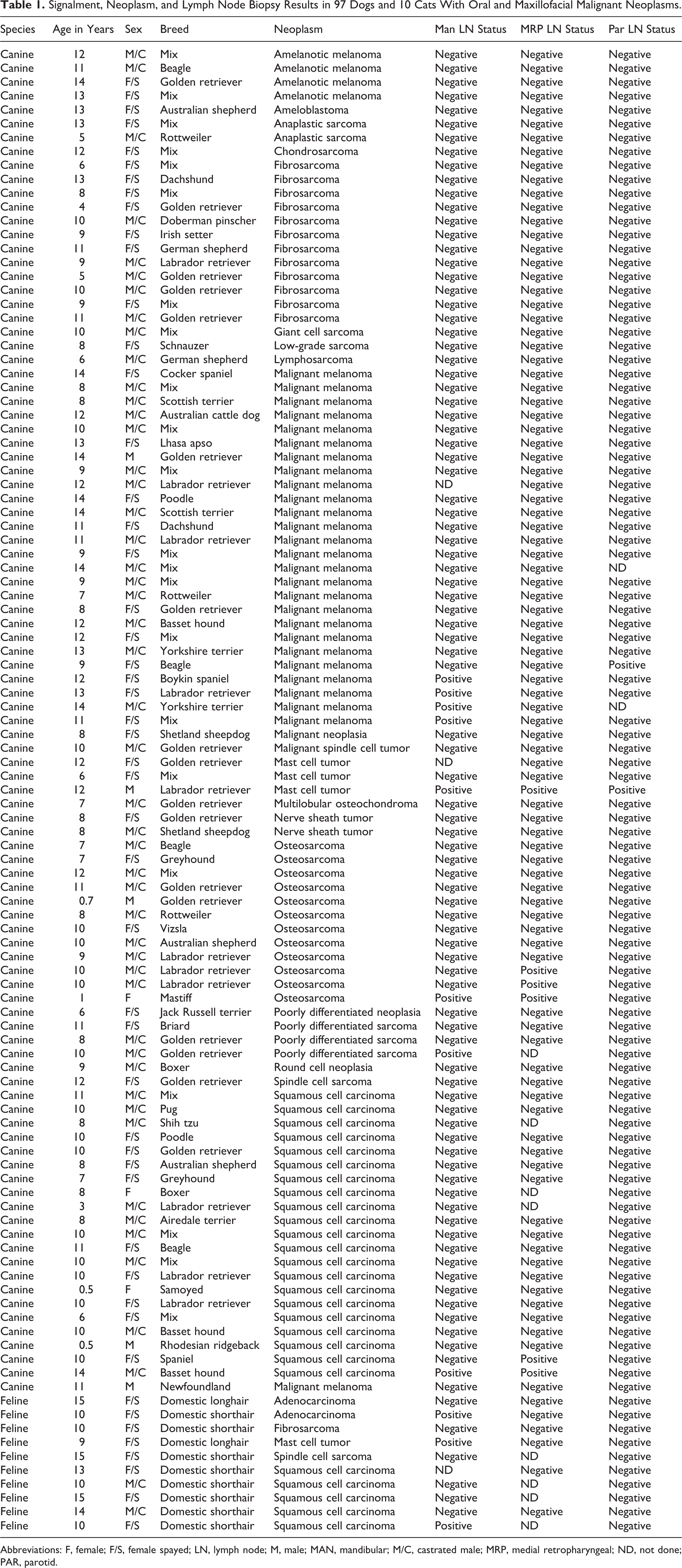
Abbreviations: F, female; F/S, female spayed; LN, lymph node; M, male; MAN, mandibular; M/C, castrated male; MRP, medial retropharyngeal; ND, not done; PAR, parotid.
Neoplasms that affected only the mandibular lymphocentrum with metastatic disease were malignant melanoma in 4 cases, mast cell tumor in 1 case, adenocarcinoma in 1 case, squamous cell carcinoma in 1 case, and poorly differentiated sarcoma in 1 case. There were 2 neoplasms that affected the medial retropharyngeal and mandibular lymphocentrums only, and these were squamous cell carcinoma and osteosarcoma with 1 case each. One case with a mast cell tumor had affected all 3 regional lymphocentrums with metastatic disease. The medial retropharyngeal lymphocentrum was the only node affected by squamous cell carcinoma in 1 case and osteosarcoma in 2 cases. The parotid lymphocentrum was the only node affected by malignant melanoma in 1 case.
Discussion
This study reports a substantially lower incidence (14.0%) of regional metastatic disease from oral and maxillofacial neoplasms compared with a previous study (35.5%). 8 Both studies had the same case selection criteria and single sectioning for each lymph node. However, in the previous study, 15 random negative lymph nodes based on a single section were reevaluated with 5 additional sections, yielding a false negative in 1 lymph node for an incidence of 6.7%. 8 In the study currently presented, only lymph nodes that appeared abnormal grossly underwent serial sectioning. Although the pathologists were different in each study, all were experienced and board certified. Although it has been shown that serial sectioning of suspect lymph nodes yields less false negatives for metastasis compared with single sectioning, no study has shown the difference to be enough to explain the disparity of metastatic disease found in the study reported here and in the previous work. 12 –14
Retrospective clinical studies in veterinary medicine, unlike those in human medicine, often lack a substantial number of cases that would contribute to the power and reliability of the results, as in the case of the previous study. In mathematics, the law of large numbers is a principal of probability that states the larger the sample size, the more likely it is to accurately represent the true population. 15 All too often, retrospective studies with limited case participation become engrained in the veterinary literature without further investigative scrutiny. Therefore, the results reported here should be considered to more accurately reflect regional lymph node metastasis for oral and maxillofacial neoplasms in dogs and cats. However, based on the same study design and single section methodology that most likely reflects histopathologic standards for commercial and academic laboratories, it is interesting to note the combined results of both studies when at least 2 lymphocentrums were biopsied. The metastatic incidence to regional lymph nodes (23/134) was 17.2%. Analysis of the combined data further supports the result reported here, providing the clinician with a reference for lymph node metastasis incidence of approximately 15%.
The parotid, mandibular, and medial retropharyngeal lymphocentrums each receive afferent lymphatics from particular regions, with some areas of overlap, including the possibility of contralateral metastasis via lymphatic anastomotic connections. 16 Therefore, metastatic disease may be diagnosed in an unexpected lymphocentrum (eg, a positive mandibular lymph node for a malignant maxillary neoplasm), as shown in both a previous study and this current study. 8 Although the mandibular lymphocentrum is the primary lymphocentrum of the head available for clinical, cytological, and surgical evaluation, the presence of metastasis from oral and maxillofacial neoplasms may not be detected if only this lymphocentrum is evaluated. 8 Since only ipsilateral major regional lymph nodes were evaluated, the prevalence of bilateral regional lymph node metastasis was not assessed in the current study.
In the current study, 4 (26.7%) of 15 cases with positive lymph nodes had no evidence of metastatic disease in the mandibular lymphocentrum. A previous study reported a 45.5% incidence of regional metastasis with a negative mandibular lymphocentrum biopsy. 8 When considering both studies, the metastatic incidence to regional lymph nodes (9/23) with a negative mandibular lymphocentrum was 39.1%. This includes cases which had at least 2 lymphocentrums biopsied, with one of them being the mandibular lymphocentrum. Analysis of the combined data provides the clinician with a reference for false-negative incidence of regional lymph node metastasis of approximately 40% if only the mandibular lymphocentrum is evaluated.
Other methods for determining metastasis to oral and maxillofacial regional lymph nodes include FNA, palpation, and computed tomography (CT). Palpation has been shown to be an unreliable indicator of metastatic disease in general, and especially in these regional lymphocentrums, since only lymph node(s) of the mandibular lymphocentrum may be reliably palpated. 8,17,18 Fine needle aspiration has been reported to be an accurate and minimally invasive method for determining metastatic disease in lymph nodes. 18 However, more recent studies have shown FNA to be less reliable in assessing metastasis, with one study reporting 77.2% accuracy in dogs and cats and another reporting an incidence of 72.8% and 85.9% diagnostic samples in dogs and cats, respectively. 19,20 The use of FNA in staging oral and maxillofacial neoplasia is limited by the regional anatomy, since only the mandibular lymphocentrum is accessible without ultrasound or CT imaging guidance, which necessitates advanced training and equipment. Depending on the level of training and experience, radiologists may be reluctant to perform ultrasound-guided needle aspiration/biopsy of the medial retropharyngeal lymph node based on the concern for iatrogenic trauma to structures coursing along its medial surface, including the terminal portion of the common carotid artery; the hypoglossal, vagus, and sympathetic nerves; and the internal jugular vein. Fine needle aspiration may document the majority of cases with oral and maxillofacial metastatic disease based on assessment of the mandibular regional lymphocentrum; however as reported here, 26.7% of cases will yield a false negative for metastatic disease if only the mandibular lymphocentrum is evaluated.
Although CT is a valuable modality in imaging pulmonary nodules associated with distant metastasis, it is only of moderate value when attempting to diagnose regional metastasis to lymphocentrums. 21 Computed tomography is able to detect abnormally large or necrotic lymph nodes, both of which are signs indicative of metastasis. However, lymph nodes found to be borderline in size without necrosis or have extracapsular spread cannot be differentiated from reactive or normal lymph nodes using CT. 21 Histology would still be required for confirmation of lymph node status in many cases with suggestive CT findings.
Another surgical technique has been described to biopsy the mandibular and medial retropharyngeal lymphocentrums bilaterally through a single midline incision. 11 However, this technique does not provide surgical access to the parotid lymphocentrum, which can be the only area affected by regional metastatic disease as reported here and in the previous study. 8 As reported in the human literature, well-lateralized malignant neoplasms are predisposed to ipsilateral lymph node metastasis, and although midline neoplasms can have bilateral metastatic lymph node disease, there is a higher incidence of metastatic disease affecting the side where the bulk of the neoplasm is located. 3 As in the previous study and this current study, excisional biopsy of the parotid, mandibular, and medial retropharyngeal lymphocentrums was performed ipsilateral to the side of origin or dominant side of the neoplasm, allowing evaluation of all 3 lymphocentrums. Bilateral excisional biopsy of all 3 lymphocentrums is recommended for true midline lesions. Our rationale for biopsy on the side of origin, even if the neoplasm encroached upon the midline, was the ipsilateral side of origin would be most likely indicative of metastatic disease, and a positive yield would warrant a similar adjunctive treatment plan.
A wide variety of dogs were included in this study, making it difficult to draw any substantial conclusions on the prevalence of oral tumors based on breed. The most prevalent pure breed represented was the golden retriever dog that accounted for 17.5% of the canine cases (n = 17). This is consistent with a study that found an increased risk for neoplasms, in general, in the Dutch golden retriever population. 22
All of the neoplasms reported in this study were malignant. The 4 most common neoplasms in dogs reported here were malignant melanoma 26.8% (n = 26), squamous cell carcinoma 21.6% (n = 21), fibrosarcoma 12.4% (n = 12), and osteosarcoma 12.4% (n = 12). These neoplasms and their rank in reported occurrence are consistent with current literature, with the exception of osteosarcoma. 23 Osteosarcoma is often reported to be less common than fibrosarcoma, but was found to be reported at the same rate in this study. The neoplasms of cats reported here were also consistent with current literature, with squamous cell carcinoma most commonly diagnosed. 23 –25
In this study, malignant oral and maxillofacial neoplasms that had metastatic disease to the regional lymphocentrums were malignant melanoma (n = 5), squamous cell carcinoma (n = 3), osteosarcoma (n = 3), mast cell tumor (n = 2), adenocarcinoma (n = 1), and poorly differentiated sarcoma (n = 1). Not surprisingly, these types of neoplasms would warrant particular evaluation of regional lymphocentrums for metastasis.
Cases selected for biopsy of the regional lymphocentrums were patients with oral and maxillofacial neoplasms who had been confirmed to be malignant based on previous biopsy or had clinical features suspicious of malignancy. These features included radiographic bone involvement, radiographic tooth destruction, and history of rapid growth. Because the definition of malignant neoplasms includes the ability to metastasize, it was appropriate to perform regional lymph node biopsy in patients with lesions, showing these characteristic signs of malignancy to rule out metastasis. 21 The treatment plan for excisional biopsy for the neoplasm and lymph nodes of suspect malignant lesions was efficient and cost-effective for owners seeking curative intent surgery despite not knowing the specific histopathologic diagnosis of the lesion. The surgical principal of obtaining a diagnosis before treatment is still the gold standard, and performing a biopsy of any tumor for histopathologic evaluation before curative surgery is justified. Distant metastasis was not noted by the referring veterinarians when these cases presented, but diagnostics to confirm this were not performed by the operating surgeon prior to the procedures.
In conclusion, the incidence of metastasis of malignant oral and maxillofacial neoplasms to the mandibular, parotid, and medial retropharyngeal lymphocentrums, albeit clinically relevant, was lower than reported previously. 8 Assessing only the mandibular lymphocentrum by FNA, although being high yield for that lymphocentrum, may be associated with a false-negative result for regional metastasis. Likewise, excisional biopsy of only the mandibular and medial retropharyngeal lymphocentrums may also be associated with a false-negative result for regional metastasis since the parotid lymphocentrum is not evaluated. The cases reviewed here confirm that excisional biopsy and histopathologic evaluation of all 3 ipsilateral regional lymphocentrums is an efficient and thorough method to determine regional metastasis of oral and maxillofacial neoplasms in dogs and cats.
Footnotes
Declaration of Conflicting Interests
The author(s) declared no potential conflicts of interest with respect to the research, authorship, and/or publication of this article.
Funding
The author(s) received no financial support for the research, authorship, and/or publication of this article.
