






Abstract
A total of 297 dogs between 8 and 12 weeks of age were examined during a 1-year period to evaluate occlusion. Dogs were categorized either as individual dogs or members of a litter and purebred or mixed breed. Occlusion was evaluated by class: normal, class 1 malocclusion (MAL1), class 2 malocclusion (MAL2), or class 3 malocclusion (MAL3). Dogs with MAL3 were also subdivided based on whether MAL3 was considered a breed standard; dogs with breed standard MAL3 were considered under normal occlusion for statistical analyses. Malocclusions were further categorized as mild, moderate, or severe. Twenty-six percent (77/297) were identified as having a malocclusion. For single dogs, purebreds had a significantly higher percentage of malocclusions compared to the mixed breeds (33.8% and 20% respectively; P = .042). For dogs in litters, there were no purebreds with malocclusion, which was significantly less than the number of mixed breeds with malocclusions (0% and 23.5%, respectively; P = .0023). No significant difference in prevalence was noted between mixed breed and purebred dogs. Occlusal evaluation is important for all dogs to allow for early recognition of malocclusion and, if necessary, intervention in a timely manner.
Introduction
Malocclusions in dogs are a common and potentially serious problem, and yet there is no published data on the prevalence of malocclusions in the deciduous teeth of dogs. In humans, the importance of identification and treatment of malocclusion in deciduous teeth is increasingly emphasized because of the relationship between malocclusions of deciduous and permanent teeth. 1 -3 Early recognition of malocclusions allows for early intervention, which has been shown to lead to more successful outcomes in humans. 4 -6 In dogs, traumatic malocclusions are caused by abnormal contact between the teeth and/or soft tissues of the mouth, resulting in pain, behavioral problems, and, in severe cases, occasional difficulty in eating. Recognition of deciduous dentition malocclusions in dogs would allow for earlier intervention to alleviate these problems. Numerous publications have described recognition and treatment of malocclusions in deciduous and permanent teeth of dogs, most of them involving purebred dogs. 7 -21 However, mixed breed dogs are a large portion of the pet dog population, with a study in the United Kingdom showing more than 35% of dogs as mixed breed. 22 There have also been several publications addressing the prevalence of congenital/genetically linked malocclusions of purebred dogs. 23 -27 None of these publications specifically address the presence of deciduous dentition malocclusions, and none have compared the prevalence of malocclusion in purebred and mixed breed dogs. The main objective of the study was to determine the incidence of deciduous dentition malocclusions in dogs. Additional objectives included recording the incidence and type of malocclusion for different breeds and determining if purebred dogs were more likely to develop malocclusions than mixed breed dogs.
Materials and Methods
All dogs included in the study were treated humanely and in accordance with state and national animal welfare laws and ethical standards. Owners gave informed consent for release of medical findings for publication. Dogs between the ages of 8 and 12 weeks admitted for a routine health examination at a private practice between June 1, 2014, and June 1, 2015, were included in this exploratory descriptive study. Healthy dogs with only deciduous teeth were included in the study. The age and the breed status were determined by examining paperwork provided by owners upon presentation. Only dogs with confirmed purebred heritage were included in the purebred category. Recognized American Kennel Club (AKC) breeds 28 were the only acceptable breeds for purebred status. Dogs with unknown heritage or whose breed was guessed based on appearance were included in the mixed breed category. When an exact birth date was not available, the examining veterinarian estimated age based on oral examination and dentition present. Dogs were excluded from the study if they had any maxillofacial deformities other than occlusal abnormalities. Anatomical differences associated with brachycephalic conformation were not considered exclusion criteria. Congenital (ie, clefts) and trauma-induced abnormalities were excluded. Additional exclusion criteria included presence of systemic illness either at the time of the first examination or prior to the first examination. Dogs were examined only once during the study period.
All dogs included in the study had complete physical and oral examinations. All data were collected by the principal investigator (N.H.) and the 6 doctors working at the private practice, after being trained by the principal investigator. Data collected from each patient included name or identification number, breed, purebred or mixed breed status, date of examination, weight at examination, occlusion, and degree of malocclusion if present. Dogs were categorized as being a single puppy or as being examined as part of a litter.
The principle investigator trained the additional 6 participating veterinarians in malocclusion recognition, categorization, and severity determination via measurement in order to standardize observations. All received training directly from the principal investigator prior to data acquisition. Occlusion and malocclusion standards of the American Veterinary Dental College (AVDC) were used for categorization. 29
Occlusion were categorized as normal occlusion (Figure 1) or malocclusion. 29 Malocclusions were recognized as neutroclusion (MAL1; Figure 2), mandibular distoclusion (MAL2; Figure 3), or mandibular mesioclusion (MAL3; Figure 4). 29 For MAL1, the direction of individual tooth malocclusion was recorded according to accepted AVDC nomenclature. All observers utilized these guidelines to categorize occlusion for all dogs.

Normal occlusion in an 8-week-old puppy. Interdigitation of the upper and lower canines and incisors. Maxillary incisor teeth are positioned rostral to the mandibular incisor teeth. The mandibular canine approximately bisects the interdental space between the maxillary third incisor and the maxillary canine.

Class 1 malocclusion (MAL1) of a 9-week-old puppy. A normal rostrocaudal relationship of the maxillary and mandibular arches with malpositioning of the left mandibular canine tooth. In these puppies, the left mandibular canine is angled in a lingual direction.

Class 2 malocclusion (MAL2) in a 9-week-old puppy. An abnormal rostrocaudal relationship between the dental arches, in which the mandibular arch is caudal to the maxillary arch.

Class 3 malocclusion (MAL3) in a young dog with mixed dentition deemed too old for inclusion in the current study. MAL3 results in abnormal rostrocaudal relationship between dental arches in which the mandibular arch is rostral to the maxillary arch. Green fabric from a plush toy is seen trapped between tooth 204 and tooth 604.
If a malocclusion was identified, an endodontic ruler was used to assess the severity (Figure 5). In the case of an MAL1, the ruler was used to measure the number of millimeters that a tooth was displaced from its expected location. If an MAL2 or MAL3 was found, the ruler was used to measure the distance (in millimeters) from the rostral aspect of the cusp tip of the mandibular first incisor teeth to the rostral aspect of the cusp tip of the maxillary first incisor teeth. Irregularities of the rostral mandibular and/or incisive bones in the dorsoventral plane were not recorded. Both MAL2 and MAL3 were categorized as mild if there was <5 mm of difference between the length of the maxilla and the mandible as measured at the level of the cusp tips of the first incisors. The categorization of mild was also used for MAL1 if there was <5 mm alteration in a tooth location in any plane. MAL1-3 was categorized as moderate if there was a 5- to 10-mm difference between the length of the maxilla and mandible at the level of the cusps of the first incisors or alteration in tooth location. Finally, malocclusions were categorized as severe if there was a >10 mm difference between the length of the maxilla and mandible or alteration in the tooth location.

Use of an endodontic ruler to measure the space between the cusp of the maxillary first incisor and the mandibular first incisor in a 10-week-old puppy with class 2 malocclusion (MAL2).
Dogs with MAL3 were divided into 2 groups based on whether the malocclusion is a breed standard according to AKC published guidelines. 28 Statistical analyses were completed for AKC-accepted MAL3 in both the normal and the abnormal category. MAL3 that were not accepted as a normal breed standard were included under MAL3 for statistical analyses.
Summary statistics were used to describe the overall study findings. Further analysis investigating variable relationships were evaluated using R software.a P values of <.05 were considered significant. Analysis of the individual dogs was performed via χ2 test in order to compare proportions. When analysis included litters, a mixed effects logistic regression was performed using the lme4 package.b
Results
A total of 297 dogs were examined during the investigative period. A total of 226 individual dogs and 71 dogs as a part of a litter were examined. Of the 297 dogs examined, 77 (25.9%) were identified as having malocclusions. MAL3 was the most common malocclusion identified, with 52 (17.5%) affected individuals. There were 9 purebred litters and 5 mixed breed litters examined. The distribution of breeds examined is shown in Table 1, along with whether MAL3 is a part of their breed standard. The distribution of malocclusions among dogs is shown in Table 2. Table 3 shows the degree of severity associated with the categories of malocclusions.
Numbers of American Kennel Club Recognized Dog Breeds That Were Examined.a
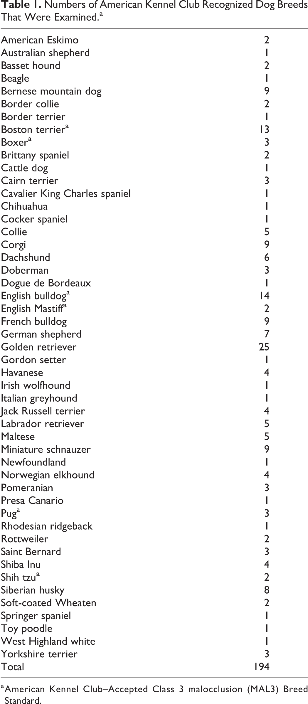
a American Kennel Club–Accepted Class 3 malocclusion (MAL3) Breed Standard.
Table of Counts (Percentages) in Each of the Occlusion Categories According to Breed Categorization.

Abbreviations: MB Single, mixed breed single; PB Single, purebred single; PB litter, purebred litter, MB litter, mixed breed litter; AKC, American Kennel Club.
Table of Counts (Percentage) Within Each Malocclusion Category According to the Degree of Severity.

Abbreviation: AKC, American Kennel Club.
In this study, the most common MAL1 recognized involved linguoversion of one or both of the deciduous mandibular canine teeth (11/12; 92%). There was a single dog with a unilateral caudal crossbite identified in this study. According to the AVDC classification system, caudal crossbite is considered an MAL1. 29
In the group where all MAL3 were considered malocclusions, there was a significant difference between the percentage of purebred dogs (47/130; 33.8%) compared to the mixed breed dogs (17/86; 20%) with malocclusions (P = .042). When only litters were considered for MAL3 abnormal standard, there was no significant difference between the percentage of purebred dogs (9/55; 16.4%) compared to the mixed breed dogs (4/17; 23.5%) with malocclusions (P = .854). In analysis combining the singles and litters, there was no significant difference between the percentage of purebred dogs (56/194; 28.8%) compared to the percentage of mixed breed dogs (21/103; 20.4%) with malocclusion (mixed logistic P value = .673).
In the group where AKC breed standard MAL3 were statistically evaluated within the normal category, for individual dogs, there was no significant difference between the percentage of purebred dogs (21/139; 15%) compared to the percentage of mixed breed dogs (17/86; 20%) with malocclusions (P value = .916).
When only litters were considered under the AKC-accepted MAL3 group, there was a significant difference between the percentage of purebred dogs (0/55; 0%) versus the percentage of mixed breed dogs (4/17; 23.5%) with malocclusions (Fisher exact test P = .0023). In analysis combining individuals and litters, there were no significant differences between the percentage of purebred dogs (21/194; 10.8%) versus the percentage of mixed breed dogs (21/103; 16.5%) with malocclusions (mixed logistic P = .828).
Discussion
When considering individual dogs with the AKC breed standard MAL3 group only, there were significantly more purebred dogs that presented with malocclusions than mixed breed dogs. When considering litter dogs with MAL3 that is not an AKC breed standard, there were significantly more mixed breed dogs with malocclusion than purebred dogs. The remainder of the analysis found no significant difference between the prevalence of malocclusion of mixed and purebred dogs.
Although this exploratory, descriptive study reports accurately the numbers of dogs observed with deciduous dentition malocclusion, there were several limitations within the study design. First, multiple observers were used to collect study data, which introduces some degree of variability in malocclusion recognition and severity measurements despite universal training. While it would be ideal to have one principal investigator compiling all the data, the number of dogs and the times of their examinations made this impossible. It was felt that the training provided by the principal investigator minimized individual observer variation, and the increased number of dogs evaluated via the additional observers added significantly to the study power, outweighing any benefit of limiting data collection to one observer. Even with multiple observers, the second limitation of the study was the small sample size for litter dogs. Because litter dogs are related and malocclusions can be inherited conditions, more litters would be needed to investigate more thoroughly the prevalence of mixed breed and purebred deciduous dentition malocclusions. The reason for performing separate evaluations of the individual dogs and dogs that were a part of litters is that an assumption of the χ2 test is that each individual observation is statistically independent. However, siblings from the same litter cannot be considered independent observations.
An additional limitation was that the distribution of dog breeds evaluated may have been skewed. Data collection was performed in a private practice with a board-certified theriogenologist on staff. Because of the challenging reproductive physiology of some brachycephalic dogs, more of these patients and their resultant dogs may have been included in the study.
When examining the severity of the malocclusion, it would have been ideal to measure the severity of the malocclusion compared to the length of the lower jaw. Because these measurements were not obtained at the time of subsequent examination, it was not possible to add them retroactively to data collection because of puppy growth.
As reported in this article, not all MAL3 are considered an abnormality by the AKC. The AVDC identifies MAL3 as a skeletal abnormality, but this characteristic is a sought-after facial characteristic for many brachycephalic dog breeds. When examining breed standards defined by the AKC 28 for the brachycephalic dogs evaluated in this population, the breed standard for bulldogs, Boston terriers, mastiffs, pugs, shih tzus, and boxers outlines that MAL3 is acceptable. Interestingly, none of the boxers or shih tzus evaluated in this population had an MAL3. There was 1 Boston terrier puppy that had not yet developed an MAL3. Also, while French bulldogs are commonly recognized as having MAL3, there is no mention of that in their breed standard.
The lack of an MAL3 in some dogs where it is expected and the overall small number of severe malocclusions may be related to the stage of development at which the dogs in the study were examined. This study did not evaluate the effect of aging on occlusion. It has been reported that mild malocclusions can resolve or worsen over time with continued maxillomandibular growth. It would be interesting to evaluate a larger number of dogs at multiple time points during skeletal maturation to determine how occlusion/malocclusion changes during growth and whether the presence of a malocclusion in the deciduous dentition correlates with malocclusion in the permanent dentition.
The bones of the maxilla and the mandible result from development of the first branchial arch, which are organizations of migrating neural crest cells in the developing embryo. 30,31 In animals, this formation is completed during embryogenesis. It was not possible to evaluate the dogs in this study at birth, but it would be interesting to know if skeletal malocclusions were present prior to the existence of dentition. Extensive research on cleft palates and lips has shown that in dogs, the major portion of maxillofacial growth occurs between week 8 and week 36. 32,33 Given the rapid rate of facial development between 8 and 12 weeks, malocclusion during this age range would have potential to be dynamic. Because the dogs in this study were examined only between 8 and 12 weeks, the number of adult malocclusions may be underestimated.
Skull growth is primarily due to intramembranous ossification, which is the direct formation of bony matrix within existing connective tissues. 31,34,35 Growth in the rostral mandible occurs very early in development, but by approximately day 50 (∼week 7), all growth is from caudal to the first molar tooth, in the area of the condyle. Maxillary growth occurs at the incisivomaxillary suture and at the palatal–maxillary suture. The contribution of the length of the palatal bones decreases with age, and the comparative length of the incisive and maxillary bone increases with age. In brachycephalic dogs, disparity of the maxilla and the mandible primarily occurs because of the chondrodystrophic arrest of the bones of the maxilla. 34 The growth of the maxilla and mandible is separately regulated but must be congruous in order for normal occlusion to result. Normal dental interlock of the canine teeth, as present in patients with normal occlusion, plays a critical role in this. 24,25,34 As the maxilla and mandible grow individually, interlock of the teeth prevents one from growing at a vastly different rate from the other. Based on the mild degree of malocclusion seen in virtually all of the AKC breed standard MAL3 dogs seen in this study, that disparity most likely develops at some point between the 8- and 12-week examination window and facial maturity. 32,33 While there is a heritable component of skeletal malocclusions that contribute to a breed’s typical appearance, there is remarkably little published data to support this. 14,25 -27
In conclusion, purebred and mixed breed dogs had similar rates of malocclusion associated with their deciduous teeth. While malocclusions are not common in deciduous dentition, they occur frequently enough that a thorough examination of the occlusion of every puppy is critical when presented for wellness examinations.
Footnotes
Acknowledgments
We could not have completed this study without the help of Ann Hess and Frank Marrs from the Department of Statistical Sciences at Colorado State University.
Declaration of Conflicting Interests
The author(s) declared no potential conflicts of interest with respect to the research, authorship, and/or publication of this article.
Funding
The author(s) received no financial support for the research, authorship, and/or publication of this article.
Materials
R Core Team. R:A Language and Environment for Statistical Computing. R Foundation for Statistical Computing. Vienna, Austria. Bales D, Machler M, Bolker B, Walker S. Fitting linear mixed-effects models using lme4. J Statis Soft. 2015;67(1):1-48.