






Abstract
Tusk fracture in elephants is a common incident often resulting in pulp exposure and pulpitis. Extensive lavage, endodontic therapy, direct pulp capping, or extraction are treatment options. In this report, the successful management of a broken tusk of a juvenile male Asian elephant (Elephas maximus) including morphological analysis of the tusk tip 2 years after surgery are presented. Treatment was carried out under barn conditions and included antimicrobial photodynamic therapy and partial pulpotomy with direct pulp capping. Immediate pain relief was reached. The fractured tusk was preserved and continued to grow. The therapeutic filling material remained intact for over 1 year but was absent 2 years after treatment. The former pulp cavity of the tusk tip was filled with reparative dentin, osteodentin, and bone, but the seal between these hard tissues and pulp chamber dentin was incomplete. Radiographs obtained 3 years after treatment showed no differences in pulp shape, pulp width, and secondary dentin formation between the treated right and the healthy left tusk. It can be concluded that in case of an emergency, the endodontic therapy of a broken elephant tusk can be attempted under improvised conditions with adequate success. Photodynamic therapy might contribute to prevent infection and inflammation of the pulp. The decision tree published by Steenkamp (2019) provides a valuable tool to make quick decisions regarding a suitable therapy of broken tusks.
Keywords
Introduction
Fractures of the tusk accompanied by pulp exposure are quite common in elephants. They are reported to occur more frequently in animals held in captivity. Causes of trauma are falling, fighting, and damage due to environmental structures.1,2 Tusks are known to have a good self-healing capability managing fissures and smaller fractures by production of new dentin sealing the pulp cavity.3,4
However, tusk fractures with pulp exposure need to be treated.2,5 The paramount aim of the veterinary dentist should always be pain management and if possible the preservation of the tusk. In the past, tusk fractures with exposed pulp were often treated by extensive lavage with different solutions such as sterile saline or hydrogen peroxide solution (3%), partial amputation of necrotic pulp, and capping of the remaining pulp. In many of these cases, the tooth dies nonetheless and has to be extracted.2,5
Unfortunately, only a few publications are available on the successful and effective treatment of tusk fractures in elephants.2,4,6,7 In a recent work, different approaches to treat tusk fractures with exposed pulp that have been used by veterinary dentists in the past were reviewed. 8 The outcome varies from continued tusk growth as a sign of a vital pulp, to lost fillings and/or to severe and painful pulpitis and the subsequent necessity for extraction.
In this report, the successful management of a broken tusk of a juvenile male Asian elephant (Elephas maximus) and the subsequent thorough morphological documentation of the therapeutic outcome are presented. With prompt treatment of the vital tooth (including photodynamic therapy [PDT] for a pronounced antimicrobial effect 9 and the execution of a partial pulpotomy with subsequent direct seal of the pulp cavity) immediate pain relief could be reached. Investigation of the tusk tip revealed massive dentinal bridge formation. Preservation of the fractured tusk and its continuous growth were accomplished.
Material and Methods
Directional terms for description of procedures and findings were used as follows: “incisal” signifying the direction towards the tusk tip and “apical” for the direction towards apex radicis dentis. 10
Animal and Surgery
The patient was a juvenile (*2011) male Asian elephant (Elephas maximus) living in the Zoo Hellabrunn in Munich (Germany). At the age of 3.5 years, after turmoil in the herd, the bull was found with a fractured right tusk. The pulp was prolapsed and bleeding. The immediate treatment was pain medication with meloxicama (15 mg/mL, dosage 0.2 mg/kg body weight) 1 × daily per os until 2 days postoperatively and antibiotic treatment with ciprofloxacin (5 mg/kg)b 2 × daily per os until 4 days postoperatively.
Anesthesia
The young male elephant (body weight ∼1500 kg) was kept with his mother in a herd of 5 animals. It was not possible to separate him from other elephants before the induction phase of anesthesia without causing too much stress. The initial dosage for the standing sedation was 0.017 mg/kg detomidine hydrochloridec and 0.027 mg/kg butorphanold in combination. The dosage was injected intramuscularly using an anesthesia blow pipe.e For the dental procedure it was necessary to have the animal in lateral recumbency. Therefore, etorphinef (0.023 mg/kg) was administered intramuscularly after the induction phase to sedate the animal. The induction time until the start of the surgery took around 50 minutes (including the positioning and preparation of the animal for the operation under barn conditions). Two intravenous cathetersg were placed in the dorsal ear vein and the vena cephalica cranially at the forelimb and fixed with tape. A permanent drip infusion was installed. The flow rate was 500 ml/h glucoseh and 1200 ml/h physiological saline solution.i Anesthesia was maintained by occasional intravenous administration of ketamine hydrochloride.j A total dosage of 0.4 mg/kg body weight ketamine hydrochloride was administered as fragmented bolus of 100 mg ketamine hydrochloride over a time interval of 75 minutes. During the anesthesia, oxygen (flow rate 6-8 l/minutes) was administered via a flexible aspiration tube (length 150 cm) in the trunk. The oxygen saturation, monitored with a portable pulse oximeterk (the sensor being positioned in the trunk) remained steady between 96% and 100% throughout the entire procedure. The heart rate was between 44 and 47 beats per minute. After a total time of 135 minutes the anesthesia was reversed by naltrexonel (dosage: 50 mg naltrexone hydrochloride per 1 mg etorphine intravenously) and atipamezolem (dosage: 1.3 mg atipamezole per 1 mg detomidine hydrochloride) injected intramuscularly. The recovery from anesthesia was very fast. The elephant positioned himself in sternal recumbency within 2 minutes. After 5 minutes he was standing up at first attempt.
Tusk Fracture Treatment
The tusk fracture was treated within 30 h after injury. The visible part of the tusk surface, the fractured surface, and the pulp were rinsed 3 times with chlorhexidine.n Then the pulp chamber was rinsed with saline solution.i Due to technical problems, the dental radiographs were of very poor quality and therefore nondiagnostic. However, a visual evaluation was possible, since the depth of the open pulp chamber was only 30 mm. After superficial anesthesia of the pulp using a lidocaine/prilocaine gel,o ∼5 mm of healthy pulp was removed with a sterile surgical blade (size 15). Hemostasis was achieved with epinephrine (1 mg/ml).p The sharp edges of the fractured tusk were smoothed and a 15 mm deep undercut was prepared for pulp capping. For this procedure a hand drillq and a sterilized sheet drill bitr were used.
Local disinfection and wound healing was supported by antimicrobial PDT. The photosensitizer phenothiazine chloride (1%)s was applied to the pulp chamber and the distal fractured surface of the tusk. After 4 minutes of contact time the area was rinsed with saline solution.i Afterwards, low-level laser light with a wavelength of 660 nmt was applied for 60 s per site. This procedure was repeated until laser light had been applied to the complete pulp chamber.
The vital pulp was covered with a collagen sponge containing gentamicinu and capped with zinc phosphate cement.v A multipurpose adhesive systemw was used to create a strong bond between the cement and the filling material. First, the etchant was applied to the pulp cavity, so that the zinc phosphate cement, the pulp chamber dentin, and the smoothed fracture surface were covered well. After 20 s, the etchant was removed by flushing with saline solution.i A light-curing bonding material was applied to the etched surface and hardened by UV-lightx for 20 s. A composite materialy was used to fill and seal the pulp cavity. The composite was also hardened by UV-light (3 times 20 s). Excess composite material was removed and the fracture area was sealed with the light-curing bonding system as described before (etching, flushing, and bonding/ hardening). The total dental treatment time was ∼45 minutes. Due to technical issues no radiographs could be obtained at the end of the procedure.
Posttreatment Period and Sample Taking
The elephant recovered quickly and was sent back to the herd within 30 minutes after the procedure. Regarding his fractured and treated right tusk, the animal never showed any signs of discomfort or pain following surgery. The tusk kept on growing and meanwhile the elephant moved from Zoo Hellabrunn (Munich, Germany) to Zoo Heidelberg (Heidelberg, Germany). Two years after treatment, a 55 mm long piece of the tusk tip was removed in its avital part. This sample was used to assess the therapy outcome anatomically and histologically. Radiographs in dorsoventral projection of both tusks of the animal were obtained 3 years after treatment.
Morphological Assessment of the Results of Treatment
Imaging
Prior to further processing, the tusk tip was imaged by microscopic X-ray computed tomography (micro-CT). The micro-CT scanz was acquired at 150 kVp with a HE#5 filter using the 0.4 × detector assembly. Projection images were recorded over a 360° rotation with 15 s exposure time per projection and an angular increment of 0.333° between projections. Virtual cross-sections (XY) were reconstructed by the softwareaa supplied with the scanner. Isotropic voxel size in the reconstructed volume was 25.38 µm. Reconstructed sections were viewed and analyzed using an open source image analysis software.11,12 Sets of XZ slices were cut from the 3D reconstruction of the tusk tip and were scrolled through for a visual exploration of the pulp chamber. In addition, standard digital lateral-medial and dorsal-ventral radiographs were obtained of the isolated tusk tip in order to allow comparison with micro-CT images.
The tusk tip and all surfaces generated by sawing were documented using a standard digital camera before further processing and after polishing with emery paper of increasing grades (240-2500). The pulp chamber dentin and its contact with the materials within the pulp chamber were recorded using a stereomicroscope (reflected light) equipped with a digital camera.
Sampling of the Tusk Tip
Three slices were cut from the sample beginning at the apical end of the tusk tip, using a water-cooled diamond band saw. The slice thicknesses were 3, 5, and 3 mm, respectively. The loss of material between the slices was ∼1 mm due to the set of the saw blade. The remaining part of the sample was cut into halves longitudinally to open the pulp chamber. From both halves, 1 slice each (thickness 2 mm) was cut for further processing.
Histology
From the 3 mm transverse and the 2 mm longitudinal slices, ground sections were prepared as described before. 13 In short, the slices were smoothed and polished with emery paper of increasing grades (240, 800, 1000, 1500, and 2500) on both sides and mounted on microscopic slides with a histological mounting medium soluble in xylene. Micrographs in transmitted and polarized light (linear polarization) as well as differential interference contrast were prepared using an appropriately equipped light microscope.ab Additionally, the complete ground sections were scanned with a slide scanner in transmitted light.ac
Electron microscopy: The 5 mm transverse tusk slice was fixed for 4 days in Lillie's buffered formaldehyde solution 14 at a negative pressure of −5 mmHg (−6,67 mBar) for enhanced penetration of the fixative to stabilize the material before decalcification. The slice was decalcified for 49 days in a formic acid solution (solution A 125 ml distilled water + 125 ml 98%-100% formic acid; solution B 50 g sodium citrate + 250 ml distilled water; mix A and B 1:1 before use). The end point of decalcification was determined by gentle probing of the dentin with a needle. After decalcification, the slice was washed in running tap water for 24 h and trimmed to make slices of <1 mm thickness of the boundary between dentin and the material filling the pulp chamber. The slices were collected in 5% phosphate buffered glutaraldehyde (pH = 7.4) and postfixed in 1% osmium tetroxide in Sörensen’s phosphate buffer. After a wash in Sörensen's buffer, they were dehydrated and routinely embedded in epoxy resin. Semi-thin sections (thickness 0.8 μm) were cut and stained with a 1% toluidine blue solution in 2% di-sodium tetraborate decahydrate. The sections were mounted with a neutral medium soluble in xylene and analyzed and documented immediately after mounting using a light microscope.ab From selected resin embedded samples, ultrathin sections were cut. They were contrasted with lead citrate and uranyl acetate and examined with an electron microscope.ad
Results
Animal
The broken right tusk of the elephant before and after treatment is shown in Figure 1. The tusk remained vital and grew. The animal did not show any signs of discomfort after treatment and used its tusk to manipulate objects within 24 h after treatment. One year after surgery the elephant was moved from Zoo Hellabrunn (Munich, Germany) to Zoo Heidelberg (Heidelberg, Germany). At this point in time, the therapeutic filling material was still in place. Two years postsurgery, at the time of sampling of the tusk tip, the filling material was noted to be lost.

Fractured right tusk of a juvenile male Asian elephant, before (A) and after (B) treatment. Note the fractured tusk with prolapsed pulp (A) and the closed pulp cavity after treatment (B).
The radiographs of the treated tusk obtained 3 years after treatment did not show any apparent differences compared to the healthy, untreated left tusk (Figure 2). The shape and width of the pulp cones were comparable throughout the entire tooth length.
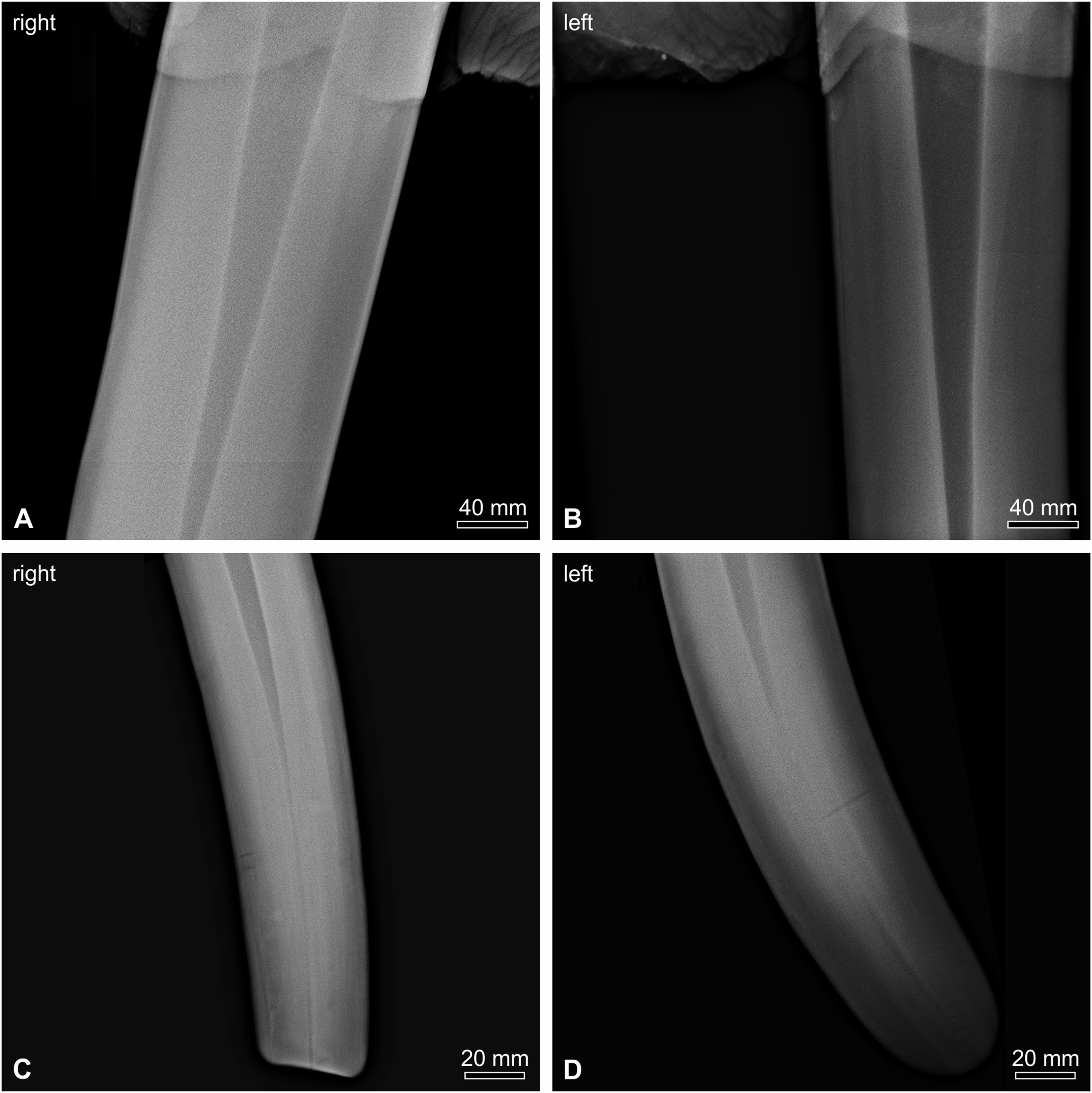
Radiographs in dorsal-ventral projection of the endodontically treated right tusk (A, C) of an Asian elephant 3 years after treatment in comparison to the healthy, untreated left tusk (B,D) of the same animal. A and B show the extraoral part of the respective tusk adjacent to the gum; C and D the incisal part. Pulp width, length, and shape are comparable in the treated and untreated tusk.
Appearance of the Tusk tip 2 Years After Treatment
At the incisal end of the separated tusk tip, an ∼8 mm deep cavity filled with debris was observed (Figure 3A and B). Remnants of the therapeutic capping and filling materials were not detected. The remaining former pulp cavity of the 55 mm long specimen was filled with material of dentin-like radiodensity with a central core visible with the naked eye on physical sections of the tooth (Figure 3A and B) as well as in radiographs (Figure 3C and D). The filling material was in close contact with one side of the pulp cavity wall, whereas an up to 1 mm wide space remained in other parts of the circumference.
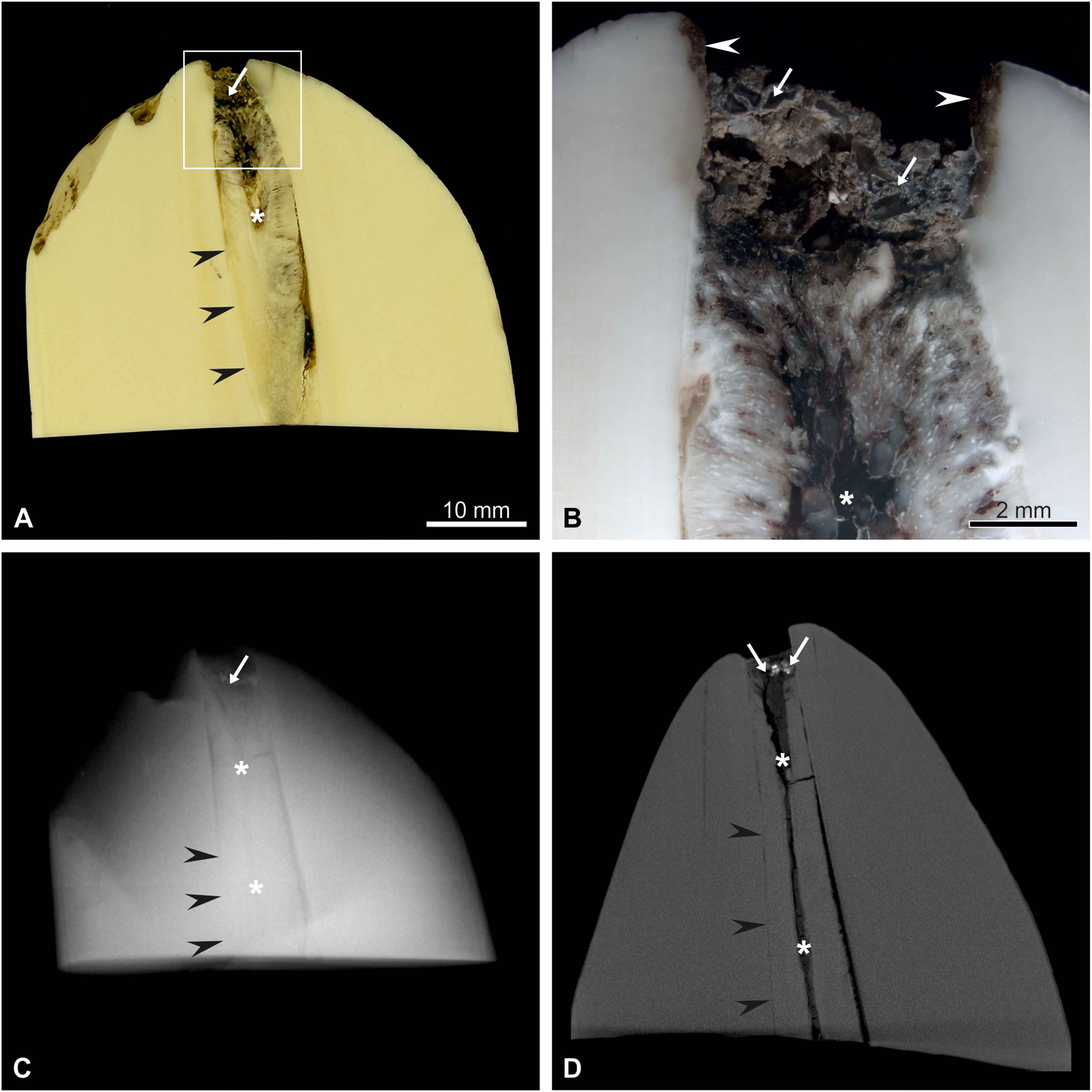
Pulp cavity of an endodontically treated elephant tusk tip. (A) Longitudinal saw-cut section, (B) detail as marked by rectangle in A after polishing; (C) radiograph, and (D) XZ virtual section of a 3D reconstructed micro-CT-series. Note the tight bonding between pulp chamber dentin and material within the pulp chamber along one side of the dental cavity (black arrowheads). Along the larger part of the pulp chamber wall, a distinct gap between dentin and filling material could be detected. Note the narrow cylinder of radiolucent material in the center of the pulp chamber (asterisks). White arrows mark debris accumulated in the incisal part of the pulp chamber, white arrowheads the empty incisal part of the pulp cavity lined with an acellular soft membrane. Remnants of restorative material were absent.
Material Filling the Pulp Chamber of the Treated Tusk tip
None of the materials used for therapeutic pulp capping could be detected in the tusk tip 2 years after treatment. The most incisal part of the former open pulp cavity contained sand and small debris, which was in direct contact with the whitish material filling the main part of the pulp chamber (Figure 3B). The “empty” part of the former pulp cavity was lined with an acellular soft membrane that could be easily removed from the dentin (Figure 3B). A similar membrane lined parts of the pulp cavity where the filling material was not in direct contact with the circumpulpal dentin.
The whitish material filling the main part of the pulp chamber was preferentially arranged radially with some parts showing a globular structure (Figure 4). Histological examination showed that the white material resembled dentin and bone-like dentin (osteodentin) (Figure 5). Cell remnants and a surrounding collagen fiber network could be clearly detected by electron microscopy (Figure 6). The central core of the material filling the pulp cavity had an irregular shape (Figure 4A and B and Figure 7). It contained lamellar bone tissue with osteons, connective tissue with blood vessels and free erythrocytes (Figure 7).

Material filling the pulp cavity of an endodontically treated elephant tusk. (A, B) Surface of transversal sawn sections of the tusk tip 50 and 40 mm from the incisal edge of the tooth; (C, D) details as marked in A. Radially arranged material is marked with black arrowheads, material with a globular appearance with black arrows. Note the central core in the middle of the filling material, the incomplete boundary formation and the amber debris (white arrows) in the slit between dentin and filling material. The clear material in the gap with the bubbles (area between the white arrows in D) is superglue used during processing of the samples.

Structure of the material filling the pulp cavity of an endodontically treated elephant tusk. (A–C) site with globular appearance resembling bone (arrow = osteon-like structure), (D–F) site resembling a composite of osteons-like (arrows) and dentin-like structures (arrowheads), and (G–I) sites resembling dentin. Bone-resembling parts were preferentially found at the periphery and the dentin-resembling parts preferentially near the center of the original pulp chamber. Unstained ground section, transmitted light (A, D, G), polarized light (B, E, H), and differential interference contrast (C, F, I); scale bar = 100 µm.
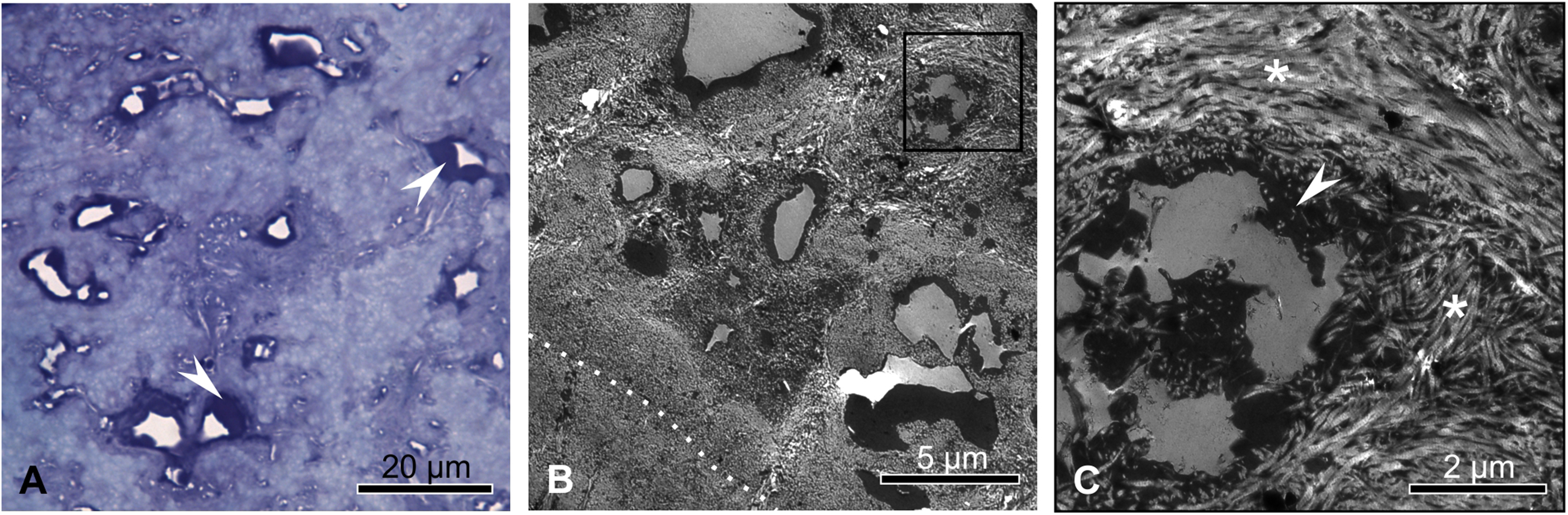
Electron-microscopic structure of the material filling the pulp cavity of an endodontically treated elephant tusk. Semi-thin section viewed by transmitted light microscopy (A) and ultrathin sections viewed by transmission electron microscopy (B, C). In (B), the boundary between pulp chamber dentin and filling material is marked with a white dotted line. The black frame indicates the detail magnification shown in (C). Small cavities with a diameter of approximately 3 to 5 µm contained cell remnants (white arrowheads). Collagen with its typical periodicity in longitudinally sectioned fibres (asterisks) and the electron-dense material decorating these fibres could be found between the cell rests.

Central core (black arrowheads) of the material filling the pulp chamber of an endodontically treated elephant tusk. (A) Ground section viewed by transmitted light microscopy; (B) polished slice viewed in reflected and (C) in transmitted light; (D) ground section viewed by polarized light microscopy and (E) with differential interference contrast. The largest blood vessel is marked with an arrow in all images, a typical osteon with white arrowheads in (B–E). In (C), lacunae for the osteocytes can be seen as rows of black dots, for example, around the marked vessel.
Interface Between Pulp Chamber Dentin and the Material Filling the Pulp Cavity
At the borderline of the pulp chamber, remnants of odontoblast bodies and their processes projecting into the dentin could be found along most of the pulp chamber circumference. Remaining odontoblast bodies did not seem to impair attachment of reparative dentin to the pulp chamber dentin. On the other hand, complete removal of odontoblast bodies did not lead necessarily to complete dentinal bridge formation without gaps between pulp chamber wall and reparative dentin. In the analyzed parts of the tusk tip, detachment of the material filling the pulp chamber was a result of splintering within the reparative hard tissue. It occurred preferentially in richly cellular regions, whereas regions with few cells and a predominance of mineralized collagen fibres seemed to be mechanically more stable (Figure 8).

Morphology of the pulp chamber dentin—reparative dentin interface (white dotted line) in an elephant tusk after endodontic treatment. Pulp chamber dentin is depicted on the left, reparative dentin on the right side. Note the varying degree of cellularization of the reparative dentin (A–C). Black arrows mark lacunae within the reparative dentin. Some of them still contain cell remnants (dark staining). Richly cellularized reparative dentin tended to crack (D, asterisk) so that reparative dentin did not adhere to the pulp chamber wall. White arrowheads mark remaining bodies of odontoblasts. Note the zone of predentin marked by white arrows and the irregular appearance of the mineralization front. Semi-thin sections, staining toluidine blue, scale bar = 20 µm.
Discussion
Tusk Structure: Chances and Challenges for a Veterinary Dentist
Tusks are elodont teeth which grow for the entire life of the animal.10,15 In elodont teeth, the apical foramen of the root tip remains open and is surrounded by structures resembling a tooth germ.3,15,16 Odontoblasts forming the outer layer of the pulp 3 move in a centripetal direction and deposit ivory along their pathway, leaving behind an odontoblast process within the forming dentinal tubules. As a result of this process, the anterior part of the conical pulp chamber fills with dentin. The lengthening of the posterior edge of the tusk coincides with its forward displacement and continuous eruption.15,17 In the human, the deposition rate of secondary dentin ranges between 4 and 8 μm per day. 18 It can be expected that in the elephant this process is even more intensive. From a dentistry point of view, elephant tusks have to be regarded as immature teeth, and appropriate treatment methods have to be chosen. The elodont characteristics of tusks also explain their exceptional healing capability.3,4
Due to its microstructure, ivory is—at least in the naturally occurring directions of load—harder than the dentin of human teeth.19,20 The pronounced microlaminar “plywood” structure of mineralized collagen fibres crossing each other and the wavy course of the dentinal tubules, resulting in the typical Schreger pattern, contribute to the toughness of the ivory,19–21 as well as other factors such as hydration of the tooth. 22 In contrast to human dentin, hypermineralized peritubular (“intratubular”) dentin18,23 does not exist. Instead, mineralized collagen fibres decorated with glycosaminoglycans immediately surround the tubules containing odontoblast processes (own results, not shown). 22 Due to these mechanical properties of ivory, debridement of the pulp chamber dentin in preparation for filling and sealing the pulp cavity can be demanding. In this specific case, the power of the chosen drillq was sufficient.
Choice of Treatment Technique
Tusk fractures with exposed pulp always need to be treated, not just because of pain experienced by the animal, but also because of the risk of pulpitis and further damage caused by sinusitis that can occur as a result of untreated tusk fractures. 8 If the treatment is unsuccessful or fractures carry a poor prognosis, the tooth often has to be extracted.2,5,7 Usually, any dental treatment of elephants requires general anesthesia and specialized dental equipment.2,10 In the case of our patient, decisions as to the treatment of the broken tusk had to be made quickly in order to preserve the vital tusk. Based on the positive experiences with a similar case concerning the father of our patient, a breeding elephant bull, in 2001 (Dr S. Heitland, Munich, Germany; personal communication), a partial pulpectomy and direct pulp capping was performed. This decision was further based on the following facts: (i) treatment of a young animal with fast growing tusks; (ii) no preexisting symptoms; (iii) proven good pulp response especially in tusks 2 ; and (iv) fresh pulp bleeding. The chosen therapy included PDT as a decontamination method, zinc phosphate cement as filling material due to our positive experiences with a similar case as mentioned above, and composite resin for sealing against leakage and intrusion of bacteria. The goal was to cause as little further traumatization of the tooth as possible and to protect the remaining vital pulp. 24 As it turned out, our choice of treatment was in line with a recently published decision making aid 8 , which was not available at the time of surgery.
PDT as an Effective Decontamination Method
Keeping the pulp vital without harming it and eliminating microorganisms effectively to protect the pulp from further infections is regarded as crucial for a successful vital pulp capping as presented in this study. Recently, PDT has been suggested as a promising effective adjunct to standard antimicrobial treatment in endodontics.9,25 In addition to its bactericidal effects, it seems to be able to disrupt biofilms. 9 It has to be taken into account that PDT can affect the bonding of filling and sealing materials to the dentin positively as well as negatively. 26 In contrast to sodium hypochlorite solutions that potentially kill cells residing in the viable pulp, PDT affects fibroblasts and osteoblasts considerably less severely. 27 In the presented case, PDT was used in addition to flushing with chlorhexidine and a sodium chloride solution, mechanical debridement and the use of antibiotics to cover the vital pulp. Since the animal did not show any pain and used its tusk continuously for the manipulation of objects after recovery from anesthesia, it can be concluded that pulp infection and inflammation were successfully prevented. This is a remarkable outcome, considering that the pulp of the broken tusk was exposed for 30 h by the time of surgery.
Filling Procedure
The aim of filling and sealing the treated pulp chamber is to generate a seal against bacterial and chemical leakage to prevent inflammation and pain as well as to promote deposition of reparative dentin and formation of a continuous dentinal bridge covering the pulp cavity.28–31 The seal should remain intact at least until complete dentinal bridge formation. In equine dentistry, where hypsodont teeth, that is, teeth with a prolonged growth period, which are exposed to severe mechanical load are treated, fillings which remain intact for 2 years are regarded as long-lasting. 32 At the time when our patient moved to Zoo Heidelberg (Heidelberg, Germany), the filling material was still in place but absent at the time of sample collection. It can be assumed that the nonshock-durable composite material fell out since the elephant might have used its tusk with higher forces as a growing elephant bull in his new enclosure.
A further requirement for filling and sealing agents for teeth with prolonged (eg, horse cheek teeth) or continuous growth (eg, elephant tusk) is their abrasion resistance—the materials should wear down at the same rate as dentin. 32 The material choice for filling the broken elephant tusk was based on our former good experiences with a similar case. Zinc-phosphate cement was used due to its high compression strength, biocompatibility, and little shrinkage. Moreover, zinc-phosphate cement does not bond to the pulp chamber dentin. Therefore, even if the cement contracted with time, the dental hard tissues would not be stressed, a feature regarded as positive when filling the large pulp chambers of teeth with a prolonged growth period. 32 On the other hand, materials bonding to dentin such as glass ionomers might provide a tighter and more durable seal (see discussion on evaluation of the therapy).
Since zinc-phosphate cement is not hard enough for permanent sealing, the finishing filling and capping material used was composite. It is both durable and able to produce a hermetic seal and thus is the material of choice for a coronal filling.2,4,33 For equine cheek teeth it is suggested not to bond bulk fill and occlusal seal to each other for safety reasons. 32 Should the occlusal seal crumble, there is still the chance that the bulk layer remains intact and prevents bacteria from reaching the pulp. In case of our patient, however, we attempted to reach the tightest bond possible between pulp chamber dentin, filling cement and resin seal by chemical bonding. Nevertheless, none of the materials were present 2 years after treatment of the broken tusk. The small incisal cavity of the tusk was filled with sand and debris instead. However, the seal seems to have remained intact for a sufficient time to prevent pulp infection and to propagate dentinal bridge formation.
Healing
The basic measure of success in tusk dentistry is the continued normal growth of the treated tooth and emergence of normal-looking dentin when the coronal filling material has been worn away, or is lost. 2 In the case of our patient, the reparative dentin visible underneath the debris had an appearance distinctly differing from ivory. In the most incisal part of the tusk (the part taken for histological examination) there was not even a complete seal between pulp chamber dentin and reparative dentin (Figure 3). However, the tusk continued to grow normally and was used by the animal to manipulate objects, signaling a vital pulp and absence of pain.
There is next to no information on the healing processes after direct pulp capping in hypsodont or constantly growing teeth. In brachyodont (human, dog) teeth, after destruction of the odontoblast layer, pulp fibroblasts in the underlying pulp migrate to the destruction site, differentiate into odontoblasts and rapidly deposit first an irregular or disorganized dentin matrix.18,24,31 These fibroblasts are assumed to stem from the cell rich zone underlying the cell-free subodontoblastic zone of human pulp.24,28,34,35 The cell-free zone is missing in the elephant, similar to rats. 3 There is no information regarding a cell-rich zone containing stem cells in the elephant. However, this stem cell zone has been shown in the constantly growing rat incisors. 36 The primary location of cells responsible for dentinal bridge formation in the elephant is unknown. From our histological results, it seems to be clear that odontoblasts themselves do not take part in reparative dentin formation (at least not in initial stages of the process), since in some regions of the dentin-pulp interface of the tusk, odontoblasts, and cells producing reparative dentin are juxtapositioned (Figure 8).
The rich vascularization of the tusk pulp is described to play an important role in mediating an immunological response, forming granulation tissue, producing reparative dentin, and in supporting other mechanisms of defense and repair. 3 A similar role of pulp vascularization in hypsodont equine teeth has been indirectly hypothesized. 37
The comparatively sparse innervation of the incisal parts of tusk pulp 3 might have an interesting impact on healing processes in the treated tooth. In the human, trauma or different substances reaching the pulp cavity can stimulate the release of inflammatory mediators from sensory nerve fibres, boosting the inflammatory processes within the pulp. 24 Fewer nerve fibres in the elephant pulp would release fewer mediators. The tendency for inflammation within the extra-oral part of the tusk pulp might therefore be lower than in animals with brachyodont teeth.
In brachyodont teeth, the clinical outcome of vital pulp capping depends on the successful prevention of bacteria and bacterial products from entering the pulp. 28 It must be possible to control pulpal bleeding within the time frame of ∼5 minutes, otherwise it is a sign that inflammation has spread to radicular pulp (hyperemic pulp) and prognosis is poor.29,38 Uninflamed and exposed dental pulps will form a dentinal bridge within 2 weeks in humans and within 9 days in monkeys. 28 In this case of an endodontically treated tusk, at least 9 cm of reparative hard tissue (new growth within vital tooth + material within tusk tip) filling the original pulp cavity was formed over a period of 3 years postsurgery. In the incisal part of the tusk, which was examined histologically, it was mostly osteodentin, a dentin-like hard tissue where cells become trapped in the matrix during early stage of formation (Figure 5, 6, and ,8). 18 A smaller amount of reparative tissue might be identified as tertiary dentin. It was characterized by radially arranged dentinal tubules which were not continuous with the ivory tubules and had a considerably larger diameter (Figure 4). 31 In the center of the former pulp cavity, bone and some remnants of necrotic tissue were found. The contact between reparative dentin and pulp chamber dentin in the incisal part of the tooth was incomplete. It is not entirely clear if this gap developed intra vitam shortly after treatment or later, when the filling and sealing material crumbled out of the pulp cavity or even after sampling of the tusk tip. The gaps occurred mostly inside the reparative hard tissue and not between reparative tissue and pulp chamber dentin, suggesting that the dentinal bridge formation was initially complete. Neither these gaps nor the central core of bone and connective tissue or remnants of blood clots seem to have impaired the eventual healing process. In contrast to the elephant, in human dentistry so-called tunnel defects developing mostly due to incompletely removed operative debris or due to unsuitable capping materials have to be avoided at all costs for therapeutic success. 24 Micro-leakage through dentinal tubules does not seem to play a role for penetration of harmful substances or bacteria to the pulp in the elephant, since even in physiological situations, the tusk dentin is exposed to the outer world without an enamel coat (which is worn away early after tusk eruption). 2
Radiographs of the treated tusk showed the typical conical shape of the tusk pulp and uniformly calcified dentin adjacent to the pulp, suggesting a regular formation of reparative tertiary or even secondary dentin in later stages of healing as already described for elephant tusks.2,4
Evaluation of the Therapy and Suggestions for Further Improvement
Due to the circumstances accompanying the elephant's accident, the therapy could not be carried out according to the state of the art. Generally, dental radiography is a prerequisite for a professional dental treatment. Due to technical problems our dental radiographs were nondiagnostic. There should always be radiographs obtained before and after treatment and continued radiographic monitoring should be performed every 3 months posttreatment. 4 Apart from the insufficient checks by radiography, the therapy presented in this case study could be improved in several ways. If the initial application of drugs for anesthesia had been intravenous, the dosages could have been reduced significantly. Unfortunately, in our case, a venous catheter could not be introduced until the animal was in lateral recumbency. Another improvement could have been the use of a tusk ring. 2 Tusk rings or composite materials used to stiffen the tooth after endodontic treatment 4 would lower the risk of longitudinal cracks, further splitting or other mechanical trauma.
The use of glass ionomer instead of zinc phosphate cement might have been considered for pulp capping. Glass ionomer cement exhibits low setting shrinkage 39 and chemically bonds to dentin, thus creating a tight (micro)mechanical lock and impeding potential microleakage39,40 It might have prevented or at least delayed the loss of the complete therapeutic filling in our patient. Furthermore, glass ionomer cement can be combined with antimicrobial components such as benzalkonium chloride, which is slowly released from the cement and thus prevents infection even more efficiently. 41 However, even with glass ionomer cement, therapeutic failure is not precluded. 42
We conclude from the case presented in this study that even if appropriate equipment is not available in case of an emergency, the endodontic therapy of a broken elephant tusk can be attempted under improvised conditions, and adequate success—the continuous growth of the treated tusk—can be reached. A recently published decision making tree provides veterinary dentists less familiar with elephant tusks with a valuable tool to make quick decisions regarding a suitable therapy. 8
Materials
a. Metacam® oral suspension, Boehringer Ingelheim GmbH, Vienna, Austria
b. Ciprofloxacin Aristo® 500 mg, Aristo Pharma GmbH, Berlin, Germany
c. Cepesedan®; CP-Pharma, Burgdorf, Germany
d. Dolorex®; MSD, Haar, Germany
e. Daninject ® J.M. SP. 25; DANINJECT Smith GmbH, Walsrode, Germany
f. M99®, Novartis, Nürnberg, Germany
g. (20G, Vaso Vet®), Braun, Tuttlingen, Germany
h. Glucose 5%, Braun, Tuttlingen, Germany
i. NaCl 0.9%, Braun, Tuttlingen, Germany
j. Ketamin 10%, WDT, Garbsen, Germany
k. OxiMax N-65, Nellcor, Tyco-Healthcare, Neustadt, Germany
l. Trexonil, Wildlife Pharmaceuticals, Windsor, CO, USA
m. Alzane, Zoetis, Berlin, Germany
n. Chlorhexamed Forte 0.2%, GlaxoSmithKline, München, Germany
o. Dentsply Detrey GmbH, Konstanz, Germany
p. Suprarenin®, Sanofi, Vienna, Austria
q. Makita 6172 D; Makita, Ratingen, Germany
r. 5-31 HSS; Ruko, Holzgerlingen, Germany
s. HELBO®, Walldorf, Germany
t. HELBO® TheraLite Laser, Walldorf, Germany
u. PARASORB® Cone Genta; Resorba Medical GmbH, Nürnberg, Germany
v. HARVARD® Cement normal setting, Harvard Dental International GmbH, Hoppegarten, Germany
w. 3 M AdperTM ScotchbondTM; 3 M, Neuss, Germany
x. Dental polymerisation light Paradigm®Deep Cure; 3 M, Neuss, Germany
y. MIRIS® 2; Coltene, Langenau, Germany
z. Zeiss XRadia MicroXCT-400, Zeiss, Vienna, Austria
aa. Leica Microsystems GmbH, Wetzlar, Germany
ab. Leica DM 2000 light microscope, Leica Biosytems, 69226 Nußloch, Germany
ac. Aperio Slide Scanner, Leica Biosytems, 69226 Nußloch, Germany
ad. Zeiss EM 900, Zeiss, Vienna, Austria
Footnotes
Declaration of Conflicting Interests
The authors declared no potential conflicts of interest with respect to the research, authorship, and/or publication of this article.
Funding
The authors received no financial support for the research, authorship, and/or publication of this article.
