
Abstract
Teeth in the mouth of vertebrates represent the modified descendants of bony dermal plates of ancestral fishes. Dental disorders, which are deviations of dental tissues origins, are derived from any or all of the dental tissues; enamel, dentin or cementum, and include dental abnormalities and diseases. These disorders can be influenced by genetic or environmental factors, or an interplay of both factors. This article reviews disorders that have been reported in both wild and domestic pigs and the frequency of occurrence of these conditions.
Introduction
Teeth represent the modified descendants of bony dermal plates that armored ancestral fishes.1,2 They are made up of tissues of varying density and hardness, such as enamel, dentin, and cementum. 3 These structures surround a pulp cavity which consists of neurovascular bundles. 4
Teeth develop within the jaws and their eruption is a complex process. During eruption, the teeth exhibit progressive movements through bone and gum, to their functional positions on the dental arch, within the oral cavity. For species with two sets of teeth (diphyodont dentition), eruption of the second set requires root resorption and exfoliation of the first. As a localized process in the jaws, tooth eruption exhibits precise timing and bilateral symmetry, involving resorption and formation of bone on opposite sides of the erupting tooth. These activities rely on a thin connective tissue investment of the developing and erupting tooth, the dental follicle.5,6 Its development, called odontogenesis, arises from sequential and reciprocal interactions between the oral epithelium and the cranial neural crest-derived mesenchyme. Its formation, therefore, involves a precisely orchestrated series of molecular and morphogenetic events, which proceed in morphologically distinct stages. 7
The processes of tooth eruption can be divided into five stages: pre-eruptive movements, intraosseous stage, mucosal penetration stage, preocclusal eruption stage, and postocclusal eruption stage. 8 Broadly, tooth eruption can be divided into two easily definable parts: intraosseous and supraosseous. Intraosseous events involve bone resorption and translocation of the tooth within bone whereas supraosseous events include the movement of teeth once part of the crown is above the alveolar crest. These stages overlap, as all teeth must have some intraosseous components for support, even after eruption. 5
Mammalian teeth function both as guides for chewing and as tools for initiating and propagating cracks through food items. Teeth tend to vary in form and structure with the mechanical properties of foods a species has evolved to eat. 9
The oral cavity is the entrance to the digestive system, therefore any abnormality, disorder or dysfunction in this region could have devastating effects on affected animals. 10 A disorder can be described as a noticeable difference or deviation from the normal architecture. Dental anomalies are deviations of dental tissues origins. They are therefore, derived from any or all of the dental tissues; enamel, dentin or cementum. 11
Developmental disorders can be genetically induced by abnormalities in the differentiation of the dental lamina and the tooth buds, by abnormalities in the formation of the dental hard tissues, or caused by traumatic, chemical, and microbial irritations. They may result in alterations in number and abnormalities in size, shape or structure of the teeth. Developmental dental disorders are well recognised clinical features in veterinary dental pathology. 12
Dental disorders, which include dental abnormalities and diseases, can be influenced by genetic factors, environmental factors or an interplay of both factors. The knowledge of the normal dental anatomy and physiology for each species is necessary, as this will help in understanding the pathogenesis of the disorders or anomalies like persistence of deciduous teeth, dental wear, dental mobility and fracture, and periodontal disease. 13
Dental disorders have been described in wild and domestic animals including, but not limited to the following: wild cats, 10 dogs,12–16 camels,17–19 alpacas, 20 donkeys, 21 horses,22–24 goats,25–28 and cattle.29–31
Abnormal tooth formulae have been reported in the impala (Aepyceros melampus), grey duiker (Sylvicapra gnimmia), Sharpe's grysbok (Raphicerus sharpei) and warthog (Phacochoerus aethiopicus), while dental abnormalities, resulting from mandibular fractures, have been described in a leopard (Panthena pandus) and in a white rhinoceros (Cenatothenium simum). 32
Pigs have been established as good research models for some biomedical and pharmacological studies due to their anatomical and physiological similarities with humans, and the availability of disease models.33–36 Miniature pig models have also been used to study regenerative processes following periodontal stem cell application, 37 dental implantation, 38 bone renewal research 39 and regeneration of teeth and jaw bones.40–42 Furthermore, they have become an important part of food security, as a good source of protein for the growing world population, as well as a source of income for pig farmers. 43 It is therefore very important to know and understand the dental disorders that affect pigs and their inter-breed similarities or differences. It has been suggested for a long time that these disorders are detrimental only to the oral cavity. However, recent studies have revealed a close association of these disorders with the general health of affected animals. The persistent infection of the oral cavity does not only cause discomfort of the affected animal, it may also cause disease in distant organs. 16
Dental Abnormalities
Dental abnormalities in animals have long aroused interest within the field of zooarchaeology, especially as certain anomalies seem to display higher frequencies in domestic species than in their wild counterparts. This may be due to the effects of artificial selection, inbreeding and general shortening of the skull.44–47 Dental abnormalities in animals are often the result of their domestication or captivity and they occur during the formation and development of a dental structure. 32
Some abnormalities result from intrinsic factors such as heredity, metabolic dysfunction or mutations; while others result from extrinsic factors including physical or chemical trauma, biologic agents, nutritional deficiencies, stress, habits or environmental conditions. In many instances, anomalies result from a combination of intrinsic and extrinsic factors. 11 Although these anomalies may be due to genetic or developmental factors, it may be difficult to ascertain which factor is responsible for a specific anomaly. 44 Congenital disorders such as polydontia (the condition of having more than the normal number of teeth), hypodontia (the genetic absence of one or more teeth, either in deciduous or permanent dentition) and malocclusion can lead to abnormal tooth wear and may also predispose to dental diseases such as dental caries and periodontitis, due to impaction of fibres between teeth. 48
Supernumerary Teeth
Although every species has a given dental formula, individuals of the species can deviate from this generic formula. 47 The presence of supernumerary teeth (Figures 1 and 2), also referred to as hyperdontia or polyodontia, occurs in most species of vertebrates, including man. 46 Two types of polydontia have been described. The first type is the reappearance of an ancestral tooth, lost in the course of phylogeny, termed atavistic polydontia, while the second type of polydontia is dental duplication, in which two or more identical teeth occur where only one should be found. As suids have the full primitive eutherian dentition of 44 teeth, this latter case seems the only possibility for pigs and boars. 47
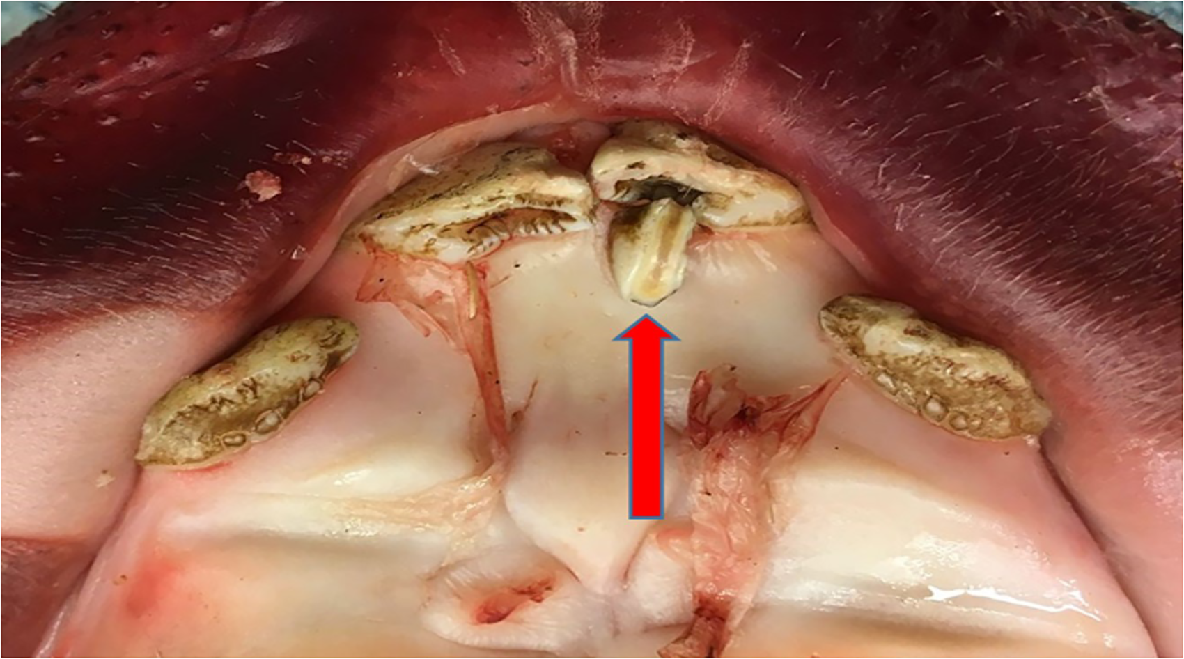
Maxilla of a domestic sow showing one supernumerary and inclined first incisor tooth (arrow). Reprinted with permission.48
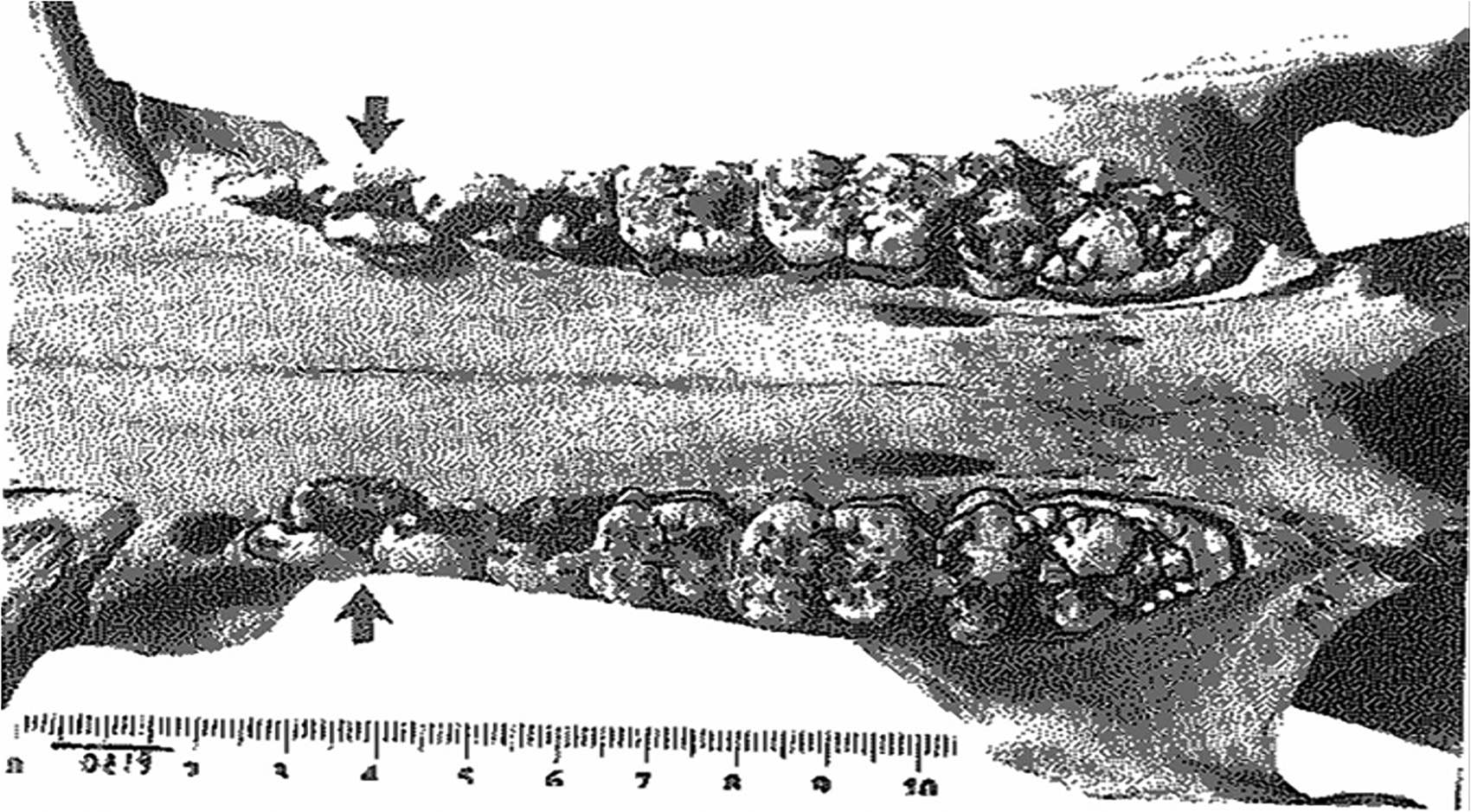
Bilateral supernumerary second premolar teeth in the maxilla of wild pig (arrows). Reprinted with permission.44
Supernumerary teeth are congenital in origin and usually result from the splitting of a bud or from local hyperactivity of the dental lamina, resulting in the formation of additional tooth germs.46,49 Polydontia could also appear in the form of a retained deciduous tooth. Normally, polydontia is unexpected in pigs, as the supernumerary dentition would exceed the primitive eutherian number, but it has been reported in both domestic and wild pigs.44,46,48,49 Due to the lack of space for a supernumerary tooth, it could be found to be rotated perpendicular to the direction of the teeth row, or displaced to a different location in the dental arch, which could result in malocclusion or crowding. 50
In a study of 58 sows (Landrace/Yorkshire-crosses) from 8 Swedish commercial pig herds, 19% of the sows were reported to have supernumerary teeth, although no information was provided on which teeth were affected. 48 However, in a previous report, incidences of polydontia in a group of wild boars were observed. 49 Supernumerary teeth were found in eight wild boars and among these, three had one extra tooth located between the mandibular third and fourth premolar, another animal had two extra teeth, bilaterally between the mandibular third and fourth premolar and one animal had an extra tooth located between the mandibular first and second premolar teeth. In two animals, a persistant maxillary second incisor tooth, was present while one animal had one extra maxillary second incisor tooth. A study also found a supernumerary canine tooth in one male wild boar from Croatia 51 while another study reported the incidence of polydontia of maxillary second premolar tooth in 107 wild pigs from Israel. 44 Similarly, another study reported four cases involving 255 skulls of both wild and domestic suids from around the world (1.6%). 47 The report from Israel showed that in one maxilla, bilateral duplication of the second premolar tooth was observed while in two maxillae it was unilateral. 44 Duplication was accompanied by rotation of both the duplicated and the original tooth in two other maxillae. The tooth most often affected appeared to be the second upper permanent premolar. Duplication of both maxillary and mandibular premolars, the third molar and the canines has also been documented. 52
Hypodontia
The term “hypodontia” generally refers to the genetic absence of one or more teeth (Figure 3), either in deciduous or permanent dentition. It is considered to be the most common craniofacial malformation in humans and may occur as part of a genetic syndrome and non-syndromic conditions.53,54 Other terms used to describe hypodontia include “congenitally missing teeth,” “tooth agenesis,” “aplasia of teeth”, “absence of teeth”, “oligodontia,” and “anodontia”.
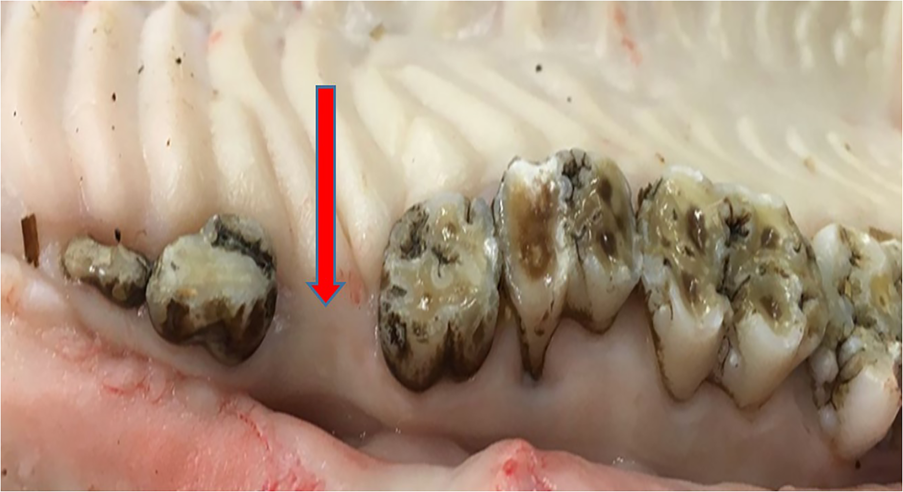
Absence (hypodontia) of maxillary third premolar tooth (arrow). Reprinted with permission.48
Congenitally missing teeth are teeth that have not erupted into the oral cavity and are not visible on a radiograph. Tooth agenesis however refers directly to the developmental failure of a tooth while oligodontia and anodontia are used to describe more severe forms of tooth agenesis, typically the absence of more than six teeth and the entire dentition, respectively.54,55
In humans, the failure of tooth bud proliferation from the dental lamina may be due to congenital absence, resulting from vertical infection from mother to foetus, medications of mother in pregnancy, intrauterine disturbances, endocrine abnormalities, local trauma, chemotherapy or radiotherapy at a young age. 54 The pathogenesis of mandibular tooth agenesis may also be related to disturbances in nerve tissue, oral mucosa and supporting tissues, all of which interact during odontogenesis. 53 Since hypodontia is often seen associated with syndromes, including Downs syndrome and ectodermal dysplasias, and in non-syndromic conditions like cleft lip/alveolus with or without cleft palate, the developmental absence of a tooth could be as a result of genetic or environmental influences. This makes it difficult to determine, by gross observation, whether missing teeth were due to “true” hypodontia (congenital absence of one or more teeth) or “false” hypodontia (periodontitis or tooth loss by other causes). 49
Hypodontia may not be evident in young, growing animals, since they only possess deciduous dentition. It however becomes noticeable in adults, when the deciduous teeth should have been replaced by the permanent dentition. It has been reported that when a deciduous tooth is congenitally missing, its permanent successor is likely to be missing too. 12
In a study on Swedish wild pigs, it was observed that absence of teeth was the most common dental anomaly, occurring in 68.7% of the examined skulls. 49 In total, 145 teeth were missing and the most commonly missing tooth was the first premolar, especially the mandibular first premolar. In one of the pigs, aged 18–21 months, all teeth except the first and second incisors, and the canine, in both the mandibles and maxillae, were missing. This condition is referred to as “Hypohidrotic Ectodermal Dysplasia” (HED) in humans. It has also been reported in dogs and cattle. 56 In a study of 58 sows (Landrace/Yorkshire-crosses) from 8 Swedish commercial pig herds, 59% of the sows were missing at least one tooth and approximately 50% of the missing teeth were premolars. 48 Another study examined the cleaned skulls of 39 wild and 30 domestic pigs and reported that oligodontia (either bilateral or unilateral) was the most common anomaly, occurring in 9 (23.1%) wild and 15 (50%) domestic pigs. Loss of the first lower premolar(s) occurred in 22 of these 24 skulls; 8 were unilateral (6 on the left side) and the others were bilateral. One domestic pig had missing left first and second premolar teeth, while two others had bilateral oligodontia of the third maxillary incisor teeth. 45 Similarly, a study found anomalous first mandibular premolars in 16 of 73 wild boars (22%) in Poland 57 while another study reported the absence of the first mandibular premolar in 6 of 14 pig skulls, as well as missing maxillary first premolar teeth in 2 of 18 skulls examined. 45 Similar observations were made in a sample of 56 Vietnamese pot-bellied pigs where 37% had one or more first premolar teeth missing. 44
The mandibular first premolars in Sus scrofa are unusual. Although a study suggested that they “erupt as permanent teeth only”, 58 they may be persistent deciduous teeth. Regardless, in most pigs, the first premolar is seen in highly variable positions. Their eruption pattern and relative isolation in the dental arcade may contribute to the high frequency with which they are missing. 45
Fractured Teeth
A fractured tooth is seen as a break in the enamel, dentin or cementum. In humans, it is considered to be a problem with increasing clinical significance and with many predisposing factors. Extensive carious lesions are associated with most fractures, with fracture incidence being higher in the first permanent molar teeth than in other tooth types, especially on the mandible. 59 Although fractures have been categorized based on type and location, such as crown fractures, crown-root fractures, split-root syndrome, enamel infraction, hairline tooth fractures, crown craze/crack type, or having craze lines and tooth structure cracks, fractures can also be classified as being either complete or incomplete. 60
he morphological and anatomical characteristics of pigs' teeth, as well as diet and behavioural habits, including fighting and bar biting, may predispose certain teeth to fractures or cracks. The possibility of root fractures secondary to tooth fractures should also be considered. 49
In a study conducted on 58 Swedish commercial sows, 41% (n=24) were found to have one or more tooth fractures. Each sow was reported to have between 1 and 6 fractures. Fractures were more common in incisor teeth and on the mandible. The most severe fractures were observed in incisors but a few sows were found to have fractured premolar and molar teeth. 48 The high incidence of fractures occurring in the incisor teeth was similar to the report obtained by a study which observed that the most affected teeth in a group of Swedish wild pigs were the first incisors. Eight wild boars (8.1%) were found to have one fractured tooth each and the teeth affected were I1 (n = 3), M1 (n = 2), P4 and M2 (n = 1, respectively). 49 Another study reported that fracture was the most common disorder observed in wild boar tusks, occurring in both the mandibles and the maxillae. 51 The wild boar uses its tusks as powerful weapons in intraspecific and interspecific fights, as well as for marking trees. The various ways in which tusks are used, in combination with the fact that they protrude from the mouth, make them particularly prone to injury. Depending on the location and the intensity of impact on a tusk, complete or incomplete intra or extra-alveolar fracture can be observed.49,51 In a group of sows either found dead or euthanized (n = 65), tooth fractures were present in 16 animals (25%), including 5 sows with one, and 11 sows with more than one affected tooth. The frequency of tooth fractures increased numerically with increasing parity, since sows with higher parities are assumed to be older. 61 Therefore, the older the sows, the more likely they are to have been exposed to factors that cause tooth fracture.
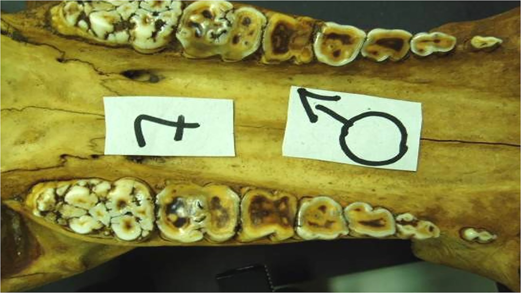
Rotation
Tooth rotation refers to the displacement of a tooth from its normal alignment on the dental arcade (Figure 4). It is usually associated with the presence of supernumerary tooth/teeth, on either the mandible or the maxilla. Tooth rotation appears to occur due to crowding of teeth in the jaws and may be due to genetic factors such as skull size and length, or to severe undernutrition. Rotation can result in malocclusion and misalignment of teeth on the mandible and the maxilla. 44 In a study involving 32 wild pig skulls, 5% exhibited rotation of the maxillary third and fourth premolar teeth, while one or both maxillary third molar teeth were rotated in 9% of the samples. In another study, 13% of 60 specimen of the bush pig (Potamochoerus spp) had rotated maxillary second, third and fourth premolar teeth. 44 Another study reported that rotation of teeth occurred in 8 wild and 3 domestic pigs and involved the first or second maxillary premolars in 10 of the 11 cases. Rotation was unilateral or bilateral and varied from slight to 90°. 45
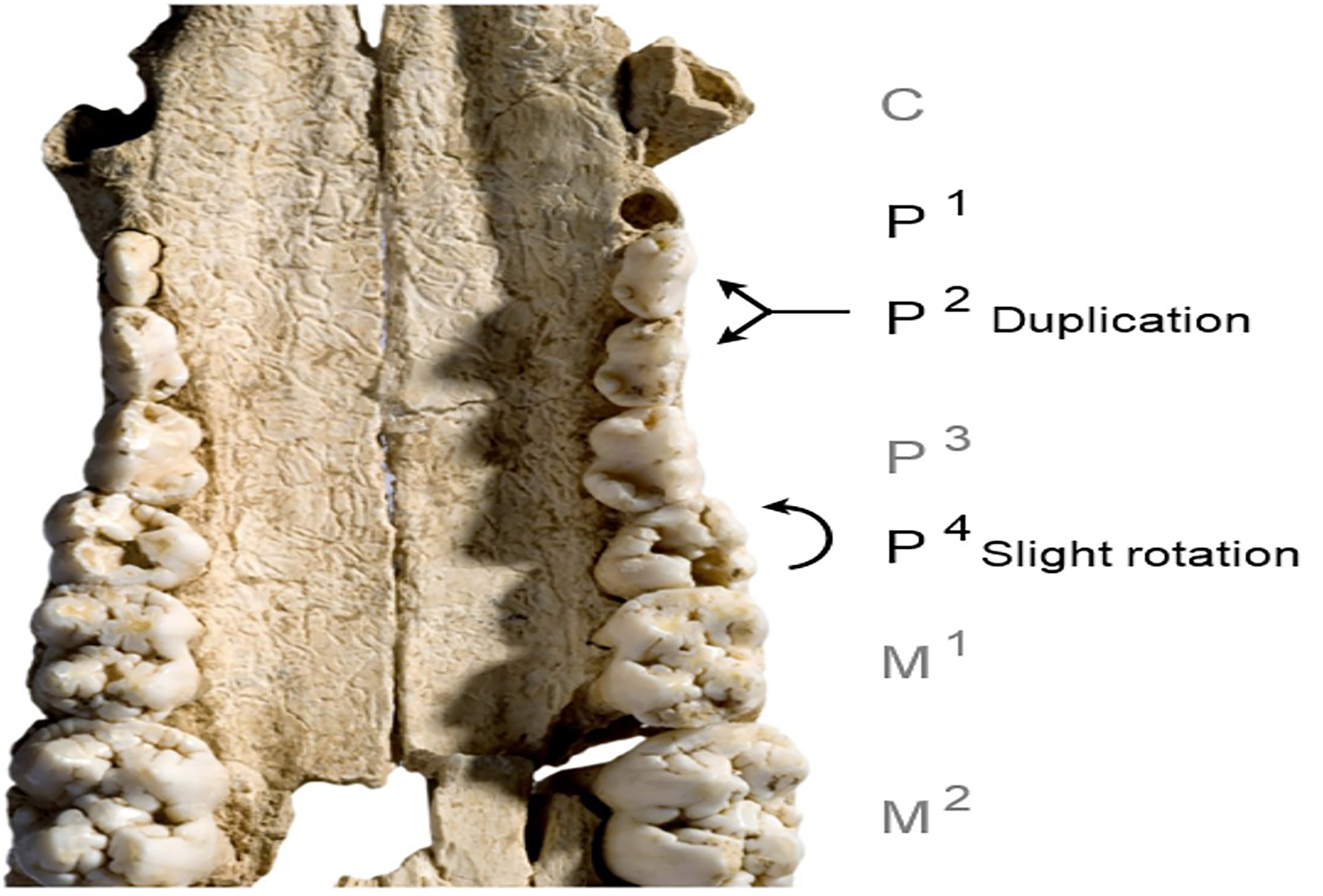
Supernumerary (Duplication) of maxillary second premolar (P2)and rotation of fourth premolar (P4) teeth of a wild pig. Reprinted with permission.47
Dental Diseases
Common dental diseases such as dental caries, periodontal disease and malocclusion are mainly influenced by environmental factors. However, even for these diseases, the genetic aspects that influence the degree of susceptibility should not be overlooked. A multifactorial etiology for all three conditions has generally been assumed, with both genetic and environmental conditions contributing to the observed variability. 62
In contrast to dental anomalies, most dental diseases are associated primarily with diet and reflect the influence of local environmental conditions. Excessive tooth wear is not primarily a dental disease but may affect oral health, as well as general health, because of impaired mastication. Moreover, severe tooth wear with pulp exposure, resulting in pulpitis, can lead to secondary periodontal lesions. This cascade of events may eventually result in tooth loss. 49
Periodontal Disease
Periodontal disease is thought to be multifactorial in nature and results from the interaction of several factors, including exposure to oral bacteria, response to bacteria (promoting or inhibiting colonization and growth), and tissue structure (facilitating or protecting against bacterial colonization and growth and the resultant tissue damage). 63 Other aetiological factors include immune status, amount of saliva, breed, age, routine prophylactic cleaning and type of food the individual feeds on. However, dental plaque is the primary etiological agent.Plaque consists predominantly of gram-positive, aerobic, non-motile bacteria in the early stages of infection, and anaerobic, gram-negative and motile bacteria in the latter stages of infection.49,64
Periodontitis, by definition, is an inflammation of the periodontium, which includes the gingiva, cementum, alveolar bone and the periodontal ligament (Figure 5). 65 Periodontitis is the result of an immune response to dental plaque and is the most common oral disease seen in small animal veterinary practice. It is a progressive disease and the age distribution in the investigated cohort may influence measurement of the prevalence of periodontitis. 49 Periodontal disease embraces everything from mild reversible gingivitis to severe tooth loss.44,66
Severe periodontitis showing uncovered root surface of the maxillary first molar tooth (arrow). Reprinted with permission.48
In a study conducted on the heads of feral and domestic pigs, periodontal lesions were observed in many of the specimens: 26 of 107 feral pigs (24%) and 12 of 52 domestic pigs (23%). In feral pigs, most of the lesions involved bone, with 14% of animals having grade two and 8% grade three lesions, whereas in domestic pigs, these figures were 8% and 6%, respectively. All the lesions involved the cheek teeth; the incisor and canine teeth appeared to be normal. 65 One study reported that severe periodontitis was found in 15.2% (n=15) of the wild boars. The study revealed that three animals had up to periodontal index (PDI) 1, seven had PDI 2 and five individuals had up to PDI 3. Eighty-six teeth were affected by severe periodontitis (PDI 1-PDI 3), with PDI 1-PDI 3 representing the severity of the disease: mild, moderate and severe respectively. 49 Periodontal disease was also found in 54% (n=107) of wild pigs from Israel. In all cases, it affected both sexes, and in most cases, the disease was present on both sides of the maxilla. 44
Dental Caries
Dental caries refers to a discoloured area of the tooth surface into which a dental explorer can be inserted with slight resistance when removed. 49 Dental caries occurs due to demineralisation of enamel and dentin (the hard tissues of the teeth) by organic acids. Acid is formed by bacteria in dental plaque through the anaerobic metabolism of sugars derived from the diet (Figure 6). 67 If allowed to continue, the process leads to formation of a cavity in the tooth, which can lead to bacterial infection of the pulp and result in severe suffering of the affected animal. 49
Ddental caries in a maxillary fourth premolar tooth of a wild pig (arrow). Reprinted with permission.44
The development of dental caries requires sugars and bacteria to be present and it is influenced by the susceptibility of the tooth, the bacterial profile, the quantity and quality of the saliva, and the time during which fermentable dietary carbohydrates are present for bacterial fermentation. 67 Although there is a close association between caries and diet, genetic factors may predispose to susceptibility to the disease. 49
In a study conducted on 99 Swedish wild boars, a total of 19 teeth were affected by dental caries (9 deciduous and 10 permanent teeth) in 11% of the boars. The most dominant tooth type affected by dental caries was the incisor (n = 12), followed by premolars (n = 3) and molars (n = 3). Dental caries was also observed in one mandibular canine. 49 In another study, 5% of wild pig jaws showed carious lesion, with 3 being males and 1 being a female. 44 Carious lesions were also observed in 9% of commercial Swedish sows. 48 The cause of these lesions may be a genetic susceptibility to the disease in combination with intense supplemental feeding (including corn, sugar beet, potatoes and carrots), which are carbohydrate rich.
Dental caries has also been reported in piglets. 68 In the study, it was observed that many piglets were seen to have discoloured teeth at birth and developed dental caries soon after birth. This could relate to conditions in utero. The occurrence of dental caries within weeks of birth indicates that enamel was not laid down sufficiently in utero, a condition referred to as “amelogenesis imperfecta”. Enamel protects underlying dentin against environmental and nutritional insults and is laid over dentin during the formation of the tooth. Defects in this tissue are permanent and can greatly reduce the integrity and longevity of the tooth. 68
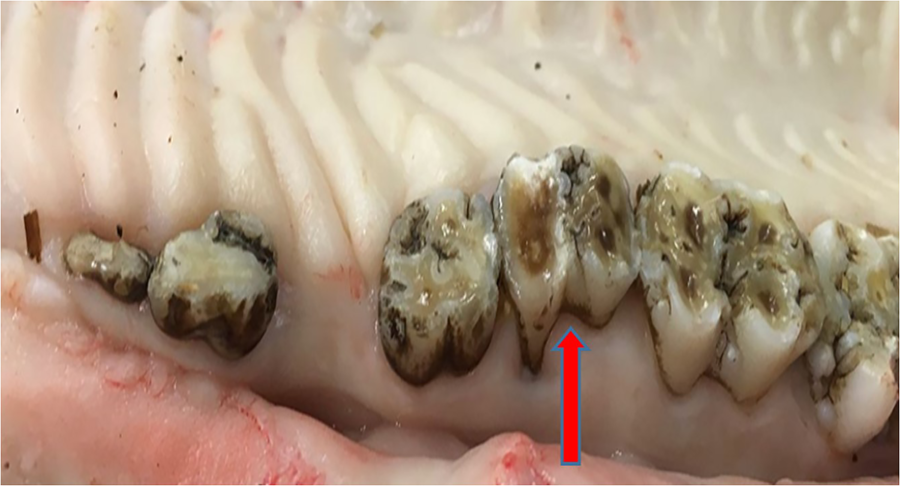
Tooth Wear
Tooth wear is considered to be a multifactorial physiological process and is commonly seen as a normal part of aging (Figure 7). It is an inevitable consequence of the evolutionary purpose of food acquisition by predation, apprehension, grasping, trituration, mastication, and ingestion. 69 Excessive tooth wear is not primarily a dental disease but may affect oral health as well as general health, due to impaired mastication. Moreover, severe tooth wear with pulp exposure, resulting in pulpitis, can lead to secondary periodontal lesions, causing tooth loss. 49
Tooth wear on the maxillary molar teeth from an adult male feral pig. Reprinted with permission.69
Tooth wear can manifest as attrition (caused by tooth to tooth contact or grinding of opposing teeth forming acquired wear facets upon pristine enamel), abrasion (caused by food and foreign body contact (eg, tooth brushing or feeding of high fibre diets) that may obliterate attrition wear patterns), or erosion (caused by acid-based leaching and dissolution of the hydroxyapatite crystals of enamel which may override previous lesions of enamel). 69 Tooth wear can affect both deciduous and permanent teeth, due to the feeding habits of affected animals. The pattern of wear is likely determined by a combination of anatomical characteristics and the function of individual teeth. 49
One study reported that in 99 wild Swedish boars examined, tooth wear affecting deciduous teeth was found in 83.6% (n = 61) of these animals, of which 16 animals had at least one tooth affected by mild lesions, 31 animals had moderate lesions and 14 had severe lesions in at least one tooth. Tooth wear affecting permanent teeth was also seen in 70% of the skulls (n = 69), with 9 wild boars having mild lesions, 40 and 20 having moderate and severe lesions, respectively. 49 A high frequency of tooth wear of 71% (n = 46) was also reported in a recent Finnish study of 65 commercial sows found dead or euthanized. 61
Another study described molar wear in 63% and incisor wear in 62% of 82 culled mature sows in USA and Canada. Tooth wear appeared to be more prevalent with increasing age in the sows. The study suggested, based on dental examination of 98 live sows, that molar wear was associated with age, whereas incisor wear was caused by some factor present in indoor housing. 70 Advanced tooth wear may lead to pulpitis and subsequent periodontitis. Severe wear has been associated with oral pain and dysfunction. 61
Dental Calculus
Dental calculus is a hard deposit that is formed by the calcification of plaque. It is composed, primarily, of calcium phosphate which is deposited on the natural tooth surface. These hard deposits may form coronal to or apical to the gingival margin and are thus referred to as supragingival and subgingival calculus respectively. 71 Bacterial plaque and calculus are accepted etiological agents in the initiation and progression of periodontal disease. Their accumulation and attachment are facilitated by a roughened root surface, and undisturbed plaque accumulation will result in gingival inflammation within a few weeks and ultimately periodontal disease.67,72 In humans, the degree of calculus and its location are population specific and are affected by oral hygiene habits, access to professional care, diet, age, ethnicity, origin, time since last cleaning, systemic disease and the use of medication. 73
In a group of sows which were found dead or were euthanized, dental calculus, with a moderate degree of severity, was present in 11% of the animals examined. 61 Calculus of varying degrees was also found in 45% of Swedish commercial sows (n = 58). 48 Another study reported the presence of calculus in Swedish wild pigs. A varying degree of calculus formation on permanent teeth was found in 40.4% (n = 40) of the individuals studied. In total, 86 teeth were affected by severe build-up of calculus and the most affected teeth were the premolars (93%, n = 80), while the remaining teeth (n = 6) were molars. Severe build-up of calculus was most prominent in the premolar teeth of older animals. 49 This suggests that calculus formation is a progressive condition and that the premolar teeth of wild boar were not excessively used during mastication, thus enabling the formation of plaque and subsequent calculus.
Conclusion
Dental disorders have been established as an important component in the oral health of wild and domestic pigs, therefore the possibility of dental disease in the oral cavities of young and adult subjects should not be overlooked. The state of the oral health of an animal can influence the general health of the animal. This is especially important for commercial pig herds, as the presence of deleterious oral conditions could result in culling of affected animals due to the discomfort of such conditions. This may ultimately result in economic losses for affected farmers. It is therefore important to know the types of disorders that may affect pigs, the susceptible age groups, the frequency of occurrence, as well as the predisposing factors of such disorders.
Pigs are omnivores and can suffer from numerous dental abnormalities, which might go unnoticed. Oral examinations may be beneficial in their routine health care management, since early detection of oral and dental abnormalities can help promote productivity and reduce losses. Factors that predispose pigs to deleterious oral conditions should be identified and reduced or removed, where possible.
Footnotes
Acknowledgements
Authors are grateful to the management of the Teaching and Research Farm, University of Ibadan, and Dr Olusoji Abiola of the Department of Veterinary Medicine, University of Ibadan for their support.
Author Contributions
Olopade JO conceived the work and the outline. Okandeji ME, Atiba FA and Lijoka AD wrote the draft. Adebiyi OA and Olopade JO corrected and edited the manuscript.
Declaration of Conflicting Interests
The author(s) declared no potential conflicts of interest with respect to the research, authorship, and/or publication of this article.
Funding
The author(s) received no financial support for the research, authorship, and/or publication of this article