
Abstract
Oral squamous cell carcinomas (OSCCs) are commonly diagnosed in animals, particularly African pygmy hedgehogs (Atelerix albiventris) (APHs). This study reviews individual hedgehog and tumor characteristics of 17 OSCCs in APHs, as well as 94 OSCC cases currently recorded in the scientific literature, to assess common trends and potential risk factors for these neoplasms in the species. A single detailed case of OSCC is described in the case series; it was submitted for autopsy with cytological and diagnostic imaging data while the remaining cases were routine histological biopsies. Histologically, OSCCs showed infiltrative trabeculae and nests of squamous epithelial cells with variable degrees of keratinization. Bone and nasal invasion were observed in some OSCCs (n = 3), consistent with the literature. Metastasis has been rarely reported and was not recorded in this case series. Inflammation was observed in examined OSCCs, as well as in literature cases, including in the mucosa adjacent to OSCCs, suggesting a potential role of chronic inflammation in tumor promotion. This study highlights that individuals older than 2 years old with chronic stomatitis are potentially at higher risk of developing OSCCs, with the maxilla (66.0%) being the most common site of tumor development. No sex predilection is evident. Recurrence and metastatic potential remain insufficiently documented, warranting further investigation.
Introduction
African pygmy hedgehogs (Atelerix albiventris) (APHs) are becoming increasingly popular exotic companion animals, 1 yet they remain relatively understudied compared with traditional domestic species. A wide range of diseases has been reported in captive hedgehogs, with oral and dental disorders representing some of the most frequently documented medical conditions.2–6 These include, among others, dental calculus accumulation, gingivitis, periodontitis, gingival recession, tooth fractures, and, less commonly, dental abscesses and periapical infections. In addition to inflammatory dental conditions, oral mass lesions are also reported in hedgehogs.6–9 Regarding nonneoplastic conditions, bacterial dermatitis and cellulitis associated with Actinomyces sp. and Corynebacterium sp., or fungal agents such as Trichophyton erinacei, have been reported with marked alteration of the face and adjacent tissues, resembling neoplastic mass lesions.10–12 As in other animals, 13 oral squamous cell carcinoma (OSCCs) are common in APHs, ranking as the third most common tumor and oral neoplasm7–9,14 in middle to old age APHs. 15 Data on the incidence of OSCCs in hedgehog species based on gender are contrasting8,16 and clinical manifestations of OSCCs encompass a spectrum of variable signs, including loose teeth and tooth loss, swelling of soft tissues and bone deviation. 9 Although OSCCs demonstrate a high propensity for local infiltration,8,17 its metastatic potential is still unclear as only rare reports recorded distant metastasis in the literature. 18 This article describes 17 OSCCs in APHs, highlighting the pathologic features and the potential biologic behavior of this neoplasm. Additionally, in the context of the case series, a review of the 94 cases published in the literature was performed to collate all the information and unravel the common clinical and pathologic features of OSCCs in APHs.
Case Series
Animal and Tumor Data
A total of 17 cases of OSCCs from APHs were collated from three veterinary pathology diagnostic services in Italy and the United Kingdom. One case of OSCC (n = 1) was submitted for autopsy with cytology and diagnostic imaging data, while the remaining OSCCs (n = 16) were submitted as biopsy specimens. Signalment data and information on the lesion sites were obtained via the biopsy submission form. Detailed age data were reported for most cases (n = 13) with a mean age of 3 years 11 months; the remaining cases (n = 4) were referred to as adults. Most animals were male (n = 11; 64.7%), while only 6 females (35.3%) were recorded. A summary of the signalment data is reported in Table 1.
Data Summary of the Oral Squamous Cell Carcinomas (OSCC) in African Pygmy Hedgehogs (Atelerix albiventris).
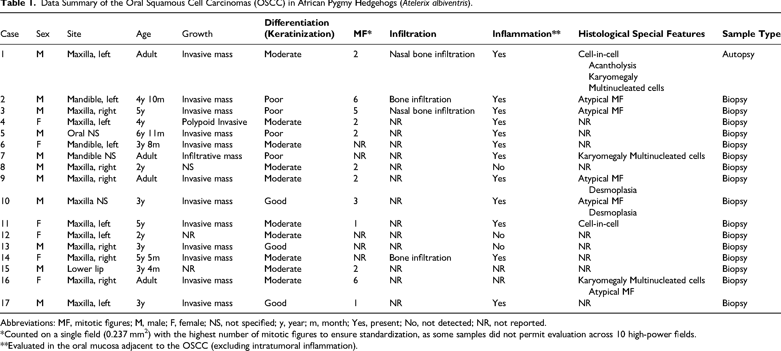
Abbreviations: MF, mitotic figures; M, male; F, female; NS, not specified; y, year; m, month; Yes, present; No, not detected; NR, not reported.
*Counted on a single field (0.237 mm2) with the highest number of mitotic figures to ensure standardization, as some samples did not permit evaluation across 10 high-power fields.
**Evaluated in the oral mucosa adjacent to the OSCC (excluding intratumoral inflammation).
Regarding the site of development, OSCCs occurred more frequently in the maxilla (n = 12; 70.6%) than in the mandible (n = 3; 17.6%). A single case (n = 1; 5.9%) was reported as developing in the lower lip, while an exact oral site was not specified in 1 case (5.9%).
Clinically, for the OSCCs the reported signs included tooth loss (n = 2), anorexia (n = 2), exophthalmos (n = 2) and tooth mobility (n = 2). Histologically, infiltrative nests and trabeculae of squamous cells arising from the mucosal epithelium were detected in all cases of OSCCs, with a varying degree of keratinization and cellular differentiation. Based on criteria of differentiation for the squamous cell carcinomas reported in the literature,19–21 many OSCCs (n = 10; 58.8%) exhibited moderate differentiation (squamous epithelial cells with absence of keratin pearl formation), while well-differentiated OSCCs with keratin pearls (n = 3; 17.7%) and poorly differentiated types were less frequently seen (n = 4; 23.5%). None of the examined OSCCs showed morphological features indicative of a viral infection (ie, koilocytes, and viral inclusion bodies). Most cases of OSCCs (n = 12), reported the presence of inflammation within the tumor and adjacent mucosa. In a few cases (n = 3) in the present study, inflammation was not seen or not reported (n = 2). Special histologic features, such as desmoplasia, cell-in-cell phenomena and presence of atypical mitoses were further assessed.22–24 A summary of all data regarding the examined cases (biopsy and autopsy specimens) with the additional special histologic features of each case is reported in Table 1.
Clinical Autopsy Case; Diagnostic Imaging and Pathology Assessment
The autopsy case was presented with left maxillary facial swelling 20 × 10 × 10 mm, local skin ulceration, oral malodor, and anorexia. A focal suppurative lesion (abscess) of the facial soft tissues was suspected on initial clinical examination. Curettage was performed under general anesthesia to clean and drain the area. Radiography revealed a soft tissue proliferation with associated left maxillary bone lysis, raising the suspicion of a malignant oral neoplasm with no other concurrent lesions in distant sites. Computed tomography scans without contrast showed the presence of a proliferative space-occupying lesion, with soft tissue density involving the left maxillary and zygomatic region plus extensive and severe maxillary osteolysis with involvement of the left palatine region and left nasal vestibule, resulting in occlusion of the nasal cavity (Figure 1a and b). No other changes were reported in other organs.

Oral squamous cell carcinoma (OSCC) in an African pygmy hedgehog (Atelerix albiventris). (a) Dorsal plane computed tomography (CT) scan of the head showing moderately radio-dense mass lesions involving the maxillary bone. (b) Transverse plane CT scan of the head showing a mass lesion (asterisk) extending into the palate and the oral cavity. (c) External, dorsal view of a cadaveric head showing the mass located on the left side of the face. (d) Transverse section of the cadaveric head showing the mass located on the left side of the face (asterisk). (e) Photomicrograph of the OSCC showing trabeculae and nests of polygonal neoplastic epithelial cells with squamous differentiation supported by thin fibrovascular septa (×200, H&E). (f) Photomicrograph of the OSCC showing central foci of degeneration with acantholytic cells, and cellular debris (×400, H&E). (g) Photomicrograph of the OSCC showing numerous mitotic figures (arrowheads) in the neoplastic trabeculae (×400, H&E); Inset: higher magnification of a mitotic figure (arrowhead). (h) Photomicrograph of the OSCC showing cell-in-cell phenomenon (cannibalism) characterized by a neoplastic cell showing the presence of an intracytoplasmic epithelial cell (×400, H&E); Inset: higher magnification of the cell-in-cell phenomenon.
A diagnosis of malignant neoplasia was favored at this stage, with differential diagnoses including bacterial (Corynebacterium sp., Actinomyces sp., Nocardia sp., Mycobacterium sp.) or fungal dermatitis (eg, T. erinacei), cellulitis, and osteomyelitis. Nonneoplastic, noninfectious (eg, posttraumatic) lesions were considered less likely. Fine needle aspirate cytology revealed individualized and clustered round-to-polygonal cells with abundant light blue cytoplasm (squamous differentiation), indistinct cell borders, frequent perinuclear vacuoles, and large central nuclei with vesicular chromatin and prominent nucleoli. Anisocytosis and anisokaryosis were marked. Neoplastic cells were mixed with a moderate neutrophilic and lymphoplasmacytic inflammatory infiltrate. No mitotic figures were seen. Cytological findings were considered compatible with an OSCC. Due to the extensive and infiltrative nature of the lesion, surgical excision was not pursued, and the owner opted for euthanasia. The animal was submitted for postmortem investigation.
Necropsy confirmed a focally extensive (20 × 10 × 10 mm) swollen area affecting the left maxilla, which on cut surface demonstrated significant extension from the maxilla to the hard palate and nasal cavity. The mass was soft, white, glistening, and bulging (Figure 1c and d). No extension to the orbit or brain were detected. No macroscopic changes or additional lesions were detected in the other organs. Samples were collected and fixed in 10% buffered formalin and embedded in paraffin. Sections 4-μm thick were stained with hematoxylin and eosin (H&E) for subsequent light microscopy.
Histopathology revealed a poorly demarcated, unencapsulated, neoplastic proliferation with infiltrative growth in the adjacent bone and muscle tissue. The neoplasm was characterized by cells organized in trabeculae and nests supported by thin fibrovascular septa (Figure 1e). The cells were polygonal, moderate to abundant, characterized by sharp margins and eosinophilic cytoplasm often with a glassy appearance (squamous differentiation). There were oval central nuclei with variably coarse chromatin and prominent, single, or multiple nucleoli. Larger islands frequently showed central degeneration with necrosis, acantholytic cells, and granulocyte karyorrhectic debris (Figure 1f). Marked anisokaryosis and anisocytosis were detected, with karyomegaly and multinucleated neoplastic cells and a mitotic count of 6 mitoses per 2.37 mm2 (Figure 1g), with the highest number of mitotic figures in a single field being 2 (0.237 mm2). Multifocally, signs of cannibalism (incorporation of cells with subsequent cell-damage), emperipolesis (incorporation of viable cells without obvious cell-damage) and apoptotic cells were observed (Figure 1h). The neoplasm showed multifocal and mild inflammatory infiltrates characterized by small mature lymphocytes and neutrophils. Only mild histologic changes were detected in other organs. Based on the histologic findings, a moderately differentiated squamous cell carcinoma was diagnosed. No microscopic evidence of detectable metastasis, within the limits of the routine histopathological evaluation of representative sections, was found in regional (mandibular and retropharyngeal) lymph nodes and in distant sites, including lungs, liver, kidneys, spleen, heart, and brain.
Literature Review
To compare the case series data with the literature data, published case reports and case series (before March 2024 and written in English, Spanish, Portuguese or Czech) were retrieved from 2 literature databases (PubMed and the first 50 pages of Google Scholar; search string: “Atelerix albiventris” AND oral AND squamous AND [tumor OR tumour]”). A total of 13 manuscripts reporting 94 OSCCs cases were retrieved and included in the literature review.7–9,14–18,25–29 Details on the characteristics of these cases are reported in Table 2. As some of the OSCCs were included in wider cancer studies, some of the literature cases lacked comprehensive details on the signalment of the single case and on crucial pathologic features such as the mitotic count, the infiltrative growth patterns, as well as the presence or absence of metastases and the specific site of tumor development. 15 A total of 20 out of the 94 literature cases clearly stated the animal's sex.8,14,16–18,25–29 Of these 20 cases, 13 (65%) were female and 7 (35%) were male. Cases were older than 2 years, except for 2 cases (Table 2). For 28 cases the site of development was clearly reported.9,16–18,25–27,29 Most of these cases were in the maxilla (n = 17; 60.7%), while a lower number was reported to be in the mandible (n = 11; 39.3%). The specific side of tumor development was reported for only 23 cases,9,17,18,25–27 with the majority developing on the right side (n = 18; 78.3%) and fewer on the left side (n = 5; 21.7%). Infiltrative growth was clearly reported in 9 studies comprising 81 of the 94 literature cases.8,9,14–18,25,26 In the remaining 13 cases, this feature was not clearly stated.7,27–29 Metastasis was clearly stated as absent for 3 cases and, for 90 cases, the presence of metastasis was not specified. In the available data, including previously reported cases, a total of four OSCC cases in APH have been evaluated by autopsy, of which one (25%) showed evidence of pulmonary metastases. 18
Data Summary of the Literature Oral Squamous Cell Carcinomas (OSCCs) in African Pygmy Hedgehogs (Atelerix albiventris).
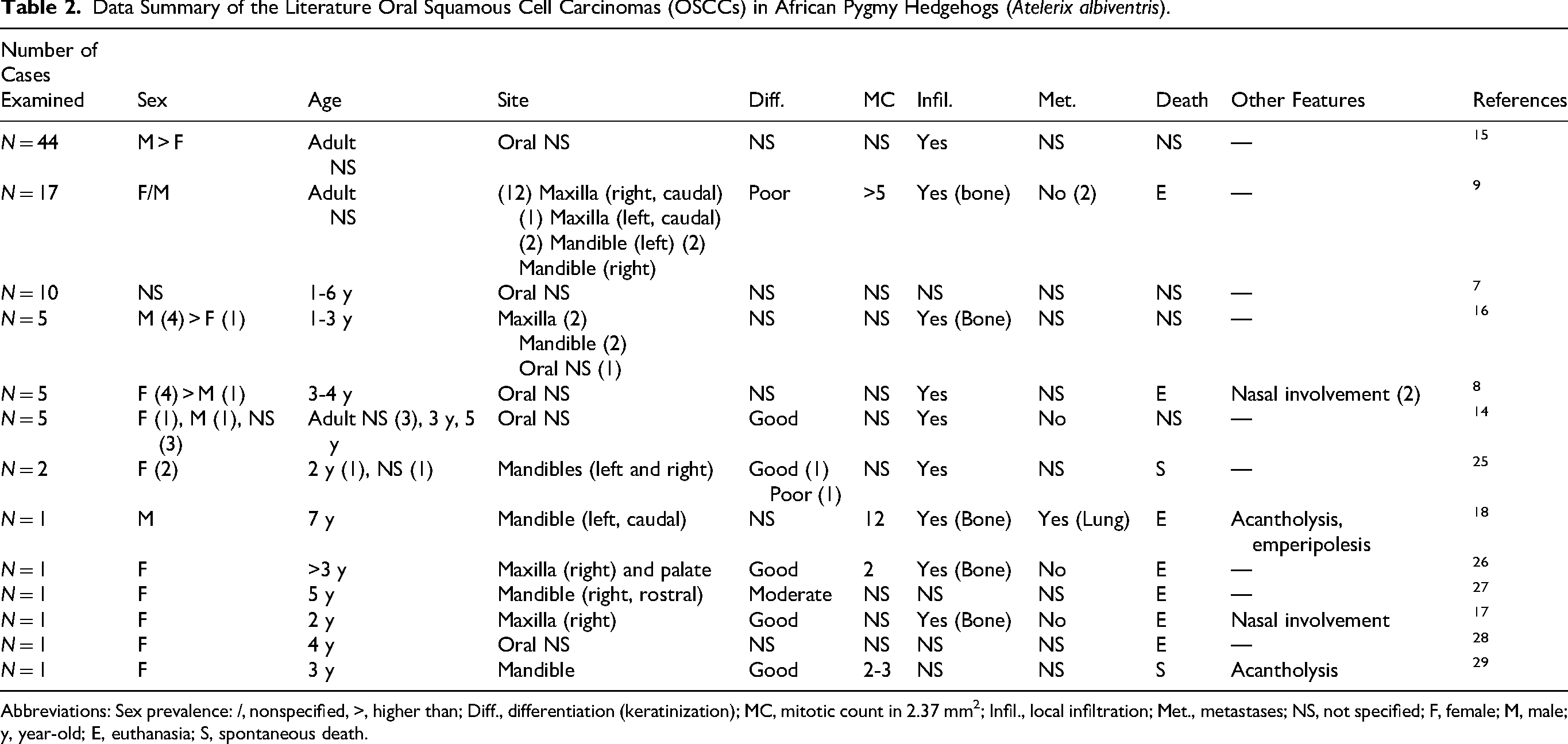
Abbreviations: Sex prevalence: /, nonspecified, >, higher than; Diff., differentiation (keratinization); MC, mitotic count in 2.37 mm2; Infil., local infiltration; Met., metastases; NS, not specified; F, female; M, male; y, year-old; E, euthanasia; S, spontaneous death.
Regarding the total data comparison (current case series and literature reports), out of the total of 111 OSCCs cases examined from the literature (n = 94) and current case series (n = 17), the sex was clearly stated in only 36 cases. An equal frequency of development was observed for males versus females. Eighteen APHs were female (50.0%) and 18 APHs were male (50.0%). A total of 44 literature and current case series cases had a well-defined site of development (maxilla, mandible, lip). Most cases involved the maxilla (n = 29; 66.0%), while the minority (n = 14; 31.8%) involved the mandible. Only a single case (n = 1; 2.2%) developed in the lower lip. Most cases, regardless of the maxillary and mandibular specific site, developed on the right side (n = 24; 54.6%), while fewer cases (n = 12; 27.2%) were located on the left side. For the remaining cases (n = 8; 18.2%) the side was not reported. Of the 44 cases, the specific side of the maxillary or mandibular involvement was clearly available for only 36. For the maxilla, 20 cases (55.5%) involved the right side, while 6 cases (16.7%) involved the left. For the mandible, 4 cases (11.1%) involved the right side, while 6 cases (16.7%) involved the left side. A graphical representation of frequency is reported in Figure 2.
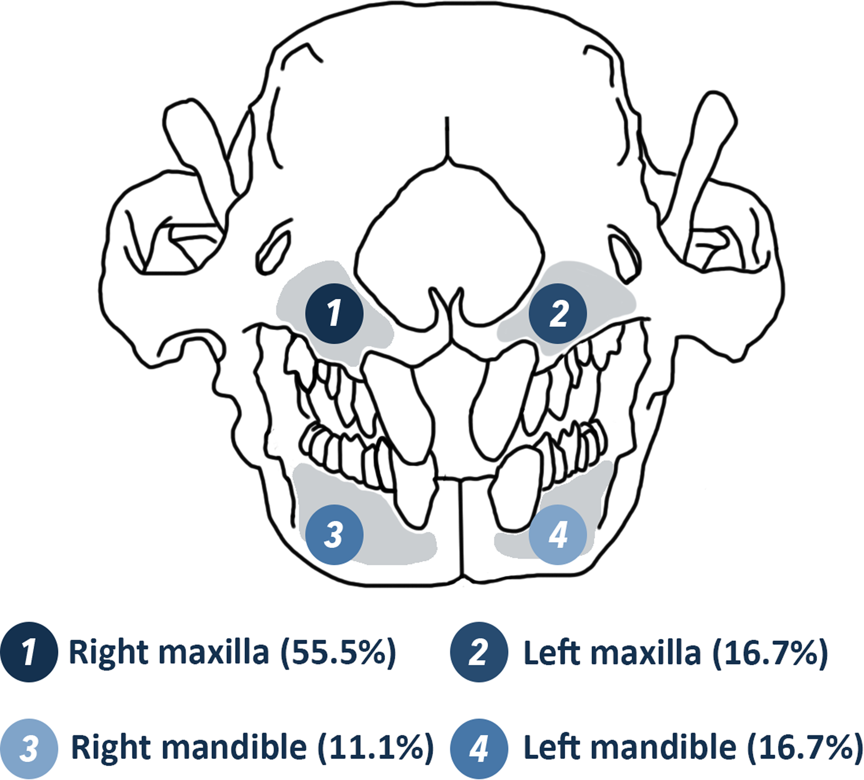
Location frequency of oral squamous cell carcinoma in African pygmy hedgehogs (Atelerix albiventris). Data are based on the available data for the case series and literature cases for the specific side of the maxillary or mandibular involvement (n = 36 cases).
Discussion
This study describes the clinical and pathologic features of OSCC in APHs 8 with a review of the literature. OSCC was the most common oral neoplasm,9,14 and in one study, the most common neoplasm for this species. 8 Approximately half of middle-aged African hedgehogs are reported to develop neoplasms. 14 In this study and in the current literature, hedgehogs developing OSCCs were typically above 2 years of age. As for the site of development, many previously published reports have reported OSCCs, similar to this report, in the maxilla, while a minority have been reported in the mandible. Unfortunately, many cases have been included in larger and multicancer studies with no specific report of the oral development site.7,8,14,15,26 Whether a specific anatomical or functional/mechanical reason (chewing) is associated with the tumor developing in the maxilla, this remains to be clarified. Additionally, the high prevalence of dental and periodontal disease in APHs may have important implications for the tumorigenesis and anatomical distribution of oral neoplasms. The literature reports calculus accumulation, gingivitis, and periodontitis as common findings, particularly affecting the maxillary dentition.5,6,30 Chronic periodontal inflammation in this region could contribute to a pro-inflammatory microenvironment that promotes epithelial dysplasia and neoplastic transformation, as suspected in humans. 31 The consistent presence of inflammation in mucosa adjacent to the tumor suggests a potential role in the tumor progression or may indicate a variable host-immune response to the presence of the tumor. Future studies are needed to better define the prevalence, severity, and anatomical distribution of periodontal disease and stomatitis in APH, as well as their possible association with OSCC development.
Furthermore, concerning the tumorigenesis, the lack of morphological features associated with a possible viral origin in all the examined cases and in the literature, makes a viral etiology less likely. PCR analysis (eg, pan-herpesvirus and pan-papillomavirus PCRs), electron microscopy or next-generation sequencing of a representative proportion of the studied tumors are ultimately required to confirm this statement, 32 given that viral etiology has been proposed in previous discussions.14,33,34
Besides the local invasion and bone lysis being frequently reported, OSCCs have also been seen extending from the primary sites (maxilla) into the nasal cavity and vice versa.8,17 OSCCs in APHs usually appear, as reported in this case, as locally invasive tumors.9,14 Low tendency to metastasize and recurrence after surgery is suspected.9,14,27 The autopsy case investigated in this study suggests, as in previous descriptions, lack of early metastatic spread, even in cases of extensive local tissue invasion. Indeed, only rare cases have been described as having lung metastasis, suggesting a potential local malignant behavior rather than a high metastatic potential for OSCCs in APHs.18,35 However, the limited number of cases undergoing complete autopsy and the lack of long-term follow-up data should be considered when interpreting these findings, as the low number of reported metastatic cases and the absence of recurrence data may also reflect incomplete evaluation of the cases reported in the literature rather than the true biological behavior of the OSCCs in APHs.7,8,15,16
Because histological subtype has been suggested to influence the biological behavior of human head and neck squamous cell carcinomas, data on this aspect should be collected and evaluated in APHs.36,37 Distinction between various histological subtypes (eg, conventional, papillary, acantholytic) has not been clearly stated in the literature for APHs, as diagnostic criteria defined in humans and domestic animals are probably not so well defined for this species. 21 All of the authors’ cases show prevalent features of a conventional subtype. 21 The lack of prognostic data highlights the need for thorough clinical documentation and follow-up evaluations to accurately assess tumor behavior in relation to the various subtypes.
Regarding the histological features, all reports describe classic features of a squamous cell carcinoma, with a variable degree of differentiation with or without formation of central aggregates of keratin in the neoplastic nests (keratin pearls).9,14,27,33 In a few previous reports on OSCCs in hedgehogs, the presence of acantholytic cells and of cell-in-cell phenomenon, as in this case, were noted.18,29,35 Similar to human OSCC, cell-in-cell phenomenon, in the form of emperipolesis and cell cannibalism, has been detected in various cases, 38 including the current report. The cell-in-cell phenomenon describes the presence of neoplastic or nontumor cells inside a neoplastic one, associated (cannibalism) or not (emperipolesis) with the degeneration of the intracellular cell. 39 In humans, the cell-in-cell phenomenon has been reported as a prognostic histologic feature related to tumor progression of various neoplastic diseases. 39 These data are lacking in animals.
The absence of specific details for some of the literature OSCC cases that were retrieved from published studies limited the depth of analysis and the ability to draw additional prognostic information. Thus, routine oral examinations, including the use of sedation and dental imaging, when necessary, should be emphasized and recommended in clinical practice.
The present study summarizes features of OSCCs in the context of the current literature. Data from this study suggests that this neoplastic disease should be high on the differential diagnosis list of adult hedgehogs with facial swelling, facial deformities, and detection of an infiltrative and/or a proliferative process in the oral cavity. Additionally, individuals greater than 2 years of age with chronic inflammation are potentially at higher risk of developing oral squamous tumors, with the maxilla and right side of the oral cavity being the most common sites of tumor development. Further studies on metastatic behavior and recurrence are needed to confirm they are rarely exhibited.
Footnotes
Acknowledgments
The authors would like to express their gratitude to S. Leto and previous/current IZVG pathology technicians for their expertise in processing histological samples. Special thanks are also made to Silvia Benali, Selina Iussich, Giorgia Mezzalira, and Veronica Patton for sharing some of the cases included in the investigation.
ORCID iDs
Funding
The authors received no financial support for the research, authorship, and/or publication of this article.
Declaration of Conflicting Interests
The authors declared no potential conflicts of interest with respect to the research, authorship, and/or publication of this article.