






Abstract
Objective
To determine the effect of subclinical synovitis on the progression of joint disease in a cohort of patients with systemic lupus erythematosus over a mean follow-up of 10 years.
Methods
A longitudinal follow-up of 96 patients diagnosed with lupus was performed. All patients were considered clinically free of joint disease or with minimal joint impairment at baseline and were studied through ultrasound study of their dominant hand to assess the prevalence of subclinical synovitis. Now, over 10 years after we contacted them and reviewed their evolution to determine the impact of had or had not been diagnosed with subclinical synovitis in their current joint condition.
Results
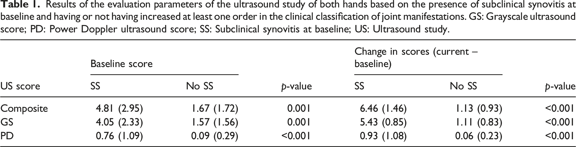
Thirty-one of the 91 reached patients developed clinical progression in their joint manifestations (at least one ordinal degree of worsening). Of these, 23 (74,9%) had demonstrated subclinical synovitis at baseline. In the group of patients who did not progress clinically, 46 (76,6%) did not have this finding at the start of follow-up (p < .01, OR 9,44 95%CI 3,46–25,74). The patients in whom clinical progression was demonstrated had worse combined ultrasound scores than the rest of the patients: 6,41 SD 1,45 vs. 1,15 SD 0,97 (p < .01).
Conclusions
The finding of subclinical synovitis in patients with systemic lupus erythematosus is associated with the development of joint disease progression both clinically and ultrasonographically.
Key messages
• Significant synovitis has been detected via ultrasound in approximately 40% of patients with systemic lupus erythematosus without joint symptoms or with very mild symptomatology. This condition has been termed subclinical synovitis. • In patients with systemic lupus erythematosus, this subclinical synovitis has been associated with a worse joint outcome compared to those patients who are asymptomatic or minimally symptomatic without subclinical synovitis. • Additionally, after an average of 10 years, patients initially identified with subclinical synovitis had worse composite ultrasound scores than those who did not originally present subclinical synovitis
Introduction
The existence of synovitis in patients with systemic lupus erythematosus (SLE) has been demonstrated in different studies.1–6 Subclinical synovitis (SS) is defined as an inflammatory finding identified in the synovial territory of patients without joint symptoms.1,5–7 Different authors have documented SS in large ultrasound studies (US) that include the entire spectrum of patients with joint manifestations, not only the asymptomatic ones.4,8,9 Besides the heterogeneity of the populations included, another problem detected in these studies is the definition of a pathological US, considering that it has been demonstrated that mild synovitis is present in healthy subjects.10–12 As a result of these limitations, the prevalence of synovitis in patients with SLE has been described in a wide range of values, from 10% 8 to 94%. 13
SS has been reported in about 40% of patients with SLE1,3,14; however, in addition to recognizing its existence, the greatest interest is determining its impact on the joint prognosis. In a study published in 2016, with a cohort of 35 patients, it was shown that the baseline presence of arthralgia and pathological ultrasound findings in the study of the hands was associated with a greater demand for immunomodulatory medication compared with patients without joint symptoms nor significant ultrasound findings. 15 However, the magnitude of SS and its effect on long-term follow-up in terms of joint prognosis were not assessed.
The objective of the present study is to determine to what extent the previous finding of SS was related to changes in joint clinical and ultrasound manifestations through a 10-year prospective study.
Method
Patients
In 2020, our group published the findings of a study of SS prevalence in the hands of 96 patients with SLE recruited between 2008 and 2016. 1 These patients had previously been diagnosed with SLE (according to the 1982/1987 American College of Rheumatology criteria)16,17 in six different centers in the city of Madrid and were under follow-up in rheumatology, internal medicine, dermatology and nephrology units. As this was a study to detect subclinical synovitis, the clinical joint profile of patients was: completely asymptomatic (20), with sporadic arthralgias without arthritis (34) and with sporadic arthritis without joint damage (42).
These three stages of musculoskeletal manifestations of SLE correspond to the Zea et al. 18 clinical classification system which includes the following ordinal grades: Grade 0: no joint symptoms; Grade I: sporadic arthralgia without arthritis; Grade IIA: sporadic arthralgia and arthritis without deformation; Grade IIB: Deforming joint disease, including Jaccoud’s disease; Grade III: Erosive arthritis; and Grade IV: Mutilating arthritis.
An attempt was made to contact all these patients for a face-to-face meeting, review of their clinical history and to perform an US of the dominant hand using the new OMERACT US definitions of 2019. 7 Also, all baseline US were reviewed again using the same definitions to avoid a bias due to use different criteria.
Data management and ultrasound study
Data related to clinical follow-up and the US study was performed by two different researchers. Both were unaware of the baseline condition (with or without SS) of each patient.
Both, the current US study and the review of baseline videos was performed by a rheumatologist with wide experience in musculoskeletal ultrasound. Both studies were performed with a Logic S8 ultrasound scanner (General Electric, USA). The protocol of current exploration and review of baseline videos included: the dorsal aspect of the wrist, second, third, and fourth metacarpophalangeal joints and second, third, and fourth proximal interphalangeal joints. The choice of this ultrasound exploration protocol was based on the results of the comparative study by Sivajumaran et al., 19 and the inclusion of proximal interphalangeal joints. The minimal definition of synovitis was a grade 1 synovial hypertrophy and/or a grade 1 power Doppler signal according to OMERACT reviewed definitions. 7 For the radiographic study of the hands, the following evaluation criteria were followed: alignment of bone structures, condition of soft tissues, state of bone matrix, and presence and recount of erosions.
The medical chart of each patient was reviewed, including radiological studies, notes from their physicians, and treatment history. In case of insufficient or inconsistent data, the responsible physician was consulted directly. Any increase in the ordinal degree of classification of the joint manifestation 18 was considered an impairment. The proportion of joint clinical impairments between patients with and without SS and the time at which they occurred were determined. US scoring results changes were compared between patients with and without SS in the baseline study.
Statistical management
To describe the characteristics of the cohort, measures of central tendency and dispersion were used for the numerical variable while proportions for the categorical variables. Comparisons of proportions of worsening were made using the chi-square test, including OR and the 95% confidence interval (95%CI). Comparisons of the US score changes were made using the Student’s t test for unpaired and paired samples, on correspondence. A joint clinical worsening-free survival curve was developed to determine the effect of SS detection at the start of follow-up on patient outcomes. For this purpose, any worsening of joint symptoms that would have moved a patient to a more advanced category of joint involvement was considered as the event of interest.
Ethical considerations
The present study was approved by the ethics committee for scientific research of the Ramón y Cajal University Hospital (File number: 141-16).
Results
Demographic aspects
From the original cohort of 96 patients, 91 finally accepted to participate in the study. Eighty-one subjects were female (89%). The mean (standard deviation) of age of the cohort was 48.9 (6.4) years with a range of 39 to 68. The mean number of years between initial contact and the current review was 9.9 (1.8) with a range of 7 to 14.
Clinical evolution
All patients contacted maintained the original diagnosis of SLE. Regarding the development of complications associated with the diagnosis of SLE, the following were observed: Twelve patients (13.2%) developed clinically significant proteinuria and in four cases there was a need for renal biopsy. Pleural effusion was recorded in two patients, one of which was clinically significant and the other was an incidental radiological finding. One patient developed headaches and seizures as a neurologic manifestation of SLE according to radiological findings on brain MRI. In 12 patients, treatment with bisphosphonates was initiated in association with a diagnosis of densitometric osteoporosis and/or exposure to corticosteroids. No major osteoporotic fracture or hip fracture was documented. In the comparative analysis, the presence of none of these manifestations was associated with the baseline diagnosis of SS.
Changes in clinical joint manifestations
In the 91 participating patients, worsening of the clinical joint classification was observed in 31 (34,0%). Of these, 23 (74,9%) had baseline SS. In the group of patients who did not worsen, 46 (76,6%) had no baseline SS (p < 0,001, OR 9,44 CI 95%: 3,46–25,74). (Figure 1). Distribution of patients based on their stage of joint involvement classification (according to the Zea et al. classification) at baseline and at review (mean observation time of 9.8 years). At baseline, a distinction is made between patients with and without subclinical synovitis (SS).
No association between sex, age at diagnosis or extension of the follow-up time and the development of clinical joint impairment was founded (data not shown).
Among patients in whom SS was detected at baseline, the mean (95% CI) time to worsening their joint clinical classification was 93,9 (79,1–108,7) months while in the rest of patients it was 153,5 (143,4–163,6) months (p < 0.01) (Figure 2). Survival curves until clinical findings of any worsening on the clinical assessment scale of joint manifestations. Patients were grouped according to whether or not they had subclinical synovitis at baseline.
Results of the evaluation parameters of the ultrasound study of both hands based on the presence of subclinical synovitis at baseline and having or not having increased at least one order in the clinical classification of joint manifestations. GS: Grayscale ultrasound score; PD: Power Doppler ultrasound score; SS: Subclinical synovitis at baseline; US: Ultrasound study.
Thirteen patients developed joint structural damage, demonstrated radiographically or by US and were considered as grade IIB in the classification of joint manifestations. Ten of these patients (76,9%) had SS at baseline. Among the rest of patients who did not reach this grade of joint impairment, 37 (47,4%) had SS at baseline (p = 0,6, OR 3,69 CI95%: 0,94 – 14,45)
Treatment modifications
All patients were under treatment with hydroxychloroquine at a dose of 200 mg/day (68 patients) or 400 mg/day (23 patients). Thirty-nine patients (44.3%) were receiving NSAIDs on demand. Forty-nine patients (53.8%) were receiving doses of up to 7.5 mg/day of prednisone or equivalent at a rate of up to two annual cycles for no more than 15 days, 40 (43.9%) received higher doses and two never received corticosteroids. No relationship was found between the baseline presence of SS and the use of corticosteroids (data not shown). Regarding the use of other therapies, 13 patients (14.3%) were started on methotrexate (MTX) at a dose of 10 mg/week or more. 10 of the 13 patients who were taking MTX belonged to the group of patients with SS at baseline who progressed to grade IIB and, according to their medical chart, were started on MTX due to this progression.
Discussion
Joint structural damage is one of the most important causes of disability in SLE patients, 20 therefore, its prediction, early detection and treatment are considered a priority.
According to our results, SS significantly increases the risk of joint impairment and this progression seems to take place faster than in patients in whom SS was not previously detected. In our cohort, specifically, worsening of joint symptoms occurred nearly halfway through the follow-up period in patients where subclinical synovitis was detected compared to those who did not have it initially. This finding likely supports the recommendation for baseline and routine ultrasound examination, as well as the treatment of those presenting with subclinical synovitis.
It is important to discriminate the type of cohort included in studies on the prevalence of ultrasound abnormalities in the study of the hands of patients with SLE. For example, in the study by Torrente-Segarra et al., 21 synovial hypertrophy was found in 25% of patients and PD signal in 14%. This cohort did not exclusively include asymptomatic patients but rather patients with arthralgia or arthritis. Similarly, Dreyer et al. 22 identified significant synovitis in the carpal region in 48% of the SLE patients they studied. Another interesting finding was the detection of erosions in 6% and tenosynovitis in 18%. However, the cohort used did not exclusively include asymptomatic patients, so these findings cannot necessarily be counted as subclinical. This lack of homogeneity in the cohorts chosen for studies on the prevalence of ultrasound findings has been recognized as a weakness when interpreting results from multiple cohorts. 23 Considering that our study includes patients from multiple centers who were completely asymptomatic or minimally symptomatic at baseline, we believe that this is where the greatest importance of our results lies after 10 years of follow-up.
In the last years, the presence of SS in SLE patients has been ratified in several studies. In 2017, Ruano et al. 5 reported a 77% prevalence of minimal synovitis in patients without joint symptoms in the hands, while the presence of a PD signal of any intensity was identified in 23%. This study included 30 patients, which could be considered its main limitation. In 2018, Salliot et al. 3 studied a consecutive series of 151 patients with SLE without overt musculoskeletal symptoms. In 85% of them, ultrasound abnormalities were identified and in this group of patients, about 75% had had a previous history of joint pain episodes. On the other hand, a PD signal was detected in 44% of the patients, however, no clinical pattern of the disease was associated with the presence of this finding. In 2019, Zayat et al. 24 proposed the ultrasound study of patients with SLE with and without musculoskeletal symptoms and detected synovitis in 68% of symptomatic patients and in 17% of asymptomatic ones. In both groups, he did not find a satisfactory association between the clinical activity indices and the existence of SS. Han et al. 2 published the largest series of patients with SLE without musculoskeletal symptoms. Of his cohort of 247 patients, 41 had at least 4 joints with synovitis, but he was unable to demonstrate any association with the clinical or analytical characteristics of the disease except for a weak correlation (r = 0.354) with the erythrocyte sedimentation rate. The conclusions of these last two studies are in line with our findings, since the fact of having or not presenting SS was not associated with any extra-articular clinical change over time.
The predictive significance of SS has been a much less studied topic. A recent study found that the presence of ultrasound abnormalities in the hands of patients with SLE and joint symptoms was associated with a greater demand for immunomodulatory therapies over a follow-up period of approximately 6 years. 15 Although this is not a study in asymptomatic patients, its results are in line with ours. Although there was an increase in immunomodulatory therapies in our cohort, we cannot conclusively state that this occurred as a result of changes in joint manifestations or as a measure in the face of other manifestations. However, the fact that a large proportion of patients with SS who progressed to stage IIB started treatment with MTX suggests that this measure was taken as a result of the joint manifestation.
This is, to our knowledge, the first prospective study aimed at assessing the impact of SS on the outcome of patients with SLE. Our results should be interpreted, however, keeping in mind two relevant considerations.
First of all, it must be recognized that patients with SLE without joint manifestations are a minority. Nevertheless, our study has included not only this peculiar group of patients but also those with mild joint symptoms. Therefore, a first limitation of our results would be their application to a selected spectrum of patients with SLE.
Secondly, we have the bias that results from images obtained 10 years ago being analyzed today. It is well known that the sensitivity of ultrasound equipment has improved over time. It is possible that, taking this factor into account, our results could have underestimated the real proportion of patients with SS. Finally, the definition of synovitis used in our study complies with that established by OMERACT in 2019, 7 not so the one used in the original study. To neutralize this potentially serious bias, all the baseline US were reevaluated with the same criteria used currently.
Conclusions
In patients with asymptomatic or minimally symptomatic SLE from an articular perspective, the ultrasound finding of synovitis in the hands increases the risk of developing clinical worsening of joint manifestations and doing so in a shorter time than in patients without subclinical joint disease. These findings would serve as the basis for recommending baseline and routine ultrasound examination of these patients, at least, while they are in this situation of low or no joint symptomatology.
Footnotes
Declaration of conflicting interests
The author(s) declared no potential conflicts of interest with respect to the research, authorship, and/or publication of this article.
Funding
The author(s) disclosed receipt of the following financial support for the research, authorship, and/or publication of this article: This work was supported by the SORCOM (Beca SORCOM) and European university of Madrid (Beca intramural).
