






Abstract
Background
Premature atherosclerosis and cardiovascular morbidity are known in adults with Systemic Lupus Erythematosus (SLE); however, there is paucity of data in Indian children with lupus nephritis (LN), who may be at higher cardiovascular risk due to ethnic differences.
Methods
This cross-sectional study was conducted at the pediatric nephrology clinic of a referral hospital in south India. Forty children with LN >1 year disease duration and 40 healthy-controls were enrolled. Brachial artery flow-mediated dilatation (FMD), carotid intima medial thickness (cIMT), and echocardiography for left ventricular (LV) mass, LV systolic and diastolic function, and global-longitudinal-strain (GLS) were done.
Results
Mean age at SLE and LN diagnosis were 10.07 years and 10.68 years respectively, with median LN disease duration of 3.2 years. Class 3 and Class 4 LN accounted for 67.5% of the cases. Overall, 6 (15%) LN cases had concentric LVH, while 4 (10%) had biplane ejection fraction≤ 55%. Mean resting brachial artery diameter was lower in cases (n = 40) than age and sex-matched healthy-controls (n = 40) (2.7 mm vs 2.9 mm, p 0.010), although median proportionate change in FMD was comparable [13.84% (8.17%, 20.31%) versus 14.49% (12.21%, 17.24%); p 0.413]. However, proportionate change in FMD <10% was more prevalent in cases vs controls (35% vs 10%, p 0.007). Cardiac assessments showed higher A-wave velocity (69.03 ± 18.0 cm/s vs 58.64 ± 11.93 cm/s, p 0.003) and lower E/A ratio (1.56 ± 0.49 vs 1.80 ± 0.44, p 0.030) in cases vs controls; with elevated medial a′ velocity (8.98 ± 2.01 vs 7.94 ± 2.12 cm/s, p 0.030). Pulmonary venous Doppler revealed shorter atrial reversal duration (85.82 ± 18.7 vs 102.25 ± 17.8 ms, p 0.001) and reduced A-wave duration (118.08 ± 19.73 vs 128.67 ± 20.27 ms, p 0.023) among cases. GLS was lower in cases (−21.59 ± 3.06% vs −22.95 ± 2.38%, p 0.030), and median LV mass was higher in cases vs controls (92.7 g vs 72.1 g, p < .001).
Conclusion
South Indian children with LN of >1 year disease duration demonstrate significant cardiovascular comorbidities, including evidence of endothelial dysfunction, as well as LV systolic and diastolic dysfunction.
Keywords
Introduction
Among adults with systemic lupus erythematosus (SLE), cardiovascular morbidities including premature atherosclerosis are an important reason for long-term morbidity and mortality,1,2 and the risk of premature end-organ dysfunction due to arterial stiffness is increased in those associated with lupus nephritis (LN). 3 Childhood-onset SLE (cSLE), in comparison to adult-onset SLE, is known to have an increased severity with higher prevalence of LN, presentation of LN earlier in the clinical course and also more aggressive clinical course. 4 Traditional risks for cardiovascular morbidities in SLE in adults include hypertension and lipid abnormalities as well as disease-specific contributors 1 ; these may be logically applicable to children too. Deranged lipid profile has been described in cSLE comparable to adults. 5 High apolipoprotein B: apolipoprotein A1 ratio (ApoB: ApoA1) may serve as an indicator of heightened cardiometabolic risk in cSLE. 6 However, while there is significant information on premature atherosclerosis in adults with SLE, 5 there have been only few previous reports on subclinical atherosclerosis in cSLE.6–8 Evidence from a recent systematic review indicates that cSLE patients have increased carotid intima medial thickness (cIMT) and pulse wave velocity (PWV) but preserved brachial artery Flow Mediated Dilatation (BA-FMD). 9 However, this review did not include Indian children; and included both SLE with and without LN. It is also known that the atherosclerotic process begins in childhood and has been documented even in asymptomatic teenagers. 10 It is therefore prudent to detect premature atherosclerosis in high-risk groups like pediatric LN. There is also scarcity of data regarding cardiovascular morbidities including left ventricular (LV) mass, LV systolic and diastolic function, and global longitudinal strain (GLS) in Indian children with LN. This aspect is also relevant since ethnic differences might affect the clinical severity of SLE, with South Asians and Black Americans being more prone to severe disease.11,12 South Asian patients with LN have a uniquely high genetic susceptibility to renal and vascular injury driven by ACE I/D and (CT)2 variants and the angiotensinogen M235 T and C-532T polymorphisms. 13
Since there is lack of information regarding the age at which atherosclerosis may occur in pediatric LN, we hypothesized that children and adolescents with LN >1 year disease duration may be at risk for endothelial dysfunction, structural atherosclerosis or abnormalities in cardiac systolic and/or diastolic dysfunction.
Material and methods
This cross-sectional analytical study was carried out between August 2023 and July 2025 after obtaining approval from the Institutional Ethics Committee. Written informed consent was obtained from the parents prior to enrolment. The primary objective of the study was to evaluate endothelial dysfunction [as assessed by brachial artery Flow Mediated Dilatation (FMD)] in patients between 5 and 18 years of age with LN of >1 year duration since diagnosis, in comparison with matched controls; while the secondary objectives were (a) to evaluate cIMT in this patient population, in comparison with matched controls, (b) to assess cardiac morbidity (by measurement of LV mass, LV systolic and diastolic function, and GLS) in this patient population, in comparison with matched controls (c) to assess determinants of brachial artery FMD in pediatric LN. Children between 5 and 18 years of age with LN >1 year duration since diagnosis were considered eligible for enrolment, while those with family history of premature coronary artery disease (≤55 years in fathers, ≤65 years in mothers), children on statin therapy, those with severe infections e.g., pneumonia, cellulitis, or sepsis, children with eGFR <60 mL/min/1.73 sq. m (attributable to CKD) and those who had experienced a lupus relapse (lupus flare) in the preceding 3 months, defined according to KDIGO 2021 guidelines 14 were excluded.
Patients (cases) were enrolled in the study if they fulfilled both the Systemic Lupus International Collaborating Clinics Classification Criteria (SLICC) criteria and the European League Against Rheumatism/American College of Rheumatology (EULAR/ACR) Classification Criteria for SLE diagnosis; and had LN.15,16 LN was defined and classified based on the revised International Society of Nephrology/Renal Pathology Society (ISN/RPS) guidelines. 17 Controls were selected to match for age and gender and were recruited from children attending the general pediatric outpatient department for minor, self-limiting illnesses e.g., common cold or diarrhea without dehydration (once fully recovered from these illnesses). Eligibility for controls required normal blood pressure levels for age, gender, and height, 18 a body mass index between 5th and 85th percentiles, 19 absence of family history of early coronary artery disease, a negative dipstick test for urine albumin, and normal lipid parameters defined as serum cholesterol <200 mg/dL and triglycerides <150 mg/dL.
Laboratory investigations
At enrolment, cases underwent routine investigations including serum albumin, lipid profile (cholesterol, low-density lipoprotein (LDL), very low-density lipoprotein (VLDL), high-density lipoprotein (HDL), triglycerides, apolipoprotein A1, apolipoprotein B), blood urea, serum creatinine, urinalysis, urine protein: creatinine ratio, cIMT, brachial artery FMD, and echocardiography, with laboratory data also retrieved from the Hospital Information System at initial diagnosis of LN. Controls were assessed with serum cholesterol, triglycerides, cIMT, FMD, and echocardiography.
Baseline demographics, anthropometry, blood pressure, age at SLE and nephritis onset, histological class of LN, treatment response, immunosuppressive drug details, lupus antibody profile, and antihypertensive use were recorded in a structured proforma. Apolipoprotein A1 and B were quantified by sandwich ELISA using Elabscience® Bionovation Inc. kits, with spectrophotometric detection, ensuring standardized, reliable, and reproducible assay performance.
Brachial artery flow mediated dilation
FMD of the brachial artery was assessed by a single radiologist specialized in vascular imaging (with 20 years’ experience in the field) using an Esaote MyLab 9eXP (Italy)TM color Doppler ultrasound scanner equipped with an 8–24 MHz linear array transducer. 20 The radiologist was blinded to the clinical and laboratory details. After a minimum three-hour fast, participants were positioned supine in a comfortable setting, and the left brachial artery was scanned longitudinally just proximal to the cubital fossa. The transducer footprint was marked to ensure reproducibility. The arterial lumen was visualized with clear delineation of the intimal layer, and baseline diameter was measured at the vessel’s center. A pneumatic cuff placed on the upper forearm was inflated to 50 mmHg above systolic blood pressure for 5 minutes. Following deflation, arterial diameter was re-measured at the same site at 30, 60, 90, 120, 180, and 300 seconds. The maximal diameter from these readings was considered the post-FMD value. All measurements were blinded to participants’ clinical and laboratory profiles. Intra-observer variability was assessed using the coefficient of variation (CV), which was 2%, indicating high reliability. FMD (Proportionate change in FMD) was expressed as the percentage change in arterial diameter using the formula: FMD = [(post-FMD diameter – baseline diameter)/baseline diameter] × 100.
Carotid intima-media thickness
cIMT was assessed by a single radiologist using an Esaote MyLab 9eXP (Italy)TM colour Doppler ultrasound scanner with an 8–24 MHz linear array transducer. The radiologist was blinded to the clinical and laboratory details. Measurements were taken between the intimal-luminal and medial-adventitial interfaces of the far wall of the common carotid artery. Electronic calipers were used at three sites, 20–40 mm proximal to the flow divider, 21 on both arteries, and the mean cIMT was calculated. The intra-observer coefficient of variation was 3%.
Measurement of left ventricular systolic and diastolic function and left ventricular mass
All echocardiographic examinations were ECG-gated and performed by a single echocardiographer (with 10 years’ experience in the field) using a Philips EPIQ 7 ultrasound systemTM (Philips Healthcare, Andover, MA, USA) with an S8-3 probe. Cine loops and images of all enrolled subjects were recorded and stored in Digital Imaging and Communication in Medicine (DICOM) and . avi formats for reference and offline analysis. Left ventricular (LV) dimensions were obtained from the parasternal long-axis view, and M-mode echocardiography was used for LV assessment. These measurements were used to calculate left ventricular ejection fraction (LVEF) by the Teicholz method and LV mass by M-mode calculations. 22
LV systolic function was evaluated by biplane Simpson’s method, and myocardial deformation was assessed by two-dimensional (2D) strain imaging. Global longitudinal strain and circumferential strain were calculated using the LV auto strain package (Tom Tec Imaging SystemsTM GmbH, Germany) incorporated in the Philips EPIQ 7. 23 LV diastolic function was assessed by measuring transmitral inflow velocities: early diastolic flow (E), late diastolic flow (A), and the E/A ratio. Tissue Doppler imaging (TDI) was performed at the mitral annulus to record systolic velocity (S′), early diastolic velocity (E′), late diastolic velocity (A′), and the E′/A′ ratio.24,25 Left atrial (LA) volume was calculated using apical four- and two-chamber views. Pulmonary venous Doppler, obtained from the apical four-chamber view, measured systolic (S) and diastolic (D) velocities, S/D ratio, atrial reversal (AR) duration, and the AR–A difference. 26
Statistical methods
Data were analyzed using SPSS 19.0 (SPSS Inc., Chicago, Illinois, 2015). Normality was tested using Kolmogorov–Smirnov test. Normally distributed data were expressed as mean (SD) and compared with Student t-test, while skewed data were expressed as median (IQR) and analyzed using the Mann–Whitney U test. Categorical variables were compared using Chi-square or Fisher exact test. Determinants of FMD were analysed by univariate analysis. A p-value <0.05 was considered significant.
Sample size
Using OpenEpi, sample size was calculated by the method for comparing two means, requiring 36 cases and 36 controls to achieve 80% power for detecting mean FMD changes of 8.4% and 15.21%, with SDs of 3.7% and 9.41% (p 0.05, 1:1 ratio).20,27 Allowing 10% attrition, the final sample size was 80 (40 cases, 40 controls).
Results
Forty-four consecutively presenting patients aged 5-18 years with LN were assessed for eligibility at the pediatric nephrology clinic. Four cases excluded from the study for reasons such as being uncooperative for the study due to neuropsychiatric manifestations (n = 1), not willing to give consent for participation in the study (n = 1) and disease duration less than 1 year (n = 2). Hence, 40 cases with LN were enrolled into the study. In addition to the 40 cases, 40 age- and gender-matched controls were recruited.
Demographic and laboratory parameters of the study subjects at enrolment into the study.

All values are depicted as Mean ± SD, Median (IQR) or n(%); SD Standard Deviation; IQR Interquartile Range. P values < 0.05 are depicted in bold font.
Characteristics at diagnosis of systemic lupus erythematosus (SLE) and lupus nephritis (LN) (n = 40).
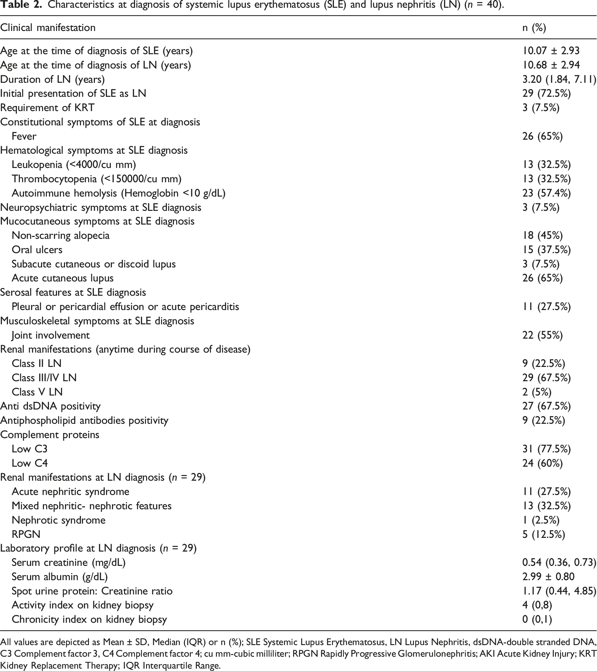
All values are depicted as Mean ± SD, Median (IQR) or n (%); SLE Systemic Lupus Erythematosus, LN Lupus Nephritis, dsDNA-double stranded DNA, C3 Complement factor 3, C4 Complement factor 4; cu mm-cubic milliliter; RPGN Rapidly Progressive Glomerulonephritis; AKI Acute Kidney Injury; KRT Kidney Replacement Therapy; IQR Interquartile Range.
Clinical profile of children with Lupus Nephritis at the time of study enrolment (n = 40).

All values are depicted as Mean ± SD, Median (IQR) or n(%); SD Standard deviation; LN Lupus nephritis; ACE Angiotensin Converting Enzyme; CNI Calcineurin Inhibitors; MMF Mycophenolate mofetil; CKD Chronic Kidney Disease, eGFR estimated Glomerular Filtration Rate, LVH Left Ventricular Hypertrophy; HDL High Density Lipoproteins, LDL Low Density Lipoproteins, VLDL Very Low Density Lipoproteins.
aChildren enrolled received more than one immunosuppressant at different points of time.
Brachial artery flow mediated dilatation (FMD) and carotid intima medial thickness (cIMT) in study subjects.
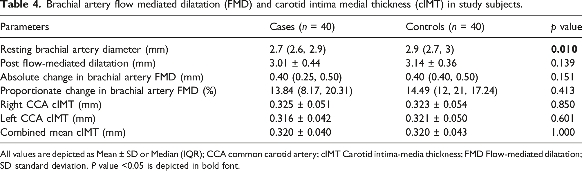
All values are depicted as Mean ± SD or Median (IQR); CCA common carotid artery; cIMT Carotid intima-media thickness; FMD Flow-mediated dilatation; SD standard deviation. P value <0.05 is depicted in bold font.
Subgroup analyses showed that median proportionate change in FMD or preclinical atherosclerosis (cIMT) in LN cases did not differ significantly between the histological class of LN, or with treatment response (complete response, partial response, no response) (p > 0.05). In order to eliminate the confounding effect of BMI on vascular parameters, subgroup analysis of LN cases with BMI <85th centile (n = 34) versus the controls (n = 40) was performed. It was observed that there was no difference in median proportionate change in FMD or cIMT between cases and controls.
Comparison of the mean cardiovascular parameters obtained on echocardiogram between cases with lupus nephritis (n = 40) and controls (n = 40) at the time of study enrolment.
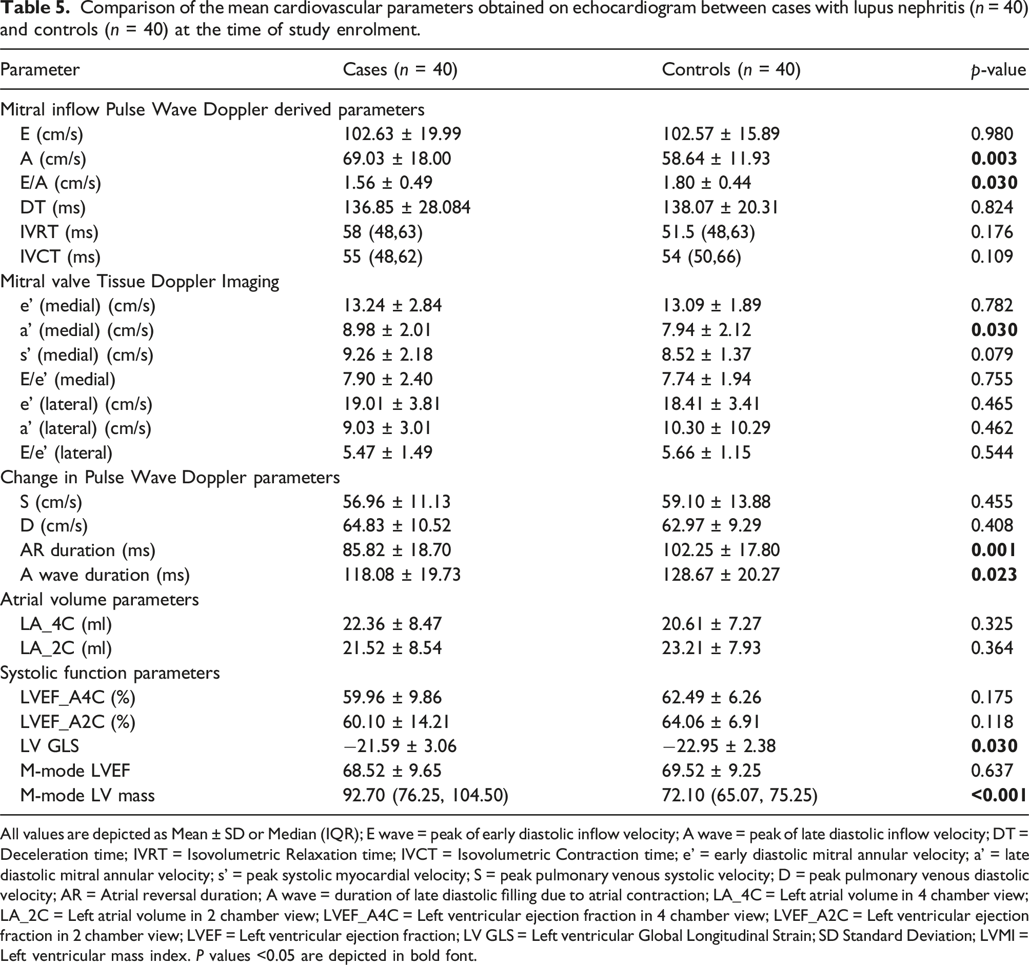
All values are depicted as Mean ± SD or Median (IQR); E wave = peak of early diastolic inflow velocity; A wave = peak of late diastolic inflow velocity; DT = Deceleration time; IVRT = Isovolumetric Relaxation time; IVCT = Isovolumetric Contraction time; e’ = early diastolic mitral annular velocity; a’ = late diastolic mitral annular velocity; s’ = peak systolic myocardial velocity; S = peak pulmonary venous systolic velocity; D = peak pulmonary venous diastolic velocity; AR = Atrial reversal duration; A wave = duration of late diastolic filling due to atrial contraction; LA_4C = Left atrial volume in 4 chamber view; LA_2C = Left atrial volume in 2 chamber view; LVEF_A4C = Left ventricular ejection fraction in 4 chamber view; LVEF_A2C = Left ventricular ejection fraction in 2 chamber view; LVEF = Left ventricular ejection fraction; LV GLS = Left ventricular Global Longitudinal Strain; SD Standard Deviation; LVMI = Left ventricular mass index. P values <0.05 are depicted in bold font.
Figure 1 demonstrates key outcome differences-higher prevalence of proportionate change in FMD <10% among cases when compared to healthy controls, reduced baseline brachial artery diameter, lower E/A ratio indicating diastolic dysfunction, and significantly increased left ventricular mass reflecting hypertrophy when compared to controls. A composite multipanel image highlighting important outcome variables. Abbreviations: FMD Flow mediated dilatation; E/A ratio E indicates early mitral filling velocity, while A indicates late atrial contraction velocity, E/A ratio indicates left ventricular diastolic dysfunction.
Determinants of brachial artery FMD <10%
On univariate analysis, diastolic hypertension was noted to be associated with Brachial artery FMD <10% (OR: 0.56; 95% CI: 0.41–0.76; p 0.034). Other variables such as age of onset of LN, male gender, duration of LN, systolic hypertension, lipid profile, degree of proteinuria, antiphospholipid antibody positivity, presentation as RPGN, class of LN, activity index and chronicity index, number of renal flares and steroid toxicity features were not found to have an association with FMD <10% on univariate analysis. Notably, all 14 children with FMD <10% had high diastolic blood pressure at enrolment.
Discussion
Cardiovascular and cerebrovascular risk factors are the leading contributor to mortality in SLE beyond the second decade.1,28,29 Studies on adults have consistently demonstrated that SLE patients exhibit impaired vascular reactivity and increased cIMT, even in the absence of overt cardiovascular disease; and LN is consistently linked to greater endothelial dysfunction and more evident structural alterations in the vasculature.3,5,30–33 cSLE has greater disease activity and more frequent kidney involvement. Additionally, survival rates in pediatric LN have improved due to advances in management strategies, leading to potential augmentation in the damage accrual in the vascular parameters over a period of time. A higher risk for premature atherosclerosis may potentially ensue. Non-invasive structural and functional assessment of premature atherosclerosis was performed in our study on 40 cases of cSLE with LN disease duration exceeding 1 year and was compared with age- and gender-matched healthy controls due to the lack of universally acceptable pediatric normative data for these measured variables.
The distribution of baseline demographic characteristics in our study was reasonably comparable between the two groups, thereby minimizing the influence of potential confounding factors. The only significant differences observed at enrolment were that LN cases were more likely to be hypertensive, proteinuric, and have hypertriglyceridemia. All the cases enrolled were also found to have an elevated Apolipoprotein B: Apolipoprotein A1 ratio predicting the increased cardiometabolic risk in these children. 34 Hypertension, as well as the biochemical and metabolic abnormalities observed in our LN patients may be attributable to the underlying disease pathophysiology, corticosteroid exposure, or complications such as nephritis and vasculitis. In a study involving cSLE (n = 30), the researchers found that these patients exhibited notably lower levels of HDL, and high cholesterol, apolipoprotein A-1; and increased levels of autoantibodies against oxidized LDL. However, these patients still had normal endothelial function and oxidative state. The authors attributed the results to younger age of the patients. 9
Our study showed that the resting brachial artery diameter had a statistically significant difference, with a lower median diameter in cases compared to controls. This finding may reflect early vascular remodelling or persistent vasoconstriction in children with LN. Also, the proportion of children with FMD less than 10% was observed more frequently in the cases compared to the controls (35% vs 10%, p 0.007), which indirectly reflects the higher prevalence of arterial stiffness and subclinical atherosclerosis among the patients with childhood-onset LN although there was no statistical significance in the proportionate change in the brachial artery reactivity and cIMT between the cases and controls. Also, it is notable that 4 controls (10% of controls) had FMD <10% even though they had no obvious risk factors like obesity, hypertension, dyslipidemia, proteinuria or family history of coronary artery disease; this hints towards a possible genetic predisposition towards atherosclerosis in these cases. The finding of no significant change in the mean cIMT in LN cases underscores the existence of a critical window for early intervention to mitigate long-term vascular risk appearing as structural remodelling on ultrasound.
Prior studies conducted in cSLE patients have evidently shown an early adaptive and adverse cardiac and vascular remodelling in the form of increased cIMT and consequent increased left ventricular mass index.8,28,35,36 In one study, 149 cases of cSLE with a median disease duration of 3.2 years were evaluated for cIMT, FMD and pulse wave velocity and compared with 178 healthy controls. 37 An inverse correlation between disease duration and FMD was noted, indicating worsening endothelial function over time. The findings were limited by the omission of LN status, a critical determinant of both treatment strategy and long-term prognosis; and by the study’s setting in Canada, where disease activity may tend to be lower due to ethnic and geographic factors, potentially limiting generalizability to populations with more severe disease phenotypes such as African-Americans and South Asians. Many previous studies on SLE8,35,37 did not exclusively involve the pediatric population and included different age groups; hence this heterogeneity in the study designs can lead to variability in the interpretation of their results. Moreover, they recruited SLE without LN too.
Several pediatric studies have also shown that cSLE frequently exhibits subclinical cardiac involvement, even when asymptomatic. A study employing radionuclide angiography, thallium myocardial perfusion scans, and echocardiography found that 5 out of 31 asymptomatic cSLE, aged 10 and 19 years, exhibited abnormalities in myocardial perfusion. 6 Likewise, echocardiographic assessments in cSLE have revealed both LV diastolic and systolic dysfunction. Renal involvement and higher disease activity have been associated with worse functional impairment, emphasizing the role of advanced techniques like speckle tracking echocardiography and TDI for early detection in cSLE.38–41
In our LN patient population, mitral inflow doppler revealed findings consistent with altered diastolic filling, while tissue Doppler showed largely preserved annular velocities except for a higher medial a′, indicating greater atrial contribution to LV filling and elevated LV filling pressures. Pulmonary venous Doppler was suggestive of changes in atrial or ventricular compliance. Similar findings have been reported earlier by Chang et al. 38
In our study, subtle systolic dysfunction was evident through a significant reduction in left ventricular GLS in cases compared to controls, despite preserved mean LVEF by both Simpson’s biplane and M-mode methods. The significantly higher LV mass in cases compared to controls as well as 15% of cases having concentric LVH likely reflects early structural remodelling that maintains ejection fraction at younger ages. These findings parallel previous cSLE reports, and emphasize the value of GLS as a sensitive marker for subclinical systolic dysfunction in cSLE.40,41
Some of the merits of our study are that it addresses an understudied population (Indian children with LN). This is one of the few cSLE studies conducted in an ethnicity (south Asians) known to have a heightened risk of severe disease, providing insights that may differ from findings in predominantly Caucasian cohorts. The inclusion of age- and gender-matched controls allowed reliable interpretation of data despite lack of normative data for these parameters. Secondly, comprehensive cardiovascular assessment including structural and functional vascular parameters (cIMT, FMD) and cardiac function across multiple modalities with sophisticated in-built packages provided an integrated picture of cardiovascular health. Also, unlike previous studies35,37 that included SLE with and without LN (making the recruited population heterogeneous), our study included LN cases only.
This study also has some limitations. The study was underpowered to explore determinants of FMD, cIMT, and cardiac dysfunction, requiring caution in interpretation. We also acknowledge that this was a single-center cross-sectional study with a median follow-up period of 3.2 years and that it cannot provide insights into the long-term cardiovascular changes that may eventually develop in these children on follow-up. The linear regression models did not fully explain the observed endothelial dysfunction, limiting causal inference. Also, cumulative corticosteroid exposure was not analyzed, and the influence of medications like ACE inhibitors on vascular reactivity could not be excluded. Although E/e′ is generally more commonly used as an age and preload-independent reliable marker of diastolic dysfunction, only E/A was significantly lower compared to controls in our cohort, making dependence on E/A alone an inherent study limitation. Finally, all imaging studies were independently evaluated by two experienced persons (an echocardiographer and a radiologist) who were blinded to clinical and laboratory data; and intra-observer agreement was assessed. Although this approach is acceptable in certain settings, the lack of an independent assessor limits the ability to evaluate inter-observer agreement.
To summarize, this study demonstrates that Indian children with LN >1 year disease duration have significant cardiovascular comorbidities compared to healthy controls. These include a lower resting brachial artery diameter as well as LV systolic and diastolic dysfunction. The proportion of LN children with arterial stiffness of <10% was higher when compared to controls. cIMT measurements between cases and controls did not reveal any differences.
Future research could be directed into longitudinal, multicentre designs to enable the assessment of temporal changes in vascular and cardiac parameters and disease progression. Larger, ethnically diverse cohorts would improve the generalizability of findings and allow for more robust exploration of determinants of endothelial dysfunction, arterial remodelling, and myocardial impairment.
Footnotes
Author Contributions
Nithya Ramaswamy, Sriram Krishnamurthy, Bobbity Deepthi and Sudarsan Krishnasamy were involved in management of the patients. Nithya Ramaswamy collected the data, reviewed the literature and drafted the first version of the manuscript. Avinash Anantharaj evaluated the patients for cardiac parameters. Ramesh Ananthakrishnan assessed the vascular parameters. Zachariah Bobby supervised the laboratory tests. Sriram Krishnamurthy conceptualized the study, collected the data, reviewed the literature and critically revised the manuscript. All authors contributed to drafting of the manuscript and approved the final version of the manuscript. Sriram Krishnamurthy shall act as corresponding author and guarantor of the paper.
Declaration of conflicting interests
The authors declared no potential conflicts of interest with respect to the research, authorship, and/or publication of this article.
Funding
The authors disclosed receipt of the following financial support for the research, authorship, and/or publication of this article: The study was supported by an intramural funding from the authors’ institution, which is gratefully acknowledged (Grant no. JIP/Res/Intramural/phs-3/2023-2024 dated 4 January, 2024).
Ethical considerations
The study was approved by the Jawaharlal Institute of Postgraduate Medical Education and Research (JIPMER) Institute Ethics Committee (JIP/IEC-OS/184/2023) dated August 9, 2023.
