






Abstract
Spindle cell epitheliomas of the vagina are rare benign tumors of uncertain histogenesis. It has been hypothesized that they originate from a pluripotent cell population. Here, we report 2 cases of spindle cell epitheliomas of the vagina. Both patients presented with a painless vaginal nodule. The resected lesions were solid, well circumscribed, and composed of a proliferation of epithelioid or epithelial cells admixed with spindle cells without atypia. The 2 cellular populations expressed epithelial and mesenchymal markers. Based on these morphologic and immunohistochemical findings, a diagnosis of spindle cell epithelioma was rendered in both cases. Furthermore, we describe for the first time the expression of WT-1 (Wilms’ tumor protein 1) and calretinin in addition to the previously defined immunohistochemical profile.
Case Report
Case 1
A 36-year-old woman presented with a nontender vaginal lump. She had no relevant personal or familial medical history. A left vestibular nodule was found on physical examination. The rest of the clinical evaluation was within normal limits. The lesion was excised and a preliminary clinical diagnosis of vestibular inclusion cyst was rendered.
Macroscopically, the lesion was described as a white to yellow solid nodule of 1.3 × 1.3 × 0.8 cm. On histological examination, the specimen consisted of a nonencapsulated, well-demarcated, and biphasic nodular lesion with an intact subepithelial zone, also called a grenz zone. It was composed of an epithelial component admixed with myxoid stromal areas. The epithelial cells formed scyncitial structures as well as glands. The latter were found predominantly at the periphery of the nodule. There was no squamous epithelium. The stroma was moderately cellular and it consisted of fibromyxoid tissue with spindle cells that were not arranged in a particular pattern. Cytologically, the epithelial cells had round nuclei with vesicular chromatin and abundant eosinophilic cytoplasm. The spindle cells exhibited oval nuclei with inconspicuous nucleoli and scant cytoplasm. None of the components had atypia or necrosis and we counted 1 mitotic figure per 10 high-power fields.
Immunohistochemically, both epithelial and spindle cells were strongly and diffusely positive for Wilms’ tumor protein 1 (WT-1), estrogen receptor (ER), progesterone receptor (PR), CD10, vimentin, smooth muscle actin (SMA), cytokeratin AE1/AE3 (CK AE1/AE3), and they were focally positive for cytokeratin 7 (CK7), epithelial membrane antigen (EMA) and membranous Bcl2. The stromal cells expressed desmin as well as calretinin weakly. Finally, none of the cellular populations were positive for S-100 and CD34 (see Figures 1 and 2).

Case 1. (A) Well-circumscribed lesion with an intact subepithelial zone, low power. (B) Transition between the epithelial component and the spindle cells, medium power. (C) Strong and diffuse WT-1 (Wilms’ tumor protein 1) expression in both epithelial and spindle cells. (D) Weak and focal calretinin expression in spindle cells only.
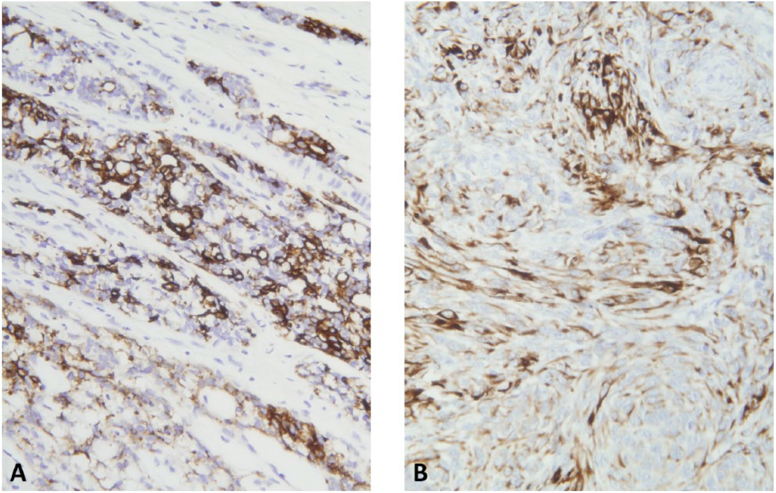
Expression of cytokeratin (CK) AE1/AE3 in case 1 and case 2. (A) Expression of CK AE1/AE3 in spindle and epithelial cells, case 1. (B) Expression of CK AE1/AE3 in spindle and epithelioid cells, case 2.
Case 2
A 40-year-old woman consulted for a nonpainful vaginal nodule that was removed surgically. She had no relevant personal or familial medical history. Macroscopically, the mass was solid, non-encapsulated, and measured 2.2 × 2.0 × 1.6 cm. Microscopically, the lesion was vaguely nodular and it consisted of a subepithelial proliferation of epithelioid and spindle cells. There was a grenz zone between the surface epithelium and the tumor. There were squamoid areas that were separated by collagen bundles but no frank squamous or glandular epithelia were found. The spindle cells were disposed around the epithelioid cells in fascicular and storiform patterns and the stroma was focally fibrotic. Cytologically, the epithelioid cells exhibited nuclei with vesicular chromatin, inconspicuous nucleoli and abundant eosinophilic cytoplasm, whereas the spindle cells showed ovoid nuclei with vesicular chromatin and scant eosinophilic cytoplasm. No atypia nor necrosis was seen and there was 1 mitotic figure per 10 high-power fields.
Immunohistochemically, both components of the tumor were strongly and diffusely positive for WT-1, ER, PR, CD10, vimentin, CK AE1/AE3, CK7, EMA, and membranous Bcl2. The spindle cells expressed SMA, desmin and CD34. Finally, S-100 and calretinin were not expressed (see Figures 2 and 3).
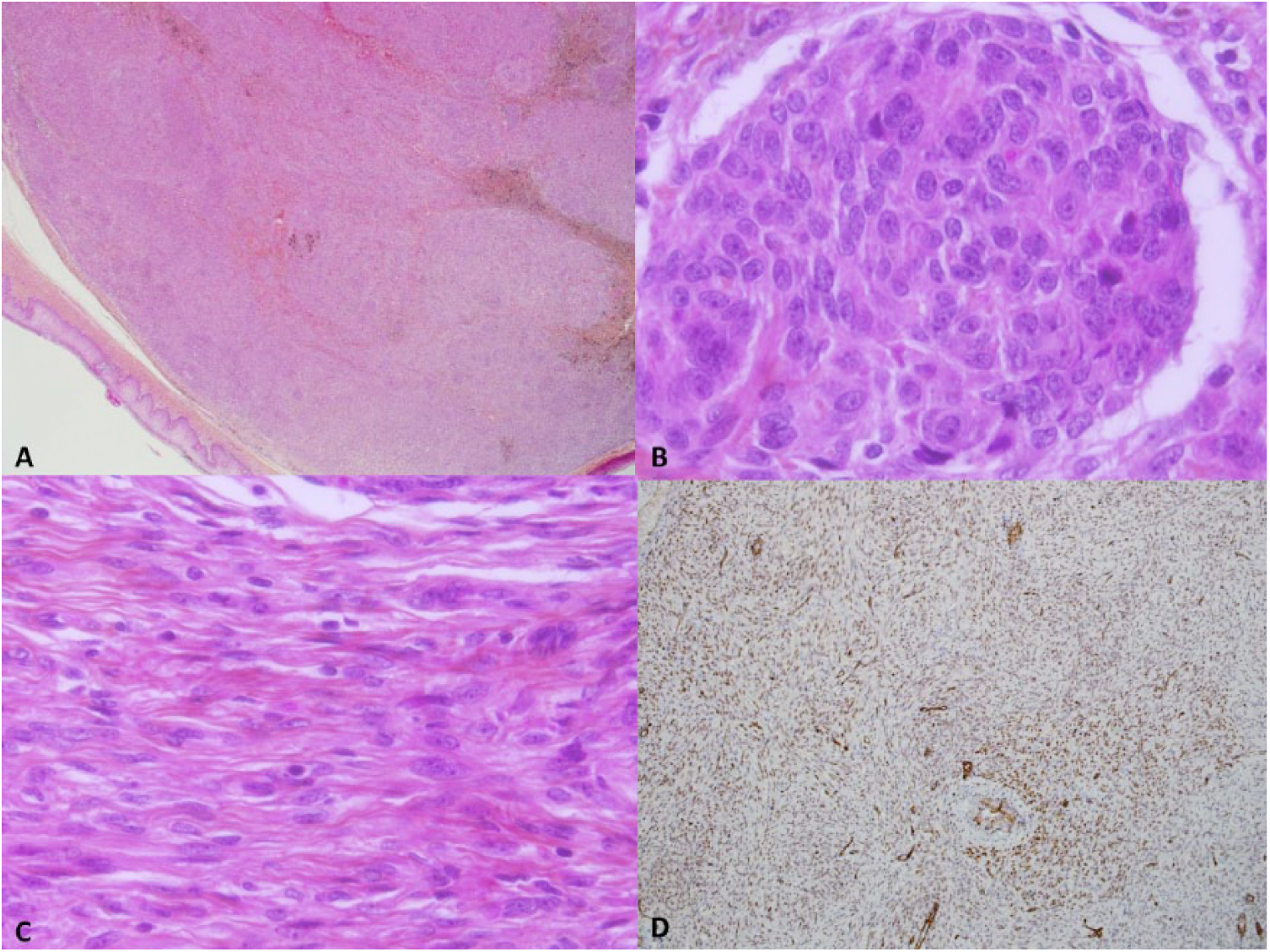
Case 2. (A) Well-circumscribed lesion with a vaguely nodular architecture, low power. (B) Nest of epithelioid cells, high power. (C) Spindle cells, high power. D. Strong and diffuse WT-1 (Wilms’ tumor protein 1) expression in both cell populations.
The morphology and immunohistochemical profile in both cases are in favor of a spindle cell epithelioma of the vagina (SCEV). There was no sign of recurrence after 1 year for the first case and after 6 months for the second case.
Discussion
Spindle cell epithelioma of the vagina, also called mixed tumor of the vagina, is a rare benign entity that was described for the first time in 1953. 1 The tumor is composed of epithelioid or well-differentiated epithelial cells admixed with spindle cells. The epithelial component may form glandular or squamous structures. 2 To our knowledge, approximately 50 cases have been described in the English literature.1-12 The results of immunohistochemical studies are summarized in Tables 1 and 2.2-8,10,11 The lesions are frequently positive for both epithelial and mesenchymal markers. The majority of the reported cases express broad-spectrum cytokeratins, CK7 and EMA. CK 20 is positive in only 3 out of 15 tumors (see Table 1). Furthermore, most lesions express SMA, desmin, CD10, CD34, Bcl2, vimentin, ER, and PR. Expression of S-100 has been described in only 1 out of 26 cases (see Table 2). However, to our knowledge, WT-1 and calretinin positivity has never been reported in the literature.
Expression of Epithelial Markers in Spindle Cell Epitheliomas of the Vagina Reported in the Literature in Addition to Our Report.
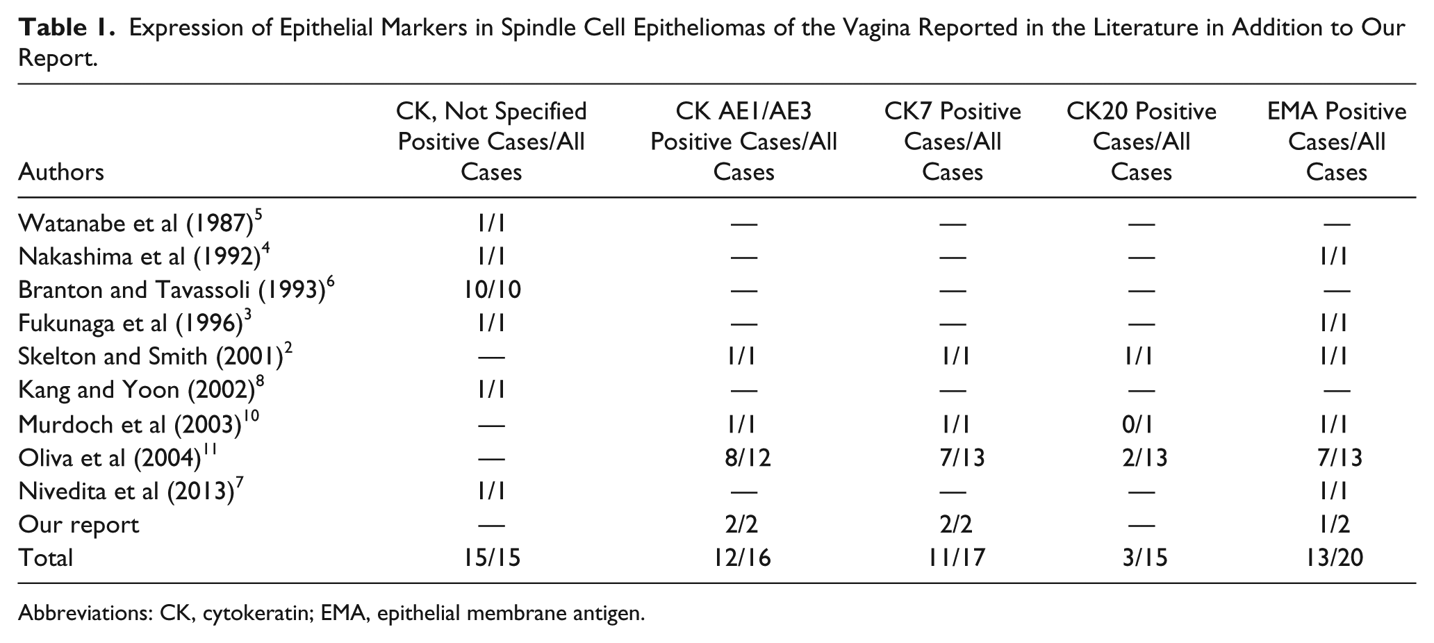
Abbreviations: CK, cytokeratin; EMA, epithelial membrane antigen.
Expression of Nonepithelial Markers in Spindle Cell Epitheliomas of the Vagina Reported in the Literature in Addition to Our Report.
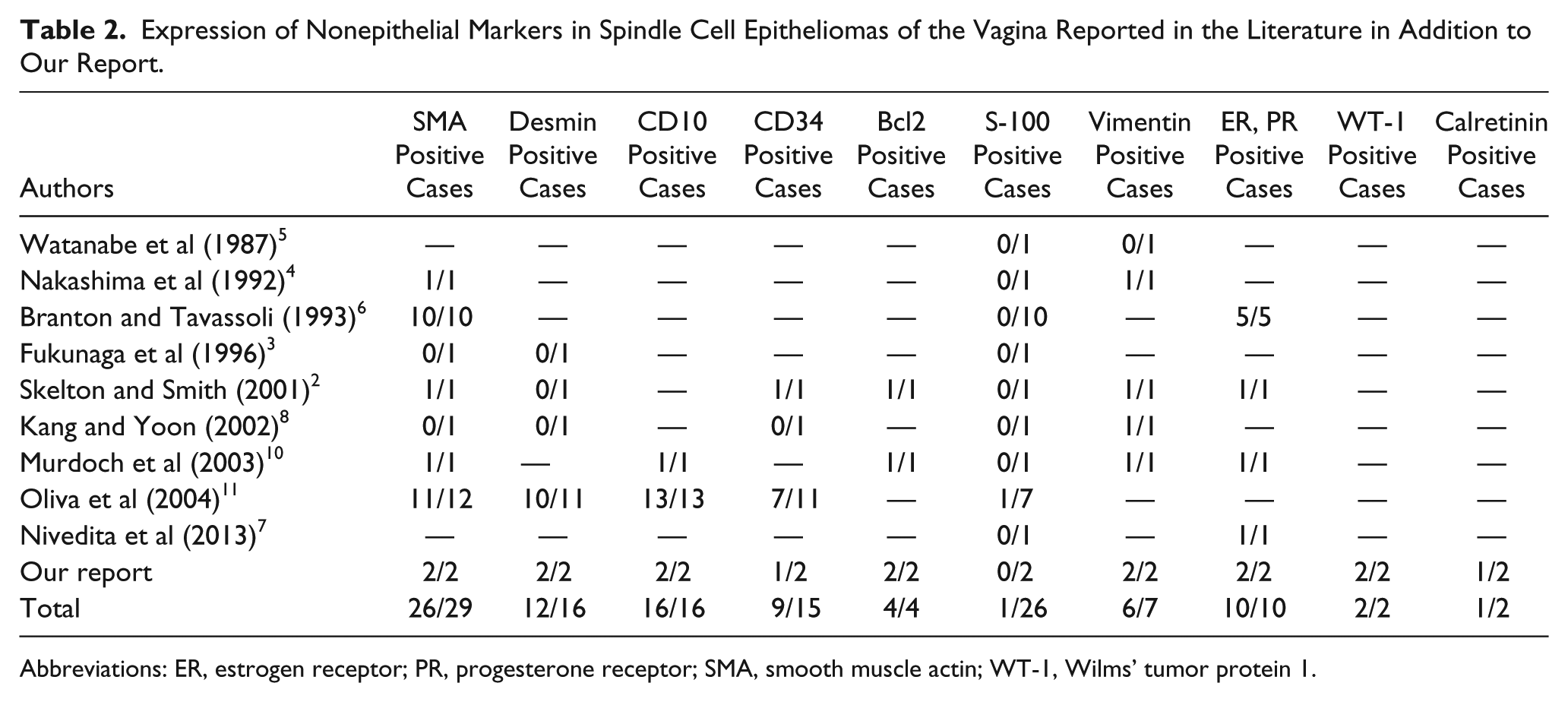
Abbreviations: ER, estrogen receptor; PR, progesterone receptor; SMA, smooth muscle actin; WT-1, Wilms’ tumor protein 1.
The origin of SCEVs is still unclear. Contrarily to mixed tumors found elsewhere in the body, such as those of the breast and salivary glands, SCEVs do not exhibit myoepithelial characteristics. 6 In fact, myoepithelial cells have not been found in the vagina. 6 Moreover, despite the expression of some myoepithelial markers such as SMA and caldesmon, SCEVs’ immunohistochemical profile argues against such an origin because of S-100 negativity. 11 Branton and Tavassoli 6 performed ultrastructural analyses on 2 of their tumors and showed that the spindle cells exhibit epithelial characteristics such as tonofilaments and desmosomes. Kang and Yoon 8 examined 1 case by electron microscopy and also observed desmosome-like structures and tonofilaments in the stromal component, but found no evidence of basal lamina and pinocytotic vesicles that are usually found in myoepithelial cells. Because of this lack of myoepithelial characteristics and since both components of the neoplasm usually coexpress epithelial and mesenchymal markers, it has been hypothesized that SCEVs originate from pluripotential cells. 11 These cells would then be able to differentiate into either epithelial or mesenchymal tissues. 11
Here, we report for the first time the expression of calretinin and WT-1 in SCEVs. Calretinin is an intracellular calcium-binding protein that is positive in glial tissue as well as in normal, reactive and malignant mesothelial cells. 13 WT-1 is a tumor suppressor gene that is expressed by normal and neoplastic mesothelial cells. 14 It is also involved in genitourinary development and in the pathogenesis of many neoplasms, including Wilms’ tumor, ovarian serous carcinoma, desmoplastic small-round-cell tumor and stromal endometrial neoplasms. 15 Calretinin was expressed in the spindle cells of our first case and WT-1 was expressed in the 2 cellular populations in both of our cases. The expression of WT-1 was nuclear, strong, and diffuse whereas calretinin was weakly and focally expressed. Two hypotheses could explain these findings, and they may eventually help determine the origin of this entity. Our first hypothesis is that the pluripotent cell, from which SCEVs are thought to originate, can confer mesothelial characteristics to the neoplastic cells. While this theory would explain why our cases express mesothelial markers, more studies would be needed to determine how common this immunophenotype is among SCEVs. Our second hypothesis is that SCEVs could be related to endometrial stromal neoplasms and particularly endometrial stromal nodules with epithelioid cells because of the strong and diffuse expression of WT-1 and CD10. 15
Clinically, SCEVs are benign tumors and simple excision is the approach that is favored in the literature, even though rare cases of recurrences have been reported.2,6-12,16 The main differential diagnoses are vulvar adnexal and spindle cell neoplasms. While eccrine tumors like spiradenomas can be biphasic, they are negative for muscle markers. 17 Other adnexal tumors have specific morphological characteristics. Tumors of sebaceous origin contain, at least partially, cells that have a vacuolar cytoplasm that indents the nucleus. 17 Pilar tumors are basaloid and exhibit primitive hair mesenchyme in tricheoepitheliomas or characteristic ghost cells in pilomatricomas. 17 Regarding spindle cell tumors such as angiomyofibroblastomas, they can be composed of epithelioid or spindle cells. However, they usually exhibit numerous thin-walled blood vessels around which the cells are aggregated. Also, they do not stain for keratins. 18 Other spindle cell lesions such as aggressive angiomyxoma are not well circumscribed, possess 1 cellular population and are moderately cellular. Therefore, SCEVs are rare tumors with an indolent clinical course and they should be included in the differential diagnosis of solid vaginal masses.
Footnotes
Declaration of Conflicting Interests
The author(s) declared no potential conflicts of interest with respect to the research, authorship, and/or publication of this article.
Funding
The author(s) received no financial support for the research, authorship, and/or publication of this article.