






Abstract
Hodgkin/Reed-Sternberg (HRS) cells of classical Hodgkin lymphoma (CHL) are of B-cell origin. In a small number of CHL cases, the tumor cells can express T-cell antigens. CD8 expression in this setting is extremely rare. We identified 5 cases of CHL with aberrant CD8 expression from our database. The patients included 3 men and 2 women with a median age of 33 years (range = 20-59 years). All the patients initially presented with lymphadenopathy and variable number of RS cells. Two cases were classified as mixed cellularity type that showed prominent vascular proliferation mimicking peripheral T-cell lymphoma. Two cases represented nodular sclerosis type. The tumor cells in all cases were positive for CD8 and negative for CD2, CD3, CD4, and CD7 and carried germline T-cell receptor genes. Molecular studies revealed T-cell receptor genes to be in germline configuration in 4 cases with available information. Given the morphologic overlap with peripheral T-cell lymphoma and the rarity of this type of CHL, identifying more cases will help our better understanding of this entity.
Introduction
Classic Hodgkin lymphoma (CHL) represents one of the common types of malignant lymphoma in the Western world. It is characterized by the presence of clonal Hodgkin/Reed-Sternberg (HRS) cells residing in an abundant heterogeneous admixture of non-neoplastic inflammatory and accessory cells. Although HRS cells usually lack or have partial expression of B lineage markers, in most instances they harbor rearranged immunoglobulin gene (Ig) with somatic hypermutations. These findings suggest that they originate from B cells at the germinal or post-germinal center stage of differentiation. There are rare reports suggesting that HRS cells carry T-cell receptor (TCR) gene rearrangements and can be of T-cell origin. However, even among the cases with aberrant expression of T-cell antigen (TCA) on HRS cells, the tumor cells usually carry somatically mutated Ig gene rearrangements, a finding indicative of a B-cell origin instead of a T-cell origin. Therefore, the expression of TCAs in CHL is aberrant. Given the low incidence of CD8 expression, the clinical significance of this finding and the biological behaviors of this subgroup are still not well understood. We describe 5 cases of CHL with CD8 expression and study their clinicopathological features.
Materials and Methods
Case Selection
We identified 5 cases with aberrant CD8 expression out of 240 cases of CHL diagnosed in 2 tertiary hospitals of Northwell Health from 2011 to 2017. Histologic, immunophenotypic, and molecular features were reviewed and correlated with clinical findings. The diagnosis of CHL was based on the criteria of the 2008 World Health Organization classification. This study was approved by the Institutional Review Board of Northwell Health.
Histology and Immunohistochemical Studies
Formalin-fixed, paraffin-embedded tissue sections were stained with hematoxylin-eosin for histological evaluation. Immunohistochemical analysis was performed using fixed, unstained slides, a panel of prediluted antibodies, and automated immunostainer (Ventana Medical System, Tucson, AZ) as per the manufacturer’s instructions. The antibodies included CD2 (Ventana), CD3 (Ventana), CD4 (Ventana), CD5 (Ventana), CD7 (Ventana), CD8 (Ventana), CD15 (Ventana), CD20 (Ventana), CD30 (Ventana), CD45 (Ventana), anaplastic lymphoma kinase-1 (Alk-1, Ventana), and Pax5 (Cellmaque, Rocklin, CA).
In Situ Hybridization Analysis
In situ hybridization was performed on formalin-fixed, paraffin-embedded tissue sections for Epstein-Barr virus–encoded small RNA (EBER) using an I View Blue Plus Detection Kit (Ventana Medical System). The assay used the Ventana INFORM EBER probe. Staining was performed according to the manufacturer’s instructions. Ventana Red Counterstain II (Ventana Medical System) was used.
Flow Cytometric Analysis
Single cell suspensions were prepared from fresh lymph node tissues and stained with a panel of fluorescence labeled monoclonal antibodies including CD2, CD3, CD4, CD5, CD7, CD8, CD10, CD19, CD20, CD23, FMC7, kappa, and lambda. The cells were washed with phosphate-buffered saline for 3 times and fixed in 4% paraformaldehyde phosphate-buffered saline solution before analysis. Routine 4-color flow cytometric analysis was performed using FACS Diva software (Becton-Dickenson Biosciences, San Jose, CA).
Molecular Analysis for TCR Gene Rearrangements
All amplification reactions were performed according to the BIOMED-2 multiplex polymerase chain reaction protocol for TCR gene rearrangements. For the amplification of TCR-β rearrangements, the Vβ family and FAM-labeled Jβ primers were used in 2 different combinations (TCR-β multiplex tubes A and B) and in one combination containing Dβ and Jβ primers (TCR-β multiplex tube C). For amplification of TCR-γ genes, we used Vγ and FAM-labeled Jγ primers in 2 multiplex combinations (tubes A and B). The polymerase chain reaction amplicons were analyzed using 6% polyacrylamide/TBE gel electrophoresis. The presence or absence of a clonal T-cell population was determined based on the overall analysis of the electrophoretic pattern.
Results
Clinical Features
The main clinicopathological characteristics of the 5 cases are summarized in Table 1. The median age at the diagnosis was 33 years (range = 20-59 years) for the patients including 3 men and 2 women. All the patients initially presented with lymphadenopathy, including 4 with cervical lymphadenopathy and 1 with axillary lymphadenopathy. In addition, patient 3 developed respiratory difficulty secondary to a large mediastinal mass. Clinical history was significant for cerebral palsy and seizure disorder (case 1) and sarcoidosis (case 4).
Clinicopathologic Findings of the 6 Classical Hodgkin Lymphoma.
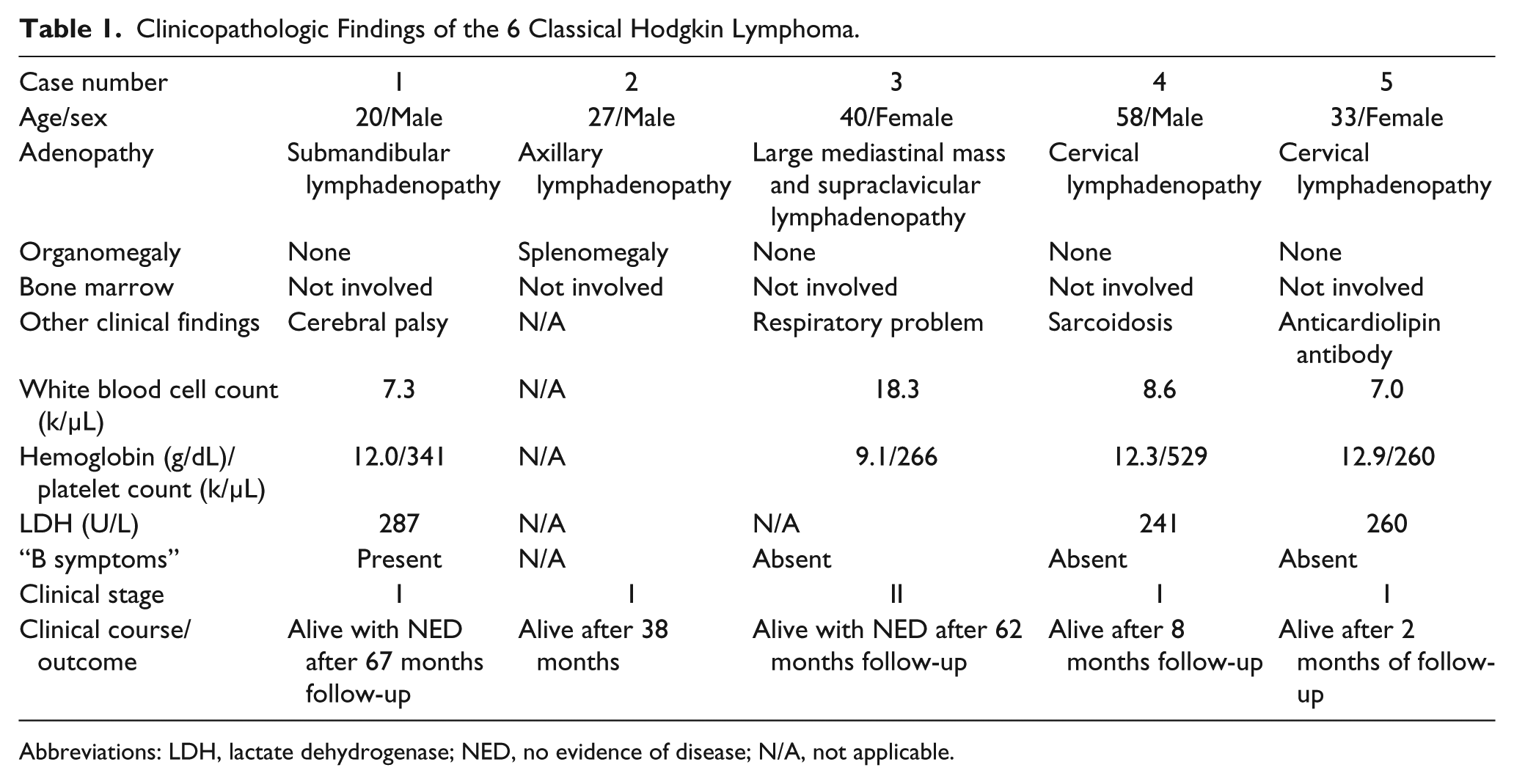
Abbreviations: LDH, lactate dehydrogenase; NED, no evidence of disease; N/A, not applicable.
Complete blood count and basic metabolic panel were within normal limits in all but case 3, which revealed anemia and leukocytosis with marked neutrophilia. Serum lactate dehydrogenase levels were elevated in 2 patients (cases 1 and 5). Anticardiolipin antibody was positive in case 5.
Morphologic, Immunophenotypic, and Molecular Findings
There were 3 cases with excisional biopsies and 2 cases with needle core biopsies. All the biopsies revealed a variable number of large cells with the features of HRS cells in a background of mixed inflammatory cells. The number of large cells varied among the cases, ranging from scattered to sheets of cells. In case 1 and case 4, the neoplastic cells were embedded in a rich background of lymphocytes and histiocytes associated with vascular proliferation (Figures 1 and 4), which mimicked peripheral T-cell lymphoma (PTCL). Two cases (cases 2 and 5) were consistent with nodular sclerosis type (syncytial variant; Figures 2 and 5). There were numerous eosinophils and neutrophils in case 3 (Figure 3).
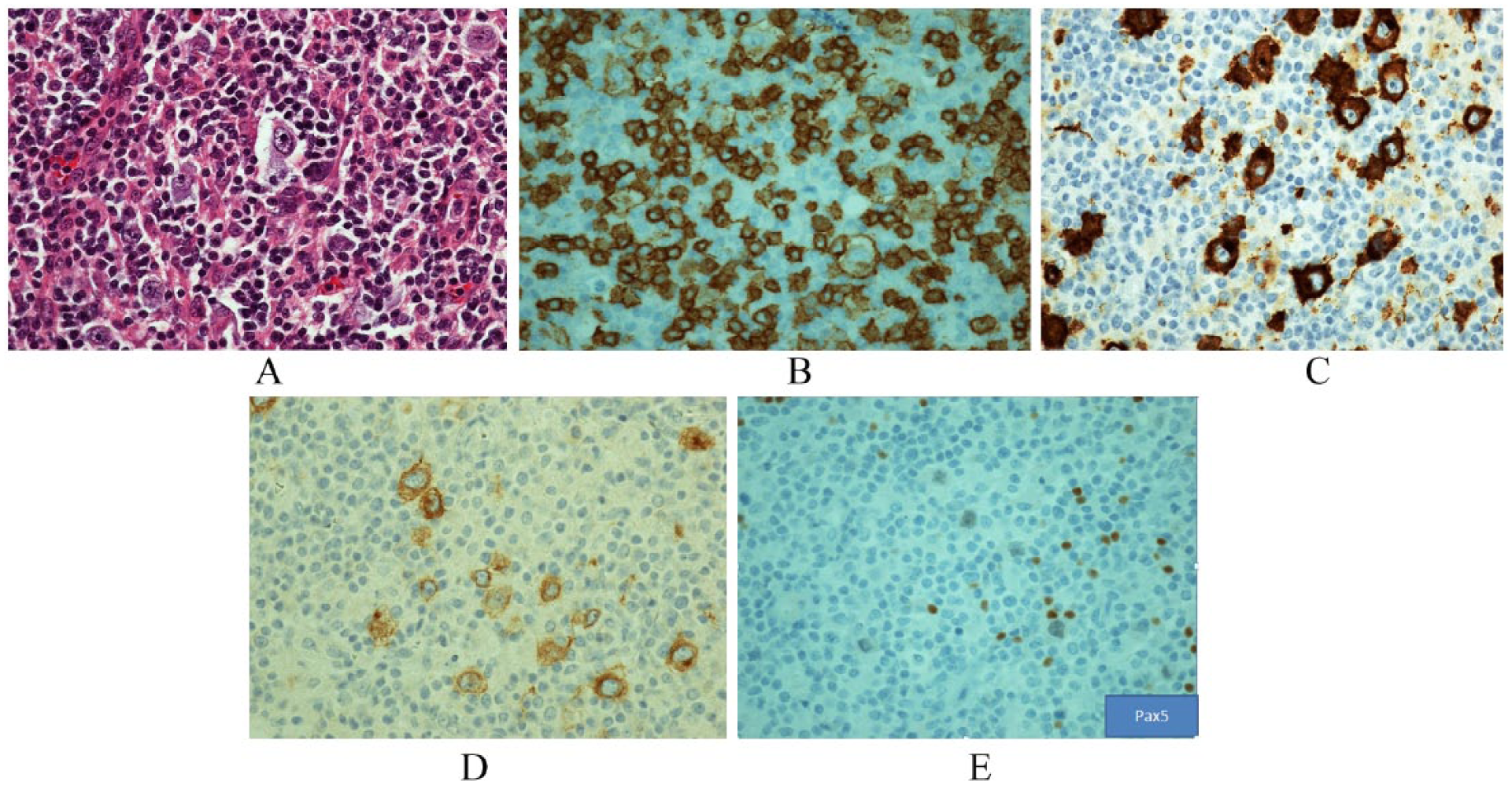
(A) Many HRS cells were embedded in a background rich in histiocytes and lymphocytes. (B-E) The neoplastic cells express uniform CD8 (×400), CD15 (×400), CD30 (×400), and dim PAX-5 (×400).
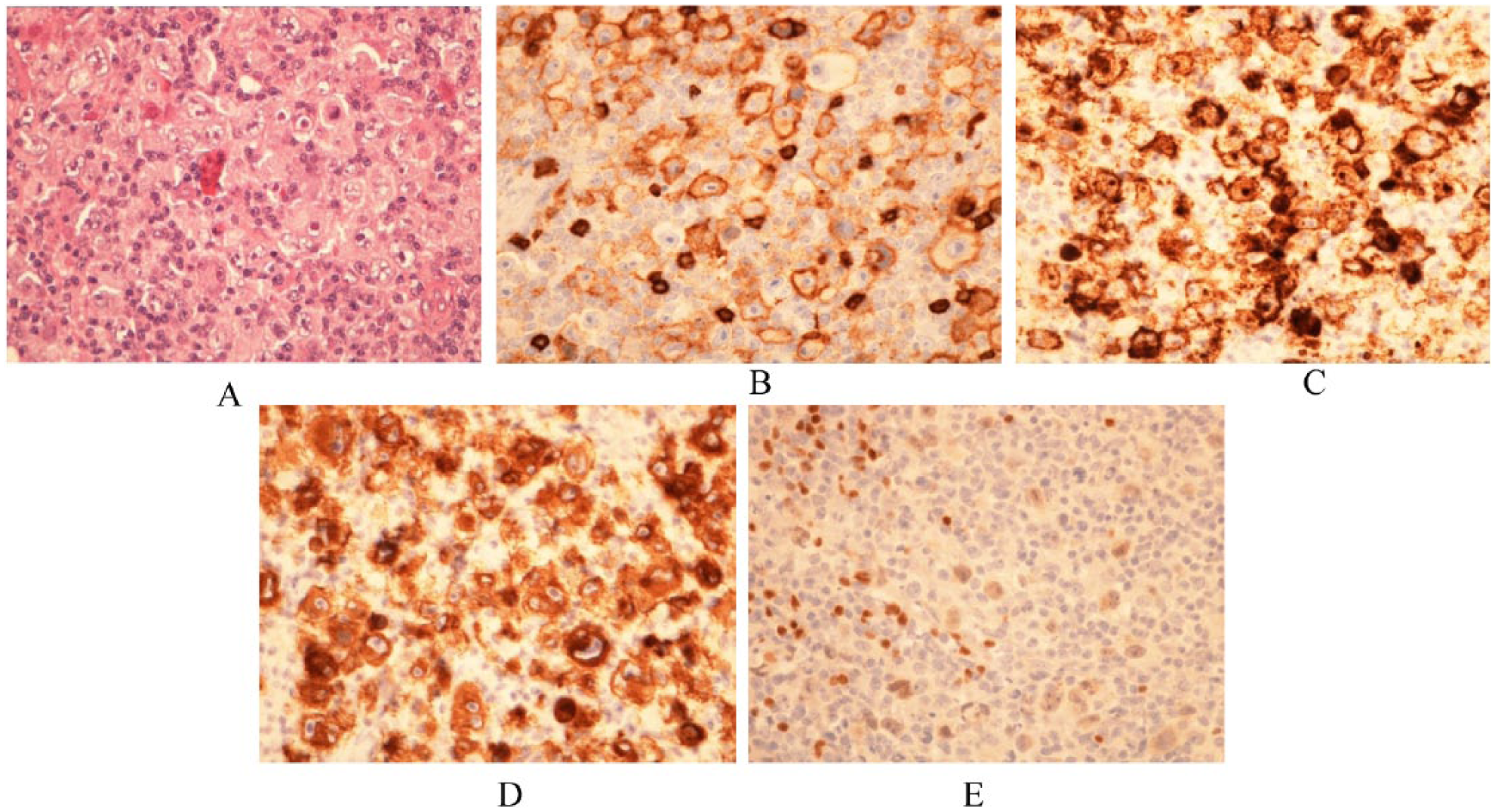
(A) Sheets of HRS cells are present. (B-E) The neoplastic cells are positive for CD8 (×400), CD15 (×400), CD30 (×400), and dim PAX-5 (×400).
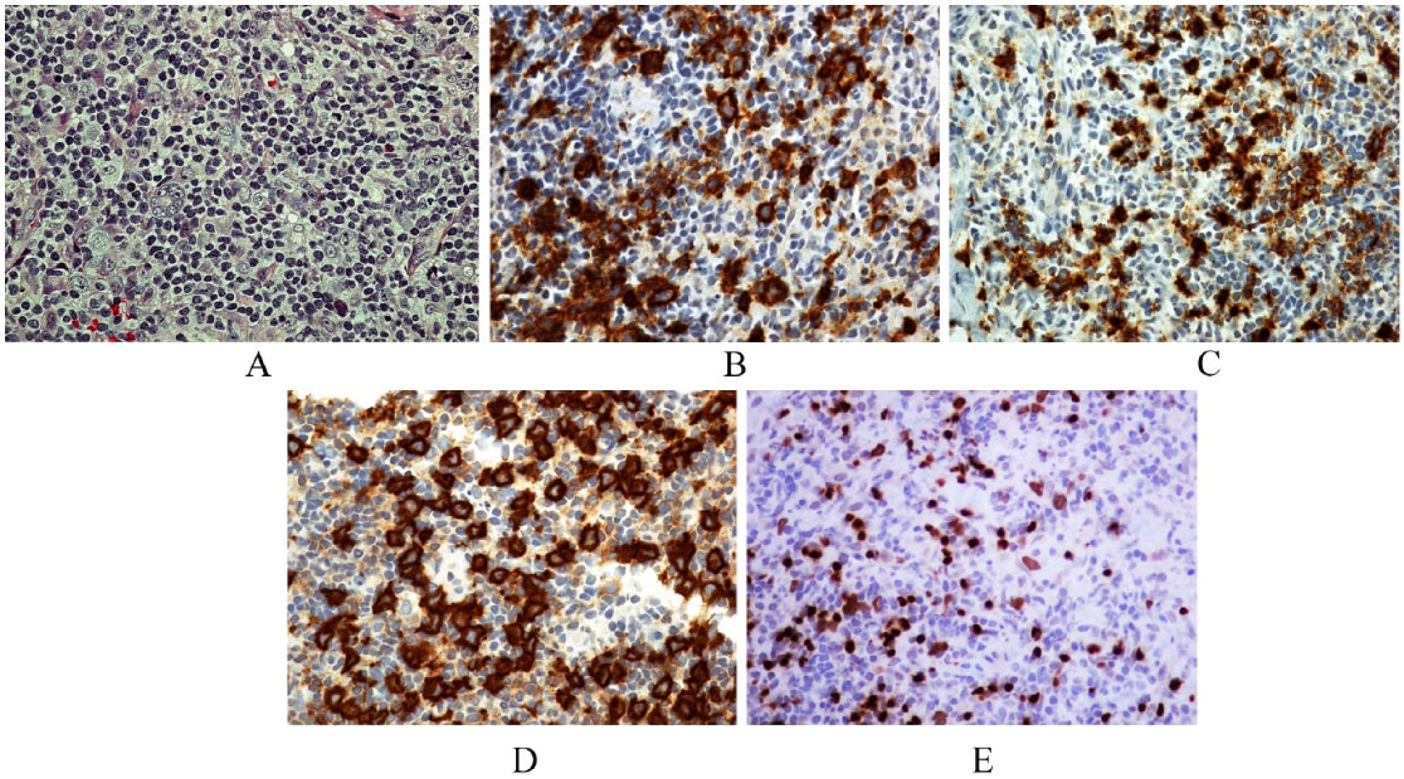
(A) Many HRS cells amid a background rich in eosinophils and neutrophils. (B-E) The neoplastic cells express CD8 (×400), CD15 (×400), CD30 (×400), and dim PAX-5 (×400).
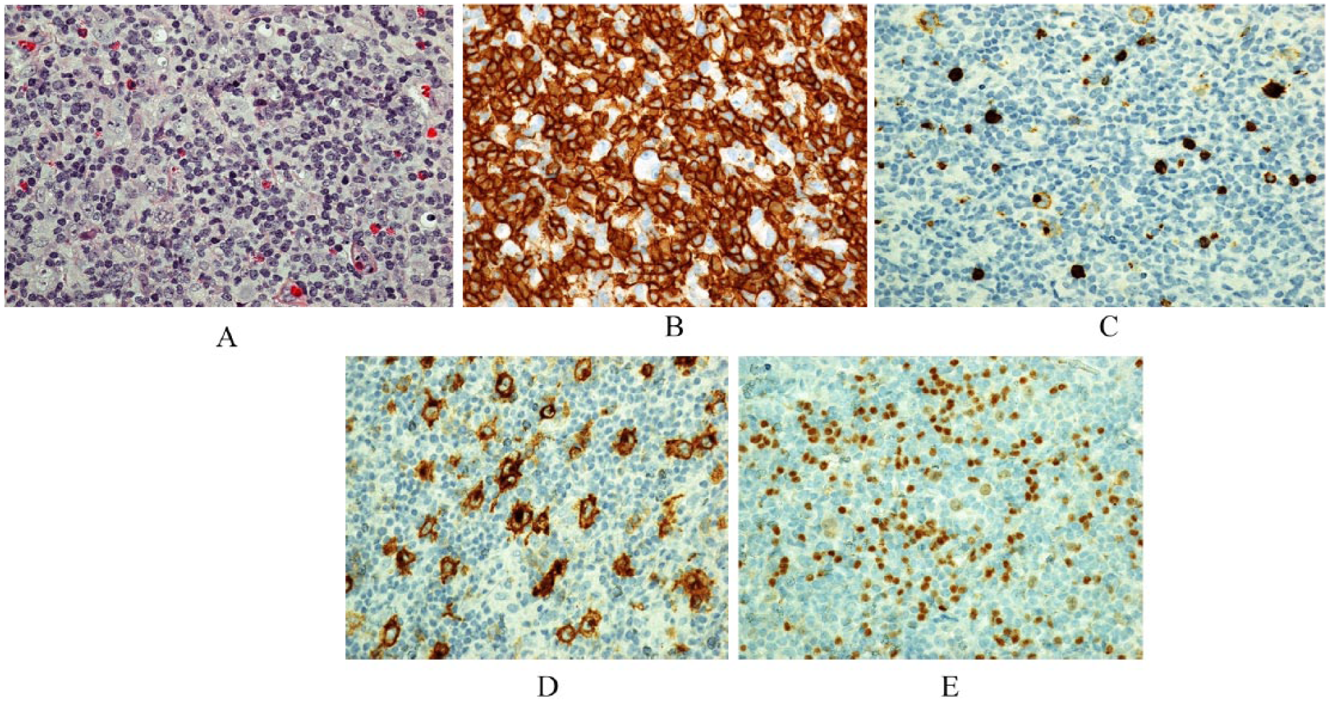
(A) Hematoxylin and Eosin stain shows many HRS cells embedded in a background rich in histiocytes and lymphocytes. (B-E) The neoplastic cells express uniform CD8 (×400), CD15 (×400), CD30 (×400), and dim PAX-5 (×400).
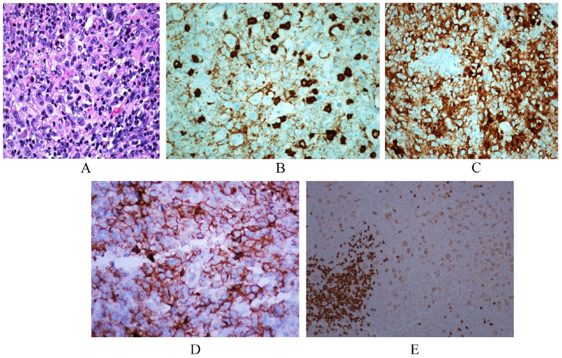
(A) Sheets of HRS cells are present. (B-E) The neoplastic cells are positive for CD8 (×400), CD15 (×400), CD30 (×400), and dim PAX-5 (×200).
Immunohistochemistry and molecular profile of 5 cases are summarized in Table 2. In all cases, the HRS cells were positive for CD30, CD15, CD8, and Pax5 (dim) and negative for CD10, ALK-1, and other T-cell markers (CD2, CD3, CD4, and CD7). One case (case 2) was partially positive for CD5. In addition, CD20 was partially positive in 3 cases (cases 1, 4, and 5). In situ hybridization study for EBER was positive in 2 cases (cases 1 and 4). Cytotoxic markers (TIA-1, granzyme B, and perforin) were negative in one case (case 5) with available materials for testing.
Immunohistochemical and Molecular Profile of the 6 Cases.
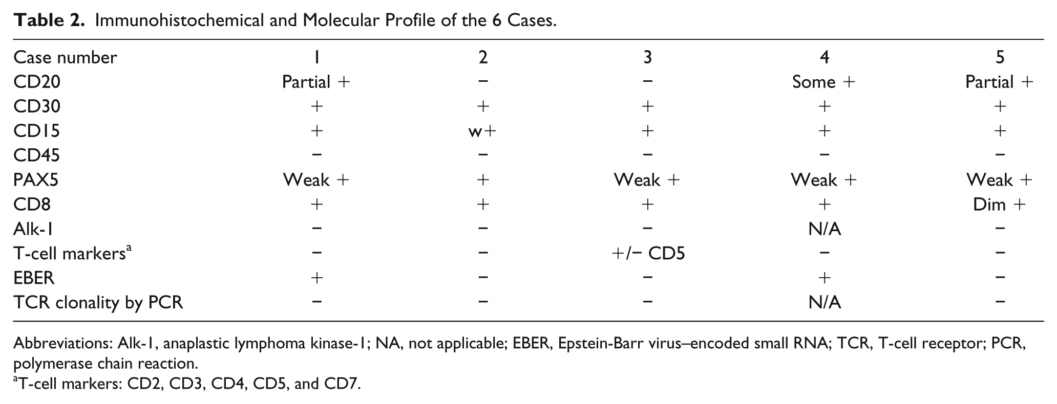
Abbreviations: Alk-1, anaplastic lymphoma kinase-1; NA, not applicable; EBER, Epstein-Barr virus–encoded small RNA; TCR, T-cell receptor; PCR, polymerase chain reaction.
T-cell markers: CD2, CD3, CD4, CD5, and CD7.
Molecular studies were negative for both TCR-β and TCR-γ gene rearrangements in 4 cases with available information.
Clinical Staging, Managements, and Follow-up
A staging bone marrow aspiration and biopsy was negative for involvement by CHL in all cases. Clinical and imaging studies revealed that 4 patients had a stage I disease and 1 patient (case 3) had stage II disease. All patients were treated with the standard regimen for CHL. With a median follow-up of 38 months (range = 2-67 months), all patients were alive without evidence of disease.
Discussion
CHL originates from mature B cells, as its tumor cells demonstrate clonal Ig heavy and light chain gene rearrangements. 1 However, it seems that the HRS cells have lost their B-cell phenotype and show co-expression of antigens of various hematopoietic cell types including T cells, B cells (weak Pax5), dendritic cells (Fascin, CCL17), NK cells (ID2), and myeloid cells.2,3 HRS cells consistently express the activation marker CD30. 4
The evaluation of T-cell-associated antigens is not used in consensus protocols for the immunophenotyping of CHL. Therefore, the exact incidence of aberrant TCA in CHL is not well established. 5 There were only few studies in the literature reporting TCA in CHL, which are summarized in Table 3. Early studies used frozen tissues for immunohistochemical study, and expression of TCA (CD2, CD3, and CD4) was found in up to 40% of CHL cases, with CD2, CD3, and CD4 being the most common. 6 Tzanko et al 7 studied expression of TCA using tissue microarray–based analysis in 259 CHL cases and identified 12 cases (5%) with expression of at least one TCA. Consistent with the previous studies, they found that CD2 and CD4 were the most common TCAs to be expressed in CHL. Only one case was found to express CD8 in their series. These findings were confirmed by a larger study with 50 cases of CHL with TCA expression (TCA-CHL). 5 Overall, among the 117 published cases of CHLs with aberrant TCA expression, only 6 cases were CD8 positive and they were all nodular sclerosis type.5-9 In our study, the tumor cells in all cases were positive for CD8 and negative for other TCAs (CD2, CD4, CD5, and CD7) in all but one case in which the tumor cells were partially CD5 positive. Four out of our cases tested for T-cell clonality were negative. These findings indicate that these cases represent CHL with aberrant CD8 expression rather than CHL of T-cell origin.
Studies on TCA Expression in Classical Hodgkin Lymphoma.
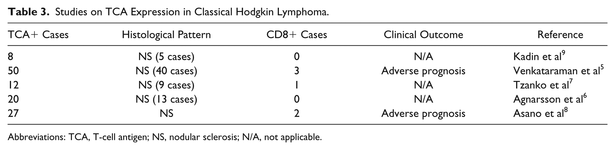
Abbreviations: TCA, T-cell antigen; NS, nodular sclerosis; N/A, not applicable.
Two out of the 5 cases reported here show a rich background of lymphocytes and histiocytes associated with vascular proliferation mimicking PTCL. T-cell markers were included in the panel to exclude T-cell lymphomas. Sometimes PTCLs can display morphological features that overlap with CHL, making it difficult to distinguish between these 2 entities. For example, angioimmunoblastic T-cell lymphomas often have Epstein-Barr virus–positive, Hodgkin-like cells in the background, and uncommonly, PTCL can express both CD30 and CD15. 10 A potential diagnostic pitfall is represented by the expression of TCAs found in CHL. Molecular studies to assess clonal TCR gene rearrangements will be helpful in differentiating the 2 entities. Furthermore, immunohistochemical staining for PAX5 is useful in this setting.11,12 In our study, HRS cells were positive for PAX-5 in all the 5 cases, partially positive for CD20 in 3 cases, and negative for ALK-1 in all the cases. Other TCAs were negative in all but 1 case (partial CD5) and cytotoxic markers were negative in 1 case tested. TCR gene rearrangements were negative in all cases with available information. These findings excluded the possibility of PTCL.
Notably, 2 out of the 5 cases in our study had the histological features of nodular sclerosis type, syncytial variant, which showed a significantly lower incidence (5% to 16%) than typical nodular sclerosis type. 13 Although some case series have suggested that syncytial variant was associated with advanced disease at presentation and comparatively aggressive course,14-16 both of our cases presented with stage I disease and had no evidence of disease during the follow-up period.
The biological behavior and clinical implication of CHL with aberrant TCA expression is unclear. Asano et al 8 identified 27 cases expressing TCAs from 324 CHL cases examined. They employed a limited panel including some T-cell markers (CD3, CD4, and CD8) and cytotoxic markers (TIA-1 and granzyme B). They found that cases expressing T-cell/cytotoxic markers presented at a younger age and were associated with worse outcome. 8 Venkataraman et al 5 studied 50 cases of CHL with aberrant TCA expression and found that the expression of any TCAs on the HRS cells was associated with adverse outcome independent of disease stage. TCA-expression status in CHL was an important and independent predictor of adverse outcome. Therefore, they concluded that a routine testing for TCAs at diagnosis may aid in the identification of CHLs with inferior survival. In our study, the patients had a median age of 33 years and 80% of the patients presented with stage I disease. With a median follow-up of 38 months, all our patients were alive without disease. Overall, our cases were characterized by an early stage at presentation, infrequent B symptoms, lower lactate dehydrogenase level, and absence of relapse or mortality. However, the small number of patient limited the ability to extrapolate our results.
Conclusion
Our data expand the spectrum of clinical, morphologic, and phenotypic features of CHL with aberrant expression of CD8. This type of CHL can show morphological and immunophenotypic features similar to PTCL and, therefore, pose a challenge to the diagnostic pathologist. Given the rarity of this disease, identifying more cases will help us better understand its pathogenesis.
Footnotes
Declaration of Conflicting Interests
The author(s) declared no potential conflicts of interest with respect to the research, authorship, and/or publication of this article.
Funding
The author(s) received no financial support for the research, authorship, and/or publication of this article.
Ethical Approval
Not applicable, because this article does not contain any studies with human or animal subjects.
Informed Consent
Not applicable, because this article does not contain any studies with human or animal subjects.
Trial Registration
Not applicable, because this article does not contain any clinical trials.
