
Abstract
Numerous surgical and non-operative approaches have been used to treat chronic recurrent subluxation of the peroneal tendons in adult athletes. There have been no published reports of surgical repair in children. In this report on a skeletally immature patient a modification of the Chrisman-Snook 3 procedure (previously described for lateral ligament reconstruction) is described to correct recurrent subluxation of the peroneal tendons. KEYWORDS
INTRODUCTION
Subluxating peroneal tendons are an uncommon but frequently diagnosable disorder of the adult ankle. It is often the result of a sudden, forced, dorsiflexion of the foot with an associated contraction of the peroneal muscles everting the foot. This injury has been reported most frequently in skiers 6 but has also been noted in patients who sustain fractures of the tip of the lateral malleolus or who have a malunited fibular fracture 5,6,14,15,17,19,23 other authors have postulated that the injury may occur by a plantar flexion-eversion mechanism in sports 2,7,8 .
If diagnosed acutely, immobilization may result in satisfactory healing. Some patients present with chronic recurrent subluxations of the peroneal tendons since initial diagnosis of this condition is often missed. This may become a painfully disabling injury even requiring operative repair. While multiple operative procedures have been described to correct this condition in adults, surgical correction of this condition in children with open physes has not yet been reported 5,7,8,12,13,17,20,22
This is a report of a skeletally immature patient requiring operative stabilization for painful, subluxating peroneal tendons. Patient follow up four years postoperatively, the child remains asymptomatic with full sports participation and radiographs confirm normal physeal growth completed at the ankle.
PATIENT AND SURGICAL PROCEDURE
K.O., a ten year old girl, presented with a history of intermittent right lateral ankle pain for approximately one year. No history of acute injury was noted by either the patient or her parents. The patient reported that if she put her foot in certain positions, she would note a painful, bulging, soft tissue mass on the lateral aspect of her right ankle. The pain would last for about one week and resolve with rest. As the patient became more active, she reported persistent right ankle pain. On physical exam she had full range of motion and a motor strength of 4/5. Resisted active eversion and palpation of the tendons at the lateral malleolus provoked pain. Radiographs were unremarkable (Fig. 1).
The child's symptoms did not improve despite ongoing physical therapy and the use of a protective Aircast for approximately eight months. Because of the frequency and severity of her symptoms, the patient underwent operative repair. The senior author performed this surgery.
A hockeystick incision was made on the lateral aspect of the right ankle over the peroneal sheath. Using care not to injure the peroneal tendons nor the sural nerve, both the superior and inferior peroneal retinaculum were identified. The peroneal tendons were found to be hypertrophied and subluxated quite easily under the retinaculum over the lateral malleolus. Given the increased size of the tendons and the inadequacy of the peroneal groove, it was felt that a retinacular repair alone would not be effective. The peroneus brevis was identified and split in half longitudinally using a tendon stripper and cut proximally at 15 centimeters, leaving it attached distally. A suture was placed in the most proximal end of the graft in a baseball stitch fashion. Using a K-wire under fluoroscopic guidance, a starting hole was made in the anterior to posterior plane, distal to the fibular epiphyseal plate and enlarged with drill bits. Additionally, two holes were made at 45 degree angles to each other in the lateral aspect of the calcaneous and then connected using a curette. The tendon graft was passed through the fibula from the anterior to posterior, inferiorly through the calcaneal tunnel and then passed upward and sutured onto itself. The superior and inferior retinaculum were reefed to the tendon graft.
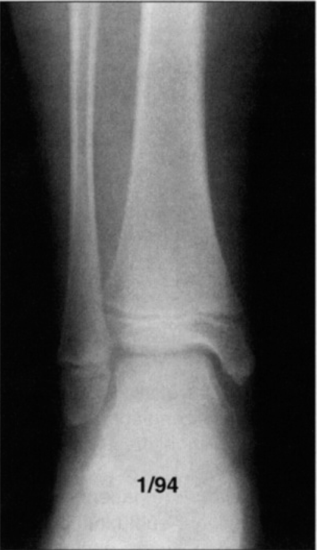
Preoperative AP view of right ankle
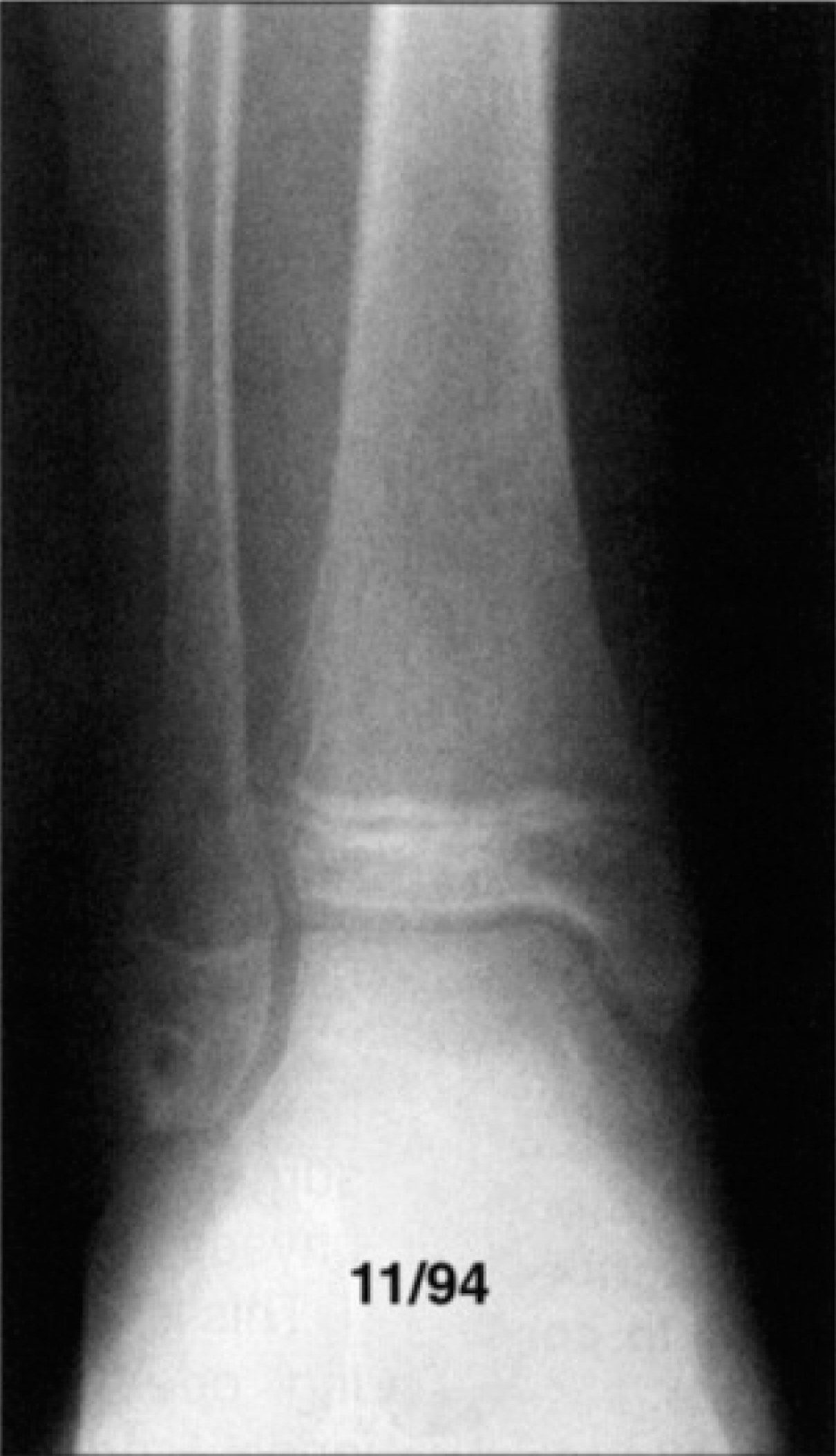
Radiograph 10 months postoperatively showing satisfactory physeal growth
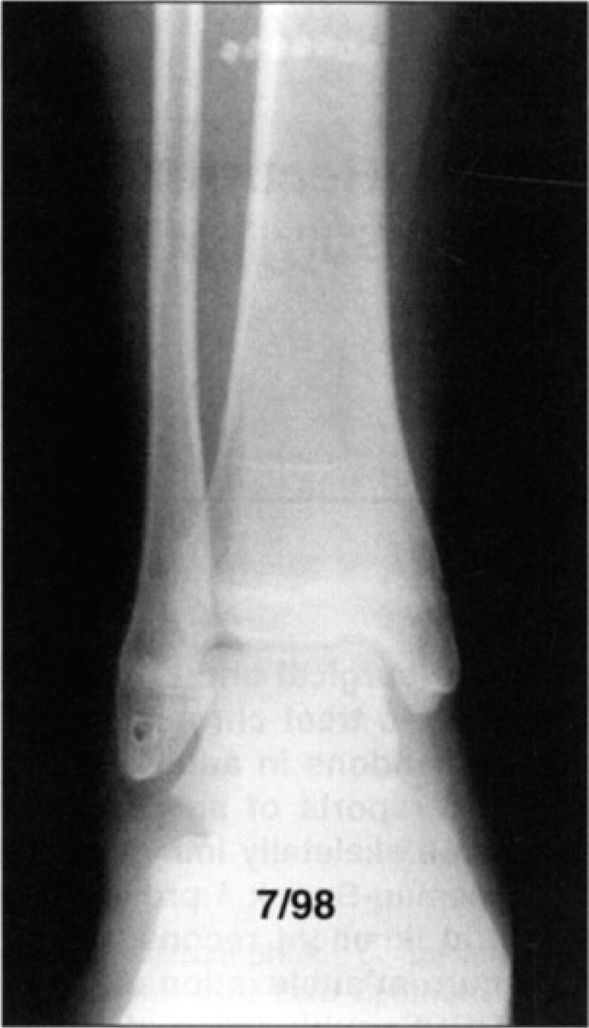
Radiograph four years postoperatively showing that the child had attained almost full skeletal maturity. There has been no physeal arrest. Some minimal physeal stimulation may have occurred.
Postoperatively the patient was placed in a short leg cast for four weeks with partial weight bearing. She was then placed in a below the knee removable cast boot for an additional four weeks during which she commenced physical therapy. By ten months postoperatively she was participating in full sports activities without pain. Radiographs confirmed uninterrupted physeal growth at 10 months postoperatively (Fig. 2). At four years postoperatively, she remains asymptomatic with full activity. Radiograph at that time confirmed almost full skeletal maturity without premature physeal arrest, and a normal relationship of the distal tibia and fibula (Fig. 3).
DISCUSSION
When treating pediatric patients with open physes, surgical procedures must be strategically planned to avoid damage to the epiphysis. Chronic recurrent subluxating peroneal tendons are an uncommon injury usually seen in adults. To our knowledge there are no reports of either acute or chronic recurrent subluxating peroneal tendons in a pediatric patient.
It is generally agreed that early recognition and immobilization or surgical repair of the torn retinaculum is the treatment of choice in adults with acute subluxations of the peroneal tendons 2,17 . Controversy still exists as to the appropriate surgical procedure. Multiple techniques have been described over the years: repair of peroneal retinaculum 1,10,20,21 ; reconstruction of the peroneal retinaculum with a periosteal flap or a tendon sling 4,7,10,13,20 ; bone block procedure 5,8,12,16 ; rerouting procedure 15,18 and deepening of the peroneal groove 12,21,22,23 .
All of these procedures were described for treatment of this condition in the skeletally mature. Needless to say, bone block procedures can not be used with open physeal plates. In addition, retinacular repair alone is rarely successful in cases of chronic subluxation with hypertrophy of the tendons, as in our case.
Stein 18 in 1987, described using a portion of the peroneus brevis tendon for reconstructing the superior peroneal retinaculum in a seventeen year old male. In our case of a pre-pubescent female, we elected to use a modification of the Chrisman-Snook 3 procedure. The Chrisman-Snook 3 procedure was described in 1969 for reconstructing lateral ligamentous structures for lateral ankle instability. In this application of the procedure we were able to simultaneously reduce the excessive size of the peroneal tendons in the groove and produce an effective augmented retinacular repair.
Subluxating peroneal tendons is a relatively uncommon condition in children. Clinical observation suggests that there is an increased tendency for this condition to occur in the ligamentously lax 11 . However, this has not yet been demonstrated in the literature.
CONCLUSION
By performing a modification of the Chrisman-Snook 3 procedure as described in this case report, we were able to successfully repair chronic subluxation of the peroneal tendons in a ten year old while avoiding injury to the distal fibular epiphyseal plate. In addition, by splitting the hypertrophied peroneus brevis tendon to use as a tendonous sling, the size of the tendon was reduced and its function was not adversely affected.