
Abstract
Tarsal navicular fractures in athletes, although rare, can present both a diagnostic and therapeutic dilemma. Failure to recognize this injury and initiate treatment early can have devastating consequences. The physician must have a high index of suspicion for the injury in any patient with midfoot pain after a direct blow. Two case reports of tarsal navicular fractures sustained by baseball players at bat in which the diagnosis was not made early are presented.
INTRODUCTION
Eichenholtz described the diagnosis of tarsal navicular fractures as “sometimes obvious, frequently difficult, and occasionally elusive.” 4 Failure to correctly diagnose and treat this injury can lead to deleterious consequences, particularly in athletes. Because of the relative rarity and often-subtle presentation of this injury, the physician must have a high index of suspicion for a navicular fracture in patients with traumatic midfoot pain.
Navicular fractures are often the result of a direct traumatic blow. Certain sports, such as baseball, expose the athlete to a speeding, hard ball that can ricochet off the player's foot. In particular, the baseball player's lead foot while batting is vulnerable.
The purpose of this paper is to present two cases of tarsal navicular fractures in major league baseball players at bat. This is a previously undescribed mechanism for this injury. Failure to accurately diagnose and treat this injury can have deleterious effects on the athlete's career.
CASE 1
A 29-year-old, left-handed, major league baseball player presented with two months of right midfoot pain after fouling a ball off the dorsum of his right (lead) foot.
Plain radiographs of the foot one day and two weeks after the injury were interpreted as negative for fracture. The player attempted to play with pain the last two months of the season. With completion of the season, the player, with a million-dollar contract, was a free agent and subsequently released by the team. At the end of the season, a computed tomography scan (CT) was obtained.
Axial view of CT scan fracture of the medial one-half of the tarsal navicular with extension into the talonavicular joint as well as into the navicular-middle cuneiform joint with bony resorption.
The patient was then referred for further treatment. Physical exam revealed a discrete area of tenderness of the tarsal navicular. The CT scan revealed a fracture of the medial one-half of the tarsal navicular with extension into the talonavicular joint as well as into the navicular-middle cuneiform joint with bony resorption (Fig. 1). The patient was placed in a short-leg, non-weight-bearing cast with a bone stimulator (EBI, Chicago IL). At five weeks after application of the cast, radiographs revealed a partial consolidation of the fracture. The cast was bivalved. Foot range-of-motion exercises and isometric calf-strengthening exercises were started. One week later, the cast was removed and the patient was placed in a mid-calf cam walker.

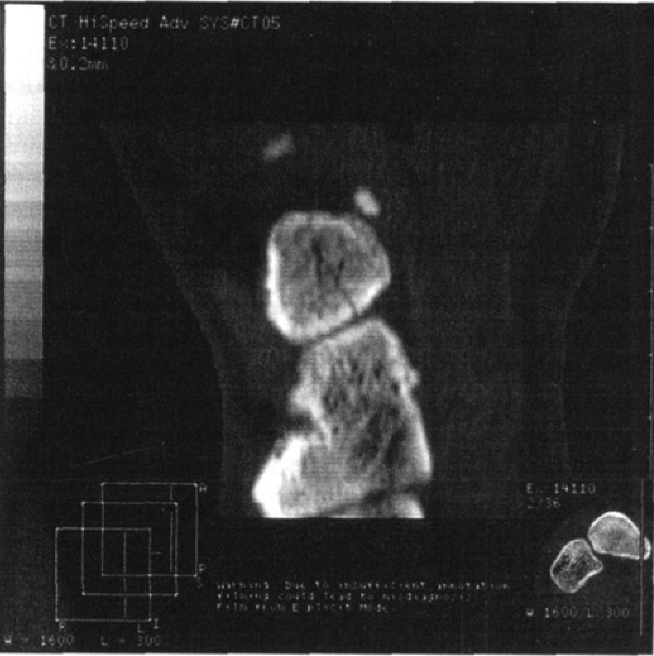
CT scan of split navicular tuberosity fracture.
Follow-up CT scan eight weeks after the start of treatment revealed complete consolidation of the fracture. Limited impact exercises with progression to running was undertaken during the next three weeks. The patient was pain free and at full activity at the following spring training, approximately eight months after the initial injury.
CASE 2
A 28-year-old, left-handed, professional baseball player was referred for treatment for right midfoot pain one week after fouling a ball off of his right (lead) foot. The patient stated that the pain was accentuated by running, weight-bearing, and inversion of the foot. The player was traded to another team two weeks after the injury. Upon his arrival to the new team, plain radiographs, bone-scan, and MRI revealed a nondisplaced fracture of the medial aspect of the navicular. The trade was cancelled due to the injury.
Upon referral, initial physical exam revealed swelling and tenderness of the medial midfoot, with focal tenderness of the medial aspect of the tarsal navicular. Plain radiographs revealed an accessory navicular. A CT scan revealed a nondisplaced, sagitally oriented fracture of the medial third of the navicular with an additional coronal split.
The patient was placed in a short-leg cast. Without notifying his physician, the patient removed his cast three weeks after it was placed. Eight weeks after presentation to us, the patient was experiencing no pain and was nontender. CT scan revealed that the fracture was healed (Fig. 2). The patient was pain-free and returned to full, competition at that time.
DISCUSSION
Tarsal navicular fractures can be classified into dorsal lip avulsion fractures, tuberosity fractures, navicular body fractures, and stress fractures. 2 Dorsal lip avulsion fractures, the most common type, 4 occur with sudden eversion or inversion of the plantarflexed foot. This fracture can be treated with four to six weeks use of a short-leg walking cast. 6 A displaced fragment of more than 25% of the articular surface should be reduced and fixed with either Kirschner wires or a small fragment screw. 6
Tuberosity fractures account for 2% to 12% of navicular fractures. They occur secondary to increased tension on the posterior tibialis tendon with an acute eversion or valgus injury to the foot 6 or as a result of direct trauma. Fractures resulting from a direct blow are usually nondisplaced due to multiple ligamentous attachments as well as the broad insertion of the posterior tibialis tendon. They have been reported to occur in association with other midtarsal joint injuries; 1,3,5,8 therefore, it is important to closely examine the entire midfoot when a navicular tuberosity fracture is diagnosed. Also, an accessory navicular is often mistaken as a tuberosity fracture. These injuries can be treated nonoperatively with use of a compressive dressing and weight bearing as tolerated or a short-leg walking cast for four to six weeks. Nonunions are usually asymptomatic; however, delayed excision is effective treatment for the occasionally symptomatic nonunion.
Sangeorzan described three types of displaced navicular body fractures. 7 For a type 1 fracture, the primary fracture line is in the coronal plane, producing dorsal and plantar fracture fragments. In a type 2 fracture, the primary fracture line is dorsal-lateral to plantar-medial. The major fragment, which is dorsal-medial, is usually displaced medially. A type 3 fracture is a comminuted fracture with disruption of the medial border of the foot. Nondisplaced navicular body fractures are treated in a short-leg weight-bearing cast for four to six weeks. Open reduction and internal fixation is suggested for displaced fractures, as attempts at closed reduction are futile. 2,4,8
Due to the rarity of tarsal navicular fractures, the clinician must have a high index of suspicion when evaluating a patient with traumatic midfoot pain. A failure to diagnose and treat navicular fractures can lead to subsequent midfoot pain and arthritis of the talonavicular joint. In the presented cases, failure to initially diagnose a tarsal navicular fracture resulted in one player's contract not being extended and the other player's trade to a World Series contender being cancelled.
Fractures of the tarsal navicular can be the result of direct or indirect trauma. Physical examination often reveals point tenderness over the navicular, or diffuse pain and swelling of the entire midfoot. The pain is accentuated by weight-bearing and activity. It is of great importance to assess the surrounding joint, particularly the talonavicular and the navicular-cuniform joints, as subluxation or dislocation as associated injuries are not uncommon.
Radiographically, navicular fractures can be diagnosed with high-quality plain film radiographs. Negative plain radiographs in the patient with persistent midfoot pain warrant further diagnostic imaging such as computed tomography, bone scintigraphy, or magnetic resonance imaging. 6 Interval computed tomography is an effective means of assessing fracture healing.
It should be noted that although the case reports involve major league baseball players, this mechanism for navicular fractures can also occur in players of lower levels where the speed of the ball coming off the bat can reach sufficient velocity to cause this injury. Protective devices such as Kevlar foot plates can be worn to protect the injured player returning to play with a foot injury or for injury prevention.
Our algorithm for evaluation and treatment of the athlete with midfoot pain is as follows. After a thorough history including the exact mechanism of injury, palpation of the foot is undertaken to identify discrete areas of tenderness. Plain film (AP, oblique, and lateral) views of the involved foot are obtained. If a fracture of the navicular is identified, a CT scan of the involved foot is obtained to better delineate the fracture. If the plain films fail to reveal a fracture or other pathology accounting for symptoms, an MRI is obtained to evaluate for bony edema indicative of fracture. In the instance of a positive MRI, a CT scan better delineates the fracture.
Our management of navicular fractures depends on the type of fracture, the amount of displacement, particular involvement, and the stability of the midtarsal joint. Unlike fractures of the proximal fifth metatarsal, there is no literature to support the use of early surgical fixation of minimally displaced navicular fractures to expedite return to play for athletes. Treatment of touchdown weight-bearing accompanied by range-of-motion and strengthening exercises to maintain calf strength and ankle range of motion is preferred. We kept the athlete with the comminuted fracture in a non-weight-bearing short leg cast and used a bone stimulator until consolidation of the fracture was noted because of the delay in treatment.
Tarsal navicular fractures in athletes, although rare, can present a diagnostic dilemma. The physician must have a high index of suspicion for the injury in any patient with midfoot pain after a direct blow. Failure to recognize this injury can have devastating consequences, and has the potential to have a great effect on the professional athlete's career, as well as the team and management's confidence in their team physician.