






Abstract
Fractures of the calcaneus with associated locked dislocation of the posterior facet have been previously described. Two patients with a calcaneal fracture with a locked dislocation of a portion of the posterior facet were treated with minimally invasive open reduction and percutaneous screw fixation of the fragment with cannulated screws. Both patients had satisfactory reductions and healed the fractures without any soft-tissue complications. This technique can be a useful addition to the armamentarium of the surgeon treating these injuries, especially in the patient at high risk for wound complications.
Introduction
Fractures of the calcaneus with associated subluxation or dislocation of the posterior facet have been previously described. 3 -5,10,12 There have been multiple reports of extrusion of the posterior facet medially with injury to the tendinous or neurovascular structures. 3,9 The treatment of patients with displaced intra-articular calcaneal fractures remains controversial with trends toward open reduction and internal fixation. 1,2,7,11,13,14 The locked fracture-dislocation of the calcaneus remains a surgical indication. 4,12 With extensile lateral approaches, severe wound complications have been described, especially in patients with risk factors such as diabetics, the elderly, or tobacco abusers. Tornetta recently revisited the role of percutaneous fixation for calcaneal fractures using the Essex-Lopresti maneuver for tongue-type fractures with encouraging results and a low complication rate. 15,16 To our knowledge, however, the use of a minimally invasive technique has not been described for a locked fracture-dislocation of the calcaneus. We report the results of two cases of patients treated with this technique.
Case #1
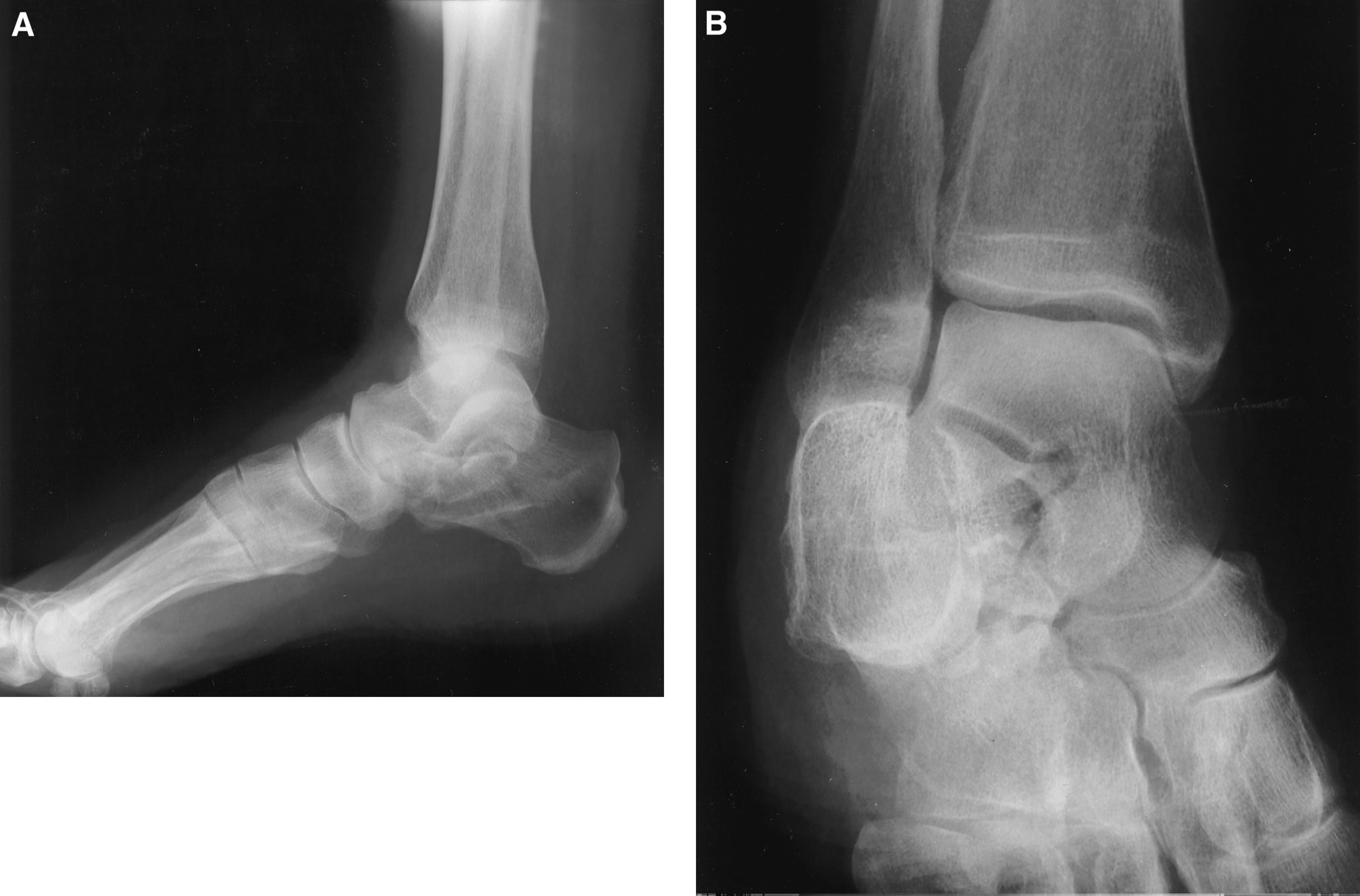
A 74-year-old female fell and sustained a closed intra-articular fracture of the calcaneus. Her past medical history was significant for hypertension. She denied diabetes or tobacco use. On physical examination she had a swollen and ecchymotic foot. Neurovascular status was normal. X-rays revealed a displaced intra-articular fracture of the calcaneus (Fig. 1, A and B). CT scan demonstrated locked displacement of a fragment of the posterior facet between the fibula and the talus (Fig. 2, A and B). Eight days after injury, after resolution of soft-tissue swelling, she was brought to the operating room. A Schanz pin was placed in the calcaneal tuberosity and longitudinal traction was applied along with manual lateral pressure over the dislocated fragment. The fragment was reduced and verified under fluoroscopy by obtaining a Bröden's view. Two 4.0-mm cannulated screws (Ace DePuy, Johnson and Johnson, Warsaw, IN) were then placed percutaneously to secure the posterior facet fragment (Fig.3, A and B). The patient was immobilized for 6 weeks in a cast and maintained nonweightbearing for 12 weeks. At last follow-up, 1 year from her injury, she had mild pain with walking on uneven ground and reported no activity restrictions. Her subtalar motion was 50% of the contralateral extremity on manual testing. Radiographs revealed maintenance of reduction and no evidence of post-traumatic subtalar arthrosis.


Postoperative lateral (
Case #2
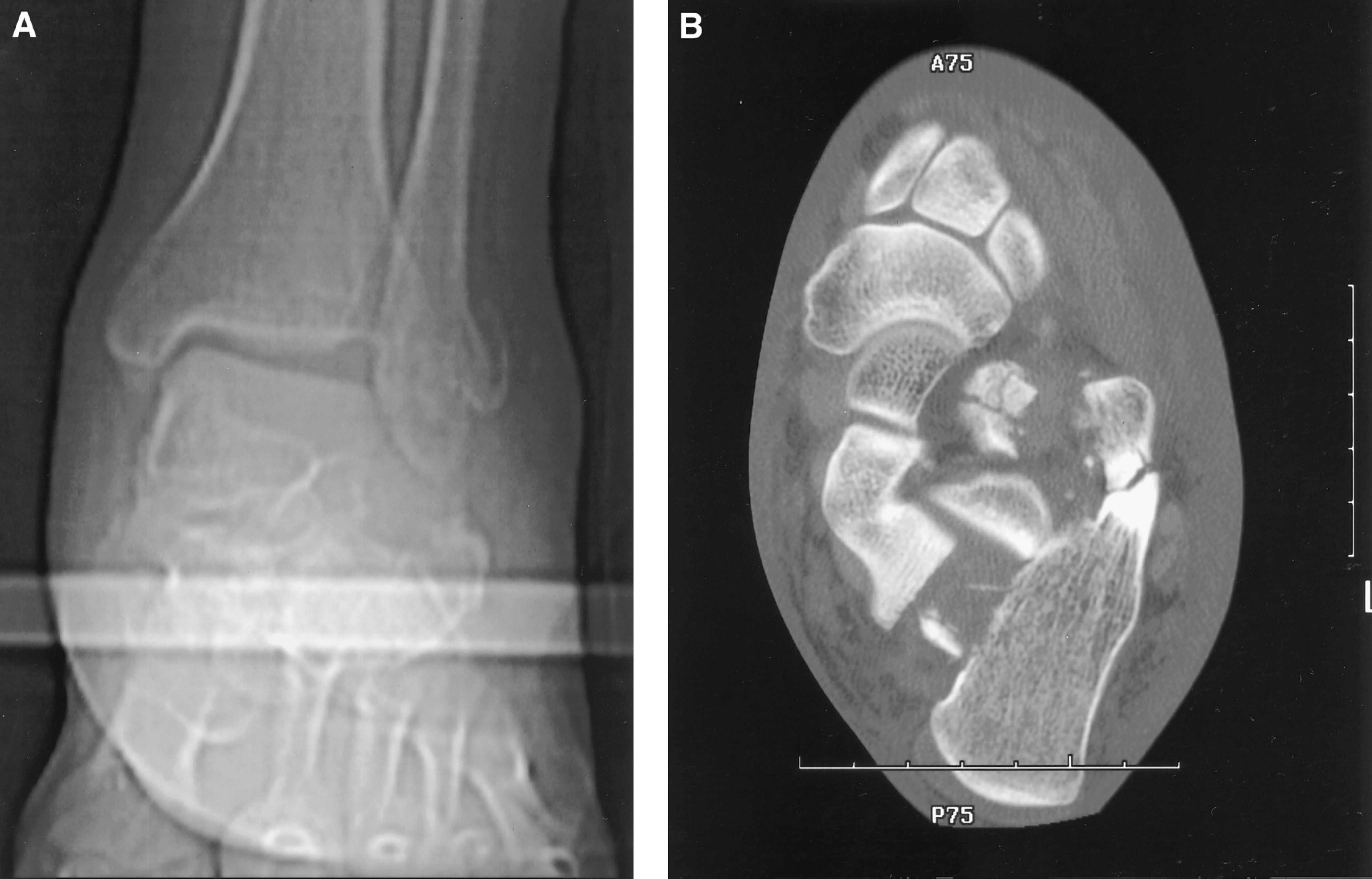
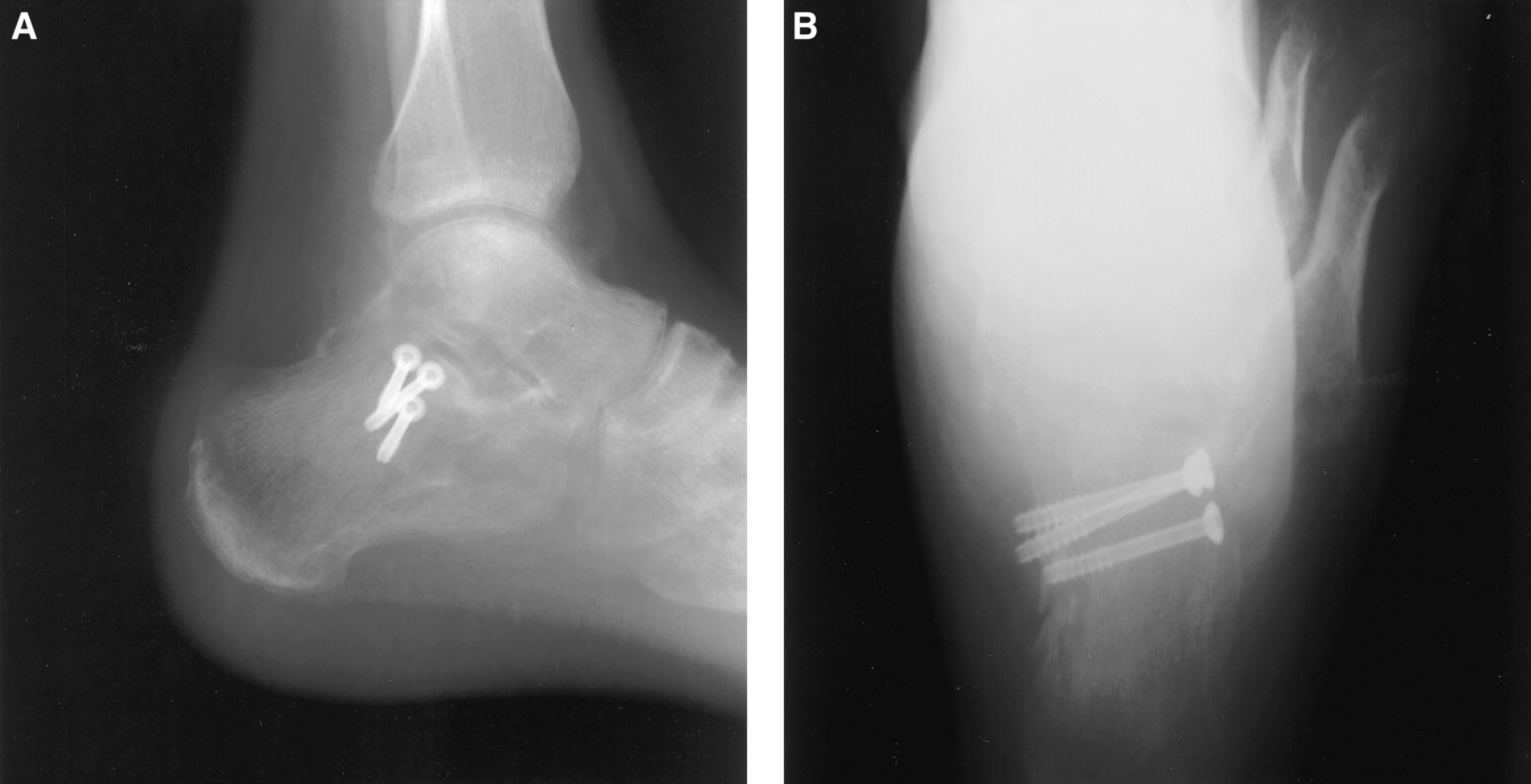
A 35-year-old male involved in a motor vehicle accident was ejected and thrown 30 feet from his car. He sustained a closed head injury, pneumothorax, nasal fracture, sacral fracture, and a closed calcaneus fracture. His calcaneus fracture was intra-articular with a locked dislocation of the posterior facet in the subfibular region (Fig. 4, A and B). His past medical history was significant for alcohol abuse, nicotine dependence, and noncompliance. Due to these risk factors a minimally invasive approach was chosen. At 12 days after injury, a Schanz pin was placed in the calcaneal tuberosity and a closed reduction was attempted, but was unsuccessful. A 3-cm sinus tarsi incision was made from the tip of the fibula and extending toward the base of the fourth metatarsal, exposing the subtalar joint. The dislocated fragment was identified and reduced with a periosteal elevator. Two 4.0-mm cannulated screws (Ace DePuy, Johnson and Johnson, Warsaw, IN) were then placed percutaneously to maintain the reduction (Fig. 5, A and B). The lateral ankle ligament complex was disrupted and repaired with nonabsorbable sutures. A smooth Steinmann pin was placed across the subtalar joint for 6 weeks due to compliance concerns. He was kept nonweightbearing for 12 weeks. His fracture united at 12 weeks without soft-tissue complications. Radiographs demonstrated maintenance of reduction and no evidence of post-traumatic arthrosis. At last follow-up 6 months after injury, he returned to manual labor and had mild pain with recreational activities.
Discussion
The mechanism of fracture-dislocation of the calcaneus has been previously reported. 3,5 The authors believed that an axial load to an inverted foot would cause rupture of the collateral ligaments with dislocation of the posterolateral portion of the calcaneus. This caused a talar tilt on radiographs. Therefore, the association of talar tilt and calcaneal fracture should alert one to the possibility of this fracture pattern. The CT scan is essential to evaluate this fracture type specifically seeking a locked, dislocated facet fragment.
The results of nonoperative treatment of calcaneal fractures have been widely reported. 2,8,11 The results are mixed and have multiple variables influencing outcomes including soft-tissue complications, workers' compensation, associated injuries, and so forth. Intra-articular fractures that have been treated nonoperatively may result in a short, wide calcaneus with post-traumatic arthritis and functional limitations. 8 In the fracture-dislocation, however, nonoperative treatment is not likely to lead to a successful result. With an unreduced dislocated fragment, it is more than likely that pain, impingement, and nonunion would result. This has led some authors to consider such a fracture a surgical indication. 4,13 When this fracture type occurs in patients at high risk for soft-tissue complications, such as diabetics, smokers, and the elderly, a minimally invasive or percutaneous technique for reduction and internal fixation may be considered.
Postoperative lateral (
Percutaneous and minimally invasive fixation for calcaneal fractures has the potential advantages of improving hindfoot alignment and joint congruity with less extensive soft-tissue dissection. In the two cases in this report, the lateral wall was not comminuted and the calcaneal height and width were normal after reduction of the facet fragment. This fracture pattern seems to be amenable to either a closed reduction or a limited open approach through the sinus tarsi for fracture reduction and screw fixation.
Previous series 7,12 have documented the association of advanced age, smoking, and diabetes mellitus with wound complications when using the extensile lateral approach. The technique described does limit soft-tissue dissection and theoretically decreases the risk of wound healing complications. We feel this approach may be considered in a select patient population with such risk factors for wound complications and a locked fracture-dislocation of the calcaneus.
In conclusion, locked fracture-dislocations of the calcaneus are uncommon, but challenging injuries to treat. Reduction of the dislocated facet fragment utilizing a closed reduction and percutaneous internal fixation, or mini-open approach if the closed reduction is unsuccessful, is a useful addition to the armamentarium of the surgeon treating these challenging injuries, especially in the patient at high risk for wound complications.