






Abstract
Background:
Irreparable peroneal tendon tears are uncommon and require complex surgical decision making. Intercalary segment allograft reconstruction has been previously described as a treatment option; however, there are no reports of the outcomes of this technique in the literature. We describe our technique and present our results using this method.
Methods:
A retrospective chart review was conducted to identify all patients who underwent intercalary allograft reconstruction of the peroneal tendons. Mechanism of injury, concomitant operative procedures, pertinent radiographic findings, pre- and postoperative physical examination, intercalary graft length, medical history, visual analog scale (VAS) score for pain, Short Form-12 (SF-12) physical health survey, Lower Extremity Functional Score (LEFS), and complications were reviewed.
Results:
Fourteen patients with peroneal tendon ruptures requiring reconstruction were identified. Mean follow-up was 17 months (range, 7-47 months; median, 12 months). The average length of the intercalary segment reconstructed was 10.8 ± 3.8 cm (range, 6-20 cm). The average postoperative VAS score decreased to 1.0 ± 1.4 (P = .0005). No patient had a higher postoperative pain score than preoperative pain score. Average postoperative eversion strength as categorized by the Medical Research Council grading scale improved to 4.8 ± 0.5 (P = .001). The average SF-12 score improved to 48.8 ± 7.8 (P = .02). The average LEFS improved to 86.4. ± 14.9 (P = .00001). Four patients experienced sensory numbness in the sural nerve distribution, and 2 of these were transient. There were no postoperative wound healing complications, infections, tendon reruptures, or reoperations. No allograft associated complications were encountered. All patients returned to their preinjury activity levels.
Conclusion:
Allograft reconstruction of the peroneal tendons can improve strength, decrease pain, and yield satisfactory patient-reported outcomes. It can be performed without incurring the deleterious effects associated with tendon transfer procedures. We believe that allograft reconstruction is a safe and useful alternative in the treatment of irreparable peroneal tendon ruptures.
Level of Evidence:
Level IV, retrospective case series.
Peroneal tendinopathy is a source of lateral ankle pain and dysfunction that is often overlooked and underappreciated.10,19 The symptoms of peroneal tendon disorders are often vague and can be misdiagnosed on initial presentation.9,10,15 Peroneal tendon dysfunction can be attributed to tendonitis, chronic tenosynovitis, subluxation, fraying, longitudinal fissures, partial tears, and complete tears.4,6,12,24,31 These abnormalities can be observed with concomitant chronic ankle instability, cavovarus foot deformities, low-lying peroneus brevis muscle bellies, superior peroneal retinacular insufficiency, and fibular bone spurs and following severe ankle sprains.5,6,13,25
Several classification systems have been described to characterize peroneal tendon tears in order to improve decision making in operative management.15,32 Acute partial thickness tears can be tubularized or repaired primarily. However, chronic tendon injuries often require other treatment options such as tendon transfers, tendon lengthening, allograft reconstructions, or synthetic graft reconstruction.11,14,17,21,23,36 Redfern and Myerson 27 proposed an algorithm for systematically addressing peroneal tendon tears based on the intraoperative findings. Within the framework of their proposed algorithm, if 1 tendon is torn and irreparable, tenodesis of the 2 peroneal muscles is recommended. Others have recommended tendon transfer, which has also been reported with satisfactory results. 7 Although both of these offer relatively simple and perhaps less technically demanding options, autologous tendon transfers suffer from the pitfalls of donor site morbidity and, in our anecdotal experience, have negatively affected normal gait mechanics.
The use of an allograft tendon for reconstruction of an intercalary segment defect is a novel technique with several advantages. Allograft reconstructions include shorter operative times and a greater availability of graft sizes compared with autograft tendon harvests. 39 Additionally, because such reconstruction restores the muscle-tendon unit, preinjury gait mechanics can potentially be restored without sacrificing the function of donor or local tendons. Numerous authors have reported satisfactory results of peroneal tendon reconstructions with autograft transfer.7,30,37 More recently, the use of an acellular dermal matrix has been reported. 26 We have previously published our operative technique for allograft reconstruction. 22 To our knowledge, there are no reports of the clinical results of single-stage allograft reconstruction of peroneal tendons. The purpose of our study was to review the clinical outcomes of this technique at our institution. We hypothesized that the use of tendon allografts in the treatment of peroneal tendon ruptures is safe, is effective, and yields favorable patient reported outcomes.
Methods
After institutional review board approval, a retrospective review of the surgical database was performed. A Current Procedural Terminology (CPT) code database identified patients with irreparable peroneal tendon ruptures (tears involving greater than 50% of the cross-sectional area) that had been reconstructed using tendon allografts between July 2007 and July 2012. Patients’ charts were reviewed and assessed for details of their mechanism of injury, concomitant operative procedures, any pertinent radiographic findings, pre- and postoperative physical examination, intercalary graft length, medical history, visual analog scale (VAS) scores for pain, Short Form-12 (SF-12) physical health survey, Lower Extremity Functional Scores (LEFS), and complications.
Fourteen patients were identified to have undergone intercalary segment peroneal tendon allograft reconstruction. Approximately 20% (3/14 patients) reported an acute inversion injury where they experienced an audible “pop” about the ankle immediately preceding presentation; the remaining 80% (11/14 patients) were presumed to have chronic tears. The acute pop associated with their injury was differentiated from a lateral ligamentous injury based on clinical examination and magnetic resonance imaging. Patient characteristics and clinical outcomes are detailed in Table 1. Their mean age was 54 years (range, 22-70 years) at the time of surgery. Mean follow-up was 17 months (range, 7-47 months; median, 12 months). Six patients were male and 8 were female. Nine of 14 surgeries were performed on the left side and 5 on the right. Twelve tears of greater than 50% of the cross-sectional area of the peroneus brevis tendon were encountered. Five patients had irreparable tears of the peroneus longus. There were 3 patients with concomitant irreparable tears of both tendons: 1 patient was amenable to reconstruction of both, and in the remaining 2 patients the peroneus brevis alone was reconstructed. Concomitant procedures included Brostrom-Gould lateral ligament reconstruction (2 patients), peroneal longus tenosynovectomy (5 patients), peroneus longus repair (2 patients), peroneus longus excision (1 patient), peroneus brevis repair (1 patient), Dwyer calcaneal osteotomy (1 patient), total ankle arthroplasty (1 patient), peroneus longus to brevis transfer (1 patient), dorsal closing wedge osteotomy of the first metatarsal (1 patient), and fibular groove deepening (2 patients). All patients underwent preoperative magnetic resonance imaging, which revealed a tear of 1 or both of the peroneal tendons (Figure 1). Average preoperative ankle eversion strength as categorized by the Medical Research Council (MRC) grading scale was 4 ± 1.0. There were 2 patients with anterior drawer translation greater than 5 mm with the ankle in a position of neutral dorsiflexion. Four patients (30%) had varus malalignment of their hindfeet. The malignment was passively correctable in three of these patients. One patient required corrective osteotomy for rigid deformity. There were no patients with neuropathic abnormalities. There was 1 patient with non-insulin-dependent diabetes mellitus; otherwise, medical comorbidities were noncontributory. Average preoperative SF-12 physical health score and LEFS were 40.1 ± 9.6 and 45.9 ± 19.4, respectively. The average preoperative VAS score for pain was 4.4 ± 2.0 (range, 1-7).
Patient Characteristics and Outcomes.

Abbreviations: ATFL, anterior talofibular ligament; CFL, calcaneofibular ligament; LEFS, Lower Extremity Functional Score; MT, metatarsal; ND, not documented; PB, peroneus brevis; PL, peroneus longus; SF-12, Short Form-12; TAR, total ankle replacement; VAS, visual analog scale.
As categorized by the Medical Research Council grading scale.
Reported as preoperative value/postoperative values.

(A) Preoperative T2-weighted magnetic resonance image (MRI) of sagittal section at the level of the fibula depicting a complete rupture of the peroneus brevis and longus tendons. (B) T2-weighted MRI axial section of the same patient’s tendon tears shown at the level of the ankle joint.
Operative Technique
Operative procedures were performed by the senior authors and an orthopedic resident or fellow. Patients were positioned in the lateral decubitus position. Preoperative antibiotics were administered. A thigh tourniquet was used to improve visualization. A longitudinal incision was fashioned over the posterolateral fibula. The incision was extended distally to the base of the fifth metatarsal (Figure 2). The lesser saphenous vein and sural nerve were identified and protected when encountered while dividing subcutaneous tissue. The peroneal tendon sheath was then opened. The contents were examined for evidence of crowding within the fibular groove, presence of an accessory peroneal muscle, presence and extent of a tenosynovitis, and status of the superior peroneal retinaculum. The proximal peroneus muscle was then identified and freed from surrounding tissue. The distal end was debrided until healthy-appearing tissue was encountered. The distal tendon stump was then identified (Figure 2). The defect length of the tendon was measured, and an appropriately sized frozen peroneal tendon or semitendinosus allograft was thawed. In 80% (10/12) of our cases, there was enough distal tendon stump of the peroneus brevis remaining to secure the tendon allograft to native tendon. Two cases involved an avulsion of the distal tendon from the fifth metatarsal in addition to a tendon defect requiring the use of suture anchors to secure the allograft distally. If the tendon was avulsed, a bleeding bone bed was prepared at its anatomic footprint. The allograft was then fixed to bone distally at the base of the fifth metatarsal with a 3.5-mm suture anchor. If there was an adequate tendon stump remaining distally, the allograft tendon was secured to it using a Pulvertaft weave and braided nonabsorbable suture (2-0 Ethibond [Ethicon, Somerville, NJ] or Hi-Fi suture [ConMed Linvatec, Utica, NY]) (Figure 3). Prior to fixation proximally, appropriate muscle-tendon unit tension of the reconstruction was approximated. This was done by placing the foot in neutral inversion and eversion as well as neutral ankle dorsiflexion. The proximal muscle stump was then pulled distally and the length of 50% of its excursion was noted. The allograft length was set from the remaining gap length when the proximal muscle and tendon were held in this position of 50% excursion. A Pulvertaft weave was also performed proximally and secured to the native muscle-tendon unit using braided nonabsorbable suture. The allograft was woven through the native muscle-tendon 3 times (Figures 4). The peroneal tendon sheath was closed with absorbable sutures. The wound was closed in layers over a drain. A sterile dressing and bulky splint were applied. Sutures were removed at 14 days, and a non-weight-bearing short-leg cast was applied. At 4 weeks, weight-bearing was progressed as tolerated in a controlled ankle motion boot, and patients were allowed to remove the boot to dorsiflex and plantar flex the ankle. Inversion and eversion were permitted after 6 weeks, and the ankle was then protected in an Aircast stirrup (DJO, Vista, CA) for ambulation. At 12 weeks, a physical therapy strengthening program was prescribed, and unprotected shoe wear was permitted.
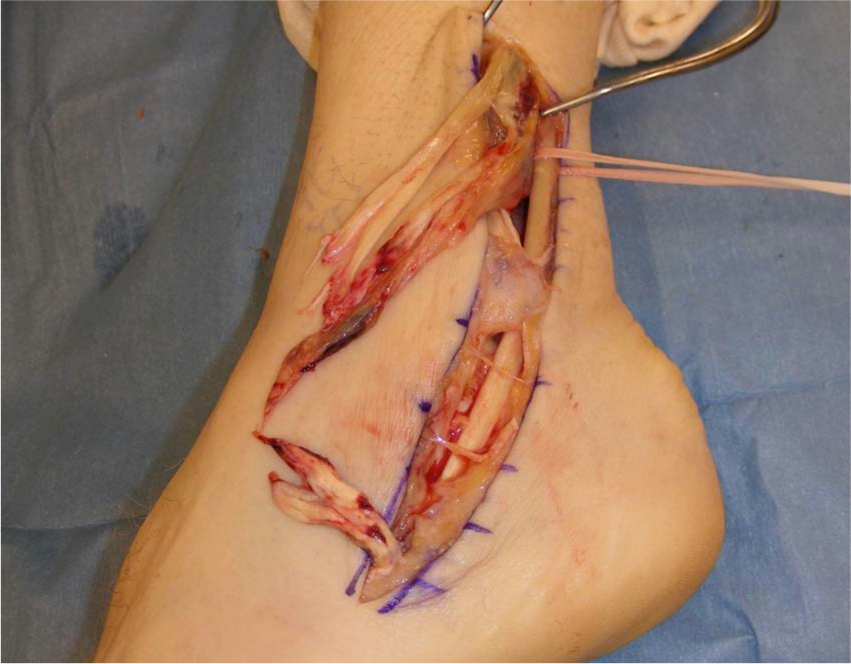
A peroneus brevis tendon rupture with an intact peroneus longus tendon and superior peroneal retinaculum.
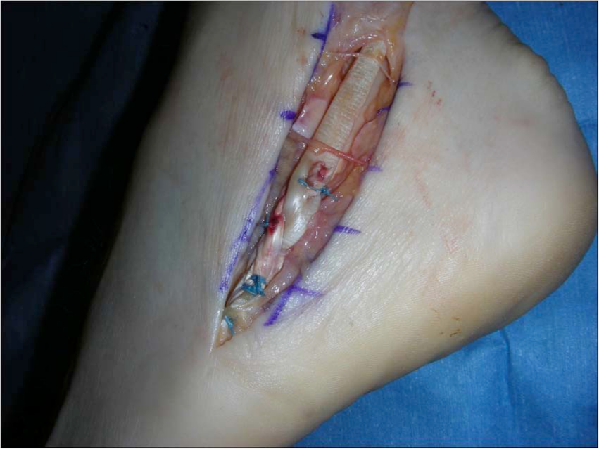
The peroneal tendon allograft fixed to the native distal tendon stump via a Pulvertaft weave.
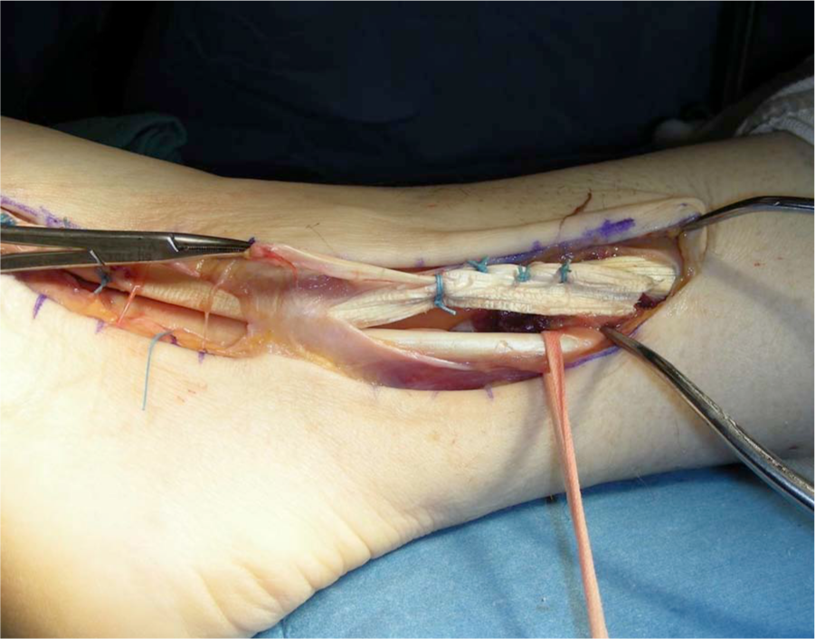
Proximal fixation of the allograft to the native tendon.
Statistical Analysis
A 2-tailed Student t test was used for comparison of pre- and postoperative VAS pain scores, strength categories, and subjective patient-reported outcomes. The criterion for significance was set at P < .05. Continuous variable averages were expressed as mean ± standard deviation when applicable.
Results
Details of individual patient results can be seen in Table 1. The average length of the intercalary segment reconstructed was 10.8 ± 3.8 cm (range, 6-20 cm). There were no differences in outcomes based on tendon graft length. Sixty-four percent (9/14) of patients had a partial tear or gross tendinosis of their peroneus longus tendon, and 5 patients had a complete rupture of the peroneus longus tendon. Three of the peroneus longus ruptures were amenable to reconstruction. The average postoperative VAS score for pain was 1.0 ± 1.4 (P = .0005). No patient had a higher postoperative pain score than preoperative pain score. All patients had improved strength postoperatively compared with their initial preoperative evaluation. Average postoperative eversion strength as categorized by the MRC grading scale was 4.7 ± 0.5 (P = .003). Sixty-four percent of patients (9/14) achieved full 5/5 eversion strength. The average postoperative SF-12 physical health score improved to 48.8 ± 7.8 (P = .02). The average lower extremity functional score improved to 86.4 ± 14.9 (P = .00001). There were no patients with postoperative lateral ankle instability. Four patients experienced sensory numbness in the sural nerve distribution, and 2 of these were transient in nature. There were no neuropathic pain complications or cases of complex regional pain syndrome. To objectively document the integrity of the reconstruction and anatomic mobility of the tendons, we evaluated a proportion of the patients with high-resolution ultrasound when they were seen in a clinic that had this capability. Six of 14 patients had postoperative high-resolution sonographic analyses of their reconstructed tendons, which all demonstrated that the allograft tendon was intact and that, when present, there was independent gliding of both tendons (Figure 5). There were no postoperative wound healing complications, infections, tendon reruptures, or reoperations. All patients returned to their preoperative activity levels. Of note, 1 patient returned to professional dancing, 1 patient returned to running 5-km races, and 1 patient returned to cycling distances of greater than 25 miles.
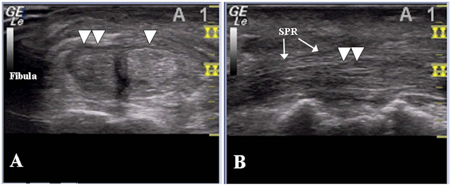
(A) Axial high-resolution ultrasound image of the intact peroneus longus tendon (single arrowhead) and reconstructed peroneus brevis tendon (double arrowheads) at the approximate level of the proximal anastomosis of the allograft and native peroneus brevis tendon 12 months after surgery. (B) Longitudinal image of the same peroneus brevis tendon (double arrowheads) at the level of the superior peroneal retinaculum (SPR).
Discussion
The literature contains numerous reports regarding the operative treatment of partial and complete tendon ruptures of the foot and ankle. When irreparable tears are appreciated, options for restoring the function of the damaged tendons include tenodesis, tendon transfer of a local tendon, and tendon reconstruction. Irreparable tears of the peroneal tendons are relatively rare, and as a consequence there is a dearth of high-level evidence to guide the management of these more complex injuries.
For tears of the peroneus brevis, Krause and Brodsky 15 were the first to propose operative treatment criteria based on the severity of the tendon tear and remaining healthy tendon. They suggested that tears that involved less than 50% of the cross-sectional area of the tendon should be treated with excision of the involved segment and tubularization of the remaining tendon. For tears involving greater than 50% of the area of the tendon, tenodesis to the peroneus longus was recommended. They presented a series of 20 patients with peroneus brevis tears, 9 of whom were treated with peroneus brevis excision and tenodesis to the peroneus longus. Eleven were treated with tubularization. 15 The investigators did not find a significant difference in outcome between groups. Although all of the patients in the tenodesis group were satisfied with their results, only 4 of the 9 had resumed unlimited activities and two-thirds had pain with activity. 15 This is not unusual for patients undergoing operative repair of peroneal tendon injuries. Steel and DeOrio 34 reported that more than 50% of their patients undergoing operative treatment of peroneal tendon tears had residual symptoms; 90% of the patients returned to work, but only 46% were able to return to sports participation.
The classification system proposed by Krause and Brodsky 15 is helpful in guiding the treatment of isolated peroneus brevis tears, but complete tears of both peroneal tendons are not addressed. Redfern and Myerson 27 have presented the largest, yet most heterogeneous, series of patients requiring operative treatment of concomitant peroneal tendon tears. Drawing on their investigation, the authors developed a treatment algorithm based on intraoperative findings. For the type I tears in which both the peroneus longus and brevis are grossly intact, tubularization is recommended following the excision of any split or fraying of the tendon. For type II injuries, characterized by the tear of 1 tendon associated with 1 tendon remaining with adequate excursion, tenodesis proximally is recommended. Finally, for type III injuries in which both tendons were torn, several options were proposed. If no proximal muscle excursion was appreciated (type IIIa), flexor digitorum longus transfer was recommended. If proximal muscle excursion was present (type IIIb) and the area of injury was without significant fibrosis, 1-stage tendon reconstruction was performed with hamstring allograft. If tissue bed scarring was present, a 2-stage reconstruction using a silicone rod placement followed by reconstruction with hamstring allograft was performed.
Under the classification listed above, 12 of 14 of our patients demonstrated type IIIb peroneal tendon tears of the peroneus brevis. Tissue bed scarring did not prompt a staged reconstruction for any our patients. Redfern and Myerson 27 also agreed with previous authors who noted that proximal muscle excursion was of utmost importance in determining the operative approach. However, Redfern and Myerson also noted that despite mobile and viable-appearing proximal muscle and a minimally invasive technique, they encountered substantially more scarring during their second procedures than anticipated. They have therefore extended their indications for single-stage transfers. This is potentially a reason contributing to the more favorable outcomes achieved in our series despite having a larger percentage of patients with more severe involvement of the peroneus brevis.
Tendon transfer procedures have also been reported with satisfactory results.7,27,29,37 Although functional outcomes have been acceptable, there are clearly some disadvantages to the described techniques. Tendon transfers can necessitate additional incisions, which can lead to increased surgical time and may also result in diminished range of motion, the development of stress fractures, and an alteration in the normal kinematics of the foot and ankle.11,23,33,35 Transfers can be performed in the setting of acute or chronic peroneal tendon tears with the use of the flexor hallucis longus, flexor digitorum longus, or plantaris tendons. Wapner and colleagues 37 reported their long-term results for the use of a staged protocol by which the diseased or ruptured peroneal tendon was excised and replaced with a silicone rod. After 3 months, the investigators transferred the flexor hallucis longus into the newly formed tendon sheath. All patients had undergone previous operative procedures prior to presentation and it was felt that their native peroneal muscle units were atrophied beyond salvage for primary reconstruction. Approximately 4 of 7 patients regained full 5/5 peroneal muscle strength and 5 of 7 achieved excellent outcomes. All patients returned to work except for the workers’ compensation patient, and 6 of 7 patients stated that they would undergo the surgery again. In their series the authors did not address concomitant ruptures of the peroneus longus tendons because the distal portion was too enmeshed in scar. They felt that the morbidity of further dissection into the plantar foot outweighed any potential benefit. We agree that the location of the peroneus longus tear is the limiting factor in determining whether it is reconstructable. We were able to reconstruct the peroneus longus in 1 patient because the tear was proximal enough that an adequate Pulvertaft weave could be performed on the exposed end of the native tendon proximal to the plantar foot. When we were unable to reconstruct the peroneus longus tendon because of the location of the tear and there was not significant fatty infiltration of the muscle belly, we tenodesed the remaining proximal musculotendinous unit to the brevis tendon to augment its function. Furthermore, Wapner et al 37 did not report any negative consequences from the loss of the dynamic peroneus longus function and felt that the results of their staged reconstruction were more favorable than the alternatives, including full-time bracing or hindfoot arthrodesis. Although the authors reported excellent outcomes, they did not perform objective biomechanical comparison of patients with and without peroneus longus function or of patients in whom the function of the flexor hallucis longus changed to that of a primary evertor.
The uses of musculoskeletal allografts have increased substantially in all orthopedic surgical subspecialties.18,20,38 Allografts offer the advantages of shorter operative times, lack of graft site morbidity or donor site complications, and an unlimited supply when compared with autografts. Their main drawbacks include the risk of disease transmission, cost, risk of inferior mechanical properties attributable to processing techniques, and potentially longer incorporation times.16,18,20 The risk of bacterial infection following allograft implantation is far greater than that of viral infection. Since 1995, approximately 30 cases of bacterial infections arising from contaminated allograft tissue have been reported.1-3,18,20 The estimated risk of transmission of human immunodeficiency virus (HIV) has been estimated to be approximately 1 in 1.6 million. 8 Although ideally any transplanted tissue would be sterilized prior to implantation, human tissue cannot be completely sterilized without altering its biomechanical properties. Sterilization of allograft tissues is not required by the US Food and Drug Administration (FDA). Nonetheless, the procurement and processing techniques of allograft tissue are closely monitored to decrease the likelihood of contamination or disease transmission. The proprietary processing techniques by which commercially available allografts are disinfected vary greatly by tissue bank. Therefore, it is critical that the surgeon be aware of the processes used to process grafts at their institution to inform their patients of the risks and benefits. The BioCleanse formula (Regeneration Technologies Inc, Alachua, FL) was used to disinfect all the allograft tendons used in this study. The manufacturer claims that treatment with this formula does not alter the biomechanical properties of their grafts and reports no instances of disease transmission in more than 1 million implants since 2004. 28 However, these claims have not been substantiated in peer-reviewed literature. We anecdotally can confirm that we have not observed any adverse events related to allograft tissue treated with this formula at our institution.
The use of autograft tendon for this reconstructive technique, potentially the ipsilateral gracilis or semitendinosus, also remains an option for patients who prefer to not have allograft tissue used or in a setting in which allograft tendons are not readily available. The surgeon and the patient must weigh the relative risks of donor site morbidity, increased operative times, allograft associated risks, and cost in these scenarios.
The use of an acellular dermal matrix (GraftJacket, Wright Medical Technology, Arlington, TN) to augment the repair of tears affecting greater than 50% of the peroneal tendons has also recently been reported. 26 Peroneal strength was returned to normal in 6 of 11 patients, and all achieved “good” or “excellent” scores on the AOFAS ankle-hindfoot measure, although the study population was quite small.
The present study was limited by its small sample size, relatively short-term follow-up, retrospective nature, and lack of a control group. It would be also be valuable to compare the objective kinematics of tendon transfer versus tendon reconstruction with an intercalary segment allograft and to determine whether these changes affect long-term outcome, which we would expect would further support intercalary reconstruction over autologous transfer.
In conclusion, this study represents the first clinical series to evaluate the outcomes of a single-stage intercalary segment allograft reconstruction for the treatment of irreparable peroneal tendon tears. This study also demonstrated that the use of allograft can restore peroneal tendon strength, decrease pain, and yield satisfactory patient-reported outcomes. It should also be noted that this can be done without causing donor site morbidity or sacrificing healthy local tendons, which can potentially alter foot function and biomechanics. Finally, with no serious complications, the study demonstrated that the use of allograft was a safe and reasonable alternative in the treatment of peroneal tendon ruptures.
Footnotes
Declaration of Conflicting Interests
The author(s) declared no potential conflicts of interest with respect to the research, authorship, and/or publication of this article.
Funding
The author(s) received no financial support for the research, authorship, and/or publication of this article.