
Abstract
Background:
The interpretation of CT scans for the evaluation of calcaneal fractures is difficult. Three-dimensional (3D) reconstruction (volume rendering technique [VRT]) has been valuable in the evaluation of irregularly shaped bones. However, their value for the analysis of calcaneal fractures is still debated. Therefore, the objective of this study was to assess the effect of additional use of 3D CTs in calcaneal fractures.
Methods:
In a prospective multicenter study, the CT data set of 5 different fractures was presented to 57 evaluators. First, the participating surgeons were asked to assess the fractures on the basis of axial, coronal, and sagittal reconstructions using a multiple-choice questionnaire. Second, 3D reconstructions (VRT) were presented. The CT scans were validated by the intraoperative findings and the results were compared to the model solution of 3 foot and ankle surgeons. Intra- and interrater reliabilities were calculated.
Results:
The proportion of intraobserver agreement was 82%, with Cohen kappa of κ = 0.748 (P < .001). Interrater agreement varied between 0.772 (P = .006) for the assessment of concomitant fractures and 0.987 (P < .001) for the suggested approach. The evaluation of several items improved after presentation of the 3D CTs (Cochrane Q test, P < .001). The benefit of 3D imaging was higher in inexperienced surgeons and complex fractures (Friedman test P < .001).
Conclusion:
The evaluation of CT scans of calcaneal fractures was improved by the additional use of 3D images (VRT).
Level of Evidence:
Level II, prospective comparative study.
Introduction
Operative fracture treatment of irregularly shaped bones like the calcaneus, scapula, or scaphoid is demanding, starting with the need for high-quality imaging not only for classification of the fracture but also for planning of the procedure: for example, which approach, how many fragments, where is the key fragment, and which implant to choose. Plain radiographs are not able to provide all this information. Therefore, CT scans have become the criterion standard for the evaluation of complex fracture pathology.16-18
Calcaneal fractures are the most common fractures of the hindfoot, and CT scans have been emphasized for the evaluation of this fracture since the early 1990s.18,23 Moreover, classification systems have been based on CT scans.18,24,26 However, none of these has reached general acceptance, as a result of low interrater reliability. 9 A recent study confirmed these results with a large number of evaluators with different levels of experience. 17 In this study, the proportion of intraobserver agreement was 82%. Cohen kappa was κ = 0.748, with P < .001. Overall interrater agreement was 61%. However, only 42% of the evaluators were able to correctly classify the fracture according to Sanders. Sixty-nine percent of the evaluators agreed on the operative procedure necessary.
For this reason, the 3-dimensional (3D) reconstruction of CT scans was proposed to improve accuracy and precision of the evaluation of fracture pathology. 25 With advancing technology in recent years, this has become available more readily. The large data sets obtained with spiral and multidetector row CT have improved the potential for creating superior 3D images (3D CT). A variety of software programs and rendering methods are available: “volume rendering,” “shaded surface rendering,” “maximum intensity projection,” and “multiplanar reformation.”
In “volume rendering,” the technique that we chose for this study, virtual light rays are emitted from a viewing point through the tissue of interest. These rays are attenuated according to their interaction with the tissues they encounter. To each tissue a color, brightness, and degree of opacity is assigned. The tissue of most interest is given the opacity 100%, the others only a fraction of it or even 0%. This process is semiautomatic. Many parameters can be modified by the user to optimize the images.4,5 Finally, the virtual rays are cast from an infinite number of positions on a virtual sphere surrounding the CT data set. This last feature enables the physician to look at the images from any desired perspective or to create a video, which shows the demonstrated organ or part of the body rotating in vertical and horizontal axes. 4 Although this technique is used more and more in daily practice, only a few systematic evaluations of the benefits of this procedural method have been published. Thus, the objective of this study was to analyze the accuracy and precision of interpretation of CT scans of calcaneal fractures with additional 3D volume rendering.
Our specific research questions were as follows: (1) How is intra- and interobserver reliability affected using volume rendering technique (VRT)? (2) Can the accuracy (validity) of the identification of number of fragments, joints involved, dislocations present, osseous defects, and additional injuries be improved? (3) How does the level of experience affect the quality of interpretation and the possible treatment decision proposed? Our hypothesis was that additional 3D reconstruction of the CT scans would be beneficial for the interpretation of calcaneal fractures.
Methods
After approval by the internal review board, the CT data set of 5 different patients/fractures (4 intraarticular and 1 extraarticular fractures; Table 1) was presented to a total of 57 evaluators. CT scans were obtained using spiral CT (Sensation 16; Siemens, Germany) with table-feed of 3 mm/s, slice collimation of 0.75 mm, pitch factor of 0.65, and slice thickness of 2 mm.
Classification of the 5 Calcanei Presented, Provided by 3 Experienced Foot and Ankle Surgeons. a
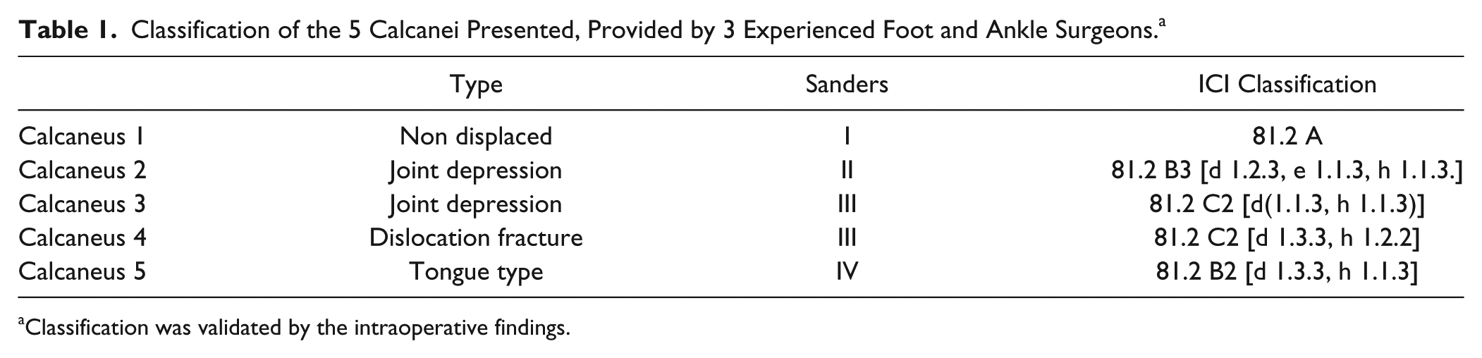
Classification was validated by the intraoperative findings.
Axial, sagittal, and coronal reconstructions were obtained of each calcaneus. Axial views were reconstructed parallel to the sole of the foot, sagittal images relative to the ankle joint, and the coronal view parallel to the calcaneo-cuboid joint (CCJ). Three-dimensional reconstructions were obtained using a VRT as described above.
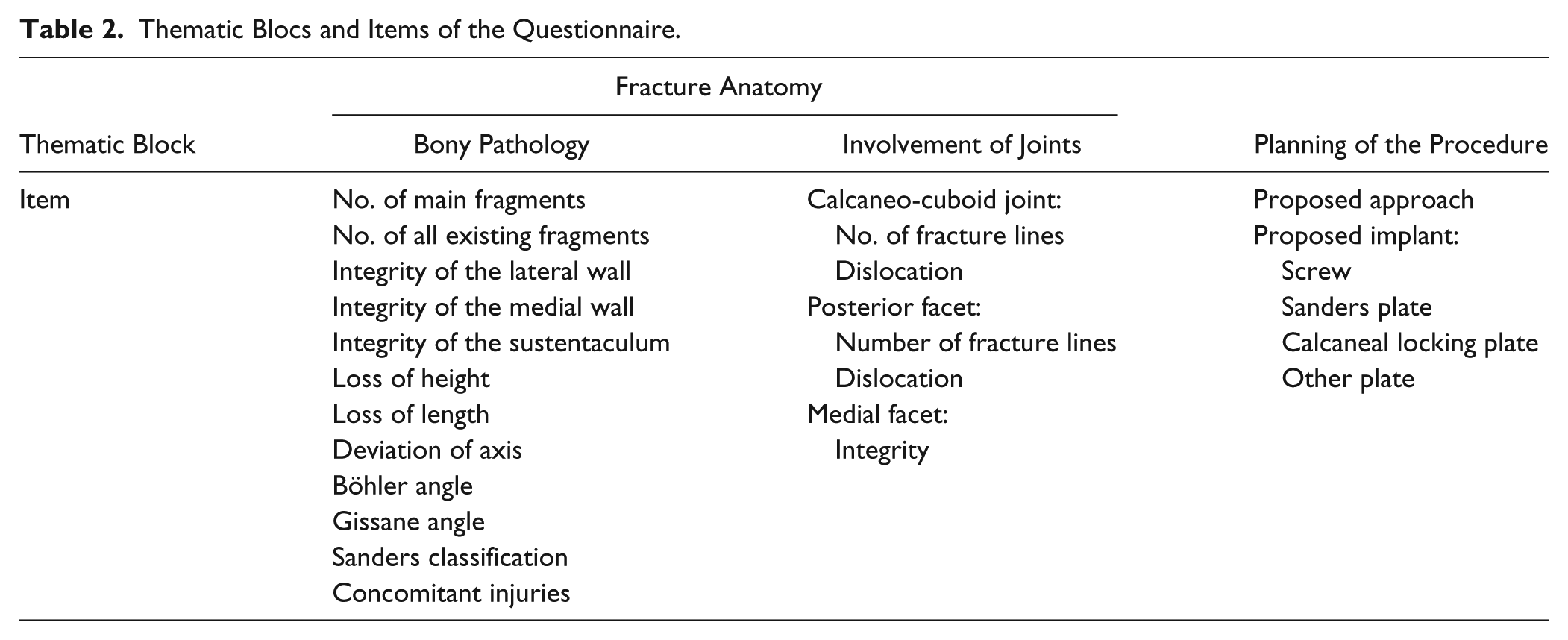
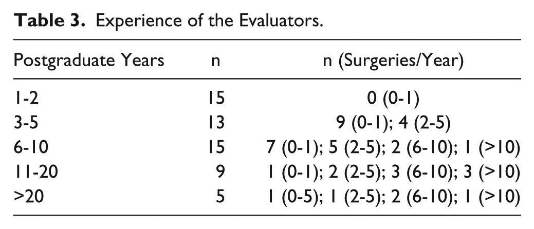
In a first run, the CT scans with the standard secondary reformations (axial, coronal, and sagittal) were presented to the evaluators in 4 different orthopedic trauma departments (2 university hospitals, 2 nonacademic level 1 trauma centers) after a short introduction into the topic and the method of the study. The participating surgeons were asked to evaluate the calcaneal fractures on the basis of a multiple-choice questionnaire, which consisted of 25 items. The questionnaire was divided into 2 sections: fracture anatomy and preoperative planning. Table 2 summarizes the items. Time for evaluation was limited to 10 minutes. In a second run, immediately following the first one, the 3D reconstructions were presented as a video, where the calcaneus was rotating around 2 axes. A second questionnaire was completed. The evaluators had different experience in their field (Table 3). No foot and ankle specialists were included.
Thematic Blocs and Items of the Questionnaire.
Experience of the Evaluators.
All patients were treated by one experienced foot and ankle surgeon. The CT scans were validated by the intraoperative findings, and discussed with 2 more experienced foot and ankle surgeons. A model answer for the questionnaire was created. According to this model solution, the answers of the evaluators were assessed correct (1) or false (0). Additionally, the evaluators were asked to mark no answer if they did not understand the question, or if they were unsure about the answer.
Statistical Analysis
Statistical analysis was performed using SPSS 21.0. Nonparametric statistical tests (Cochrane Q test) were used to determine a meaningful difference between the parameters and groups (2D vs. 3D). To assess intrarater reliability, symmetric cross-tables were constructed and Cohen kappa was calculated. Interrater reliability of the 57 raters was calculated using intraclass correlation coefficient (1-way random single measure).
After the presentation of the 3D images, a 20% positive correction of the answers of the questionnaire was regarded as clinically relevant. Statistical significance was tested using a nonparametric 1-way analysis of variance (Friedman test). Statistical significance was assumed with P <.05.
Results
The participating evaluators were asked to subjectively judge image quality. Overall image quality of the presented CT scans was found to be good or excellent in 97%.
One item of the questionnaire asked whether the 3D projection could help to more exactly analyze the fracture. Fifty-seven percent of the inexperienced evaluators (≤6 years of experience) and 30% of the experienced evaluators (>6 years of experience) stated that the 3D reconstructions were helpful or very helpful for evaluation of the fracture (Spearman rank correlation coefficient ρ = −0.291, P < .01). Fracture type had no significant influence on whether the 3D CT was regarded as helpful or not (P = .224).
Intra- and Interobserver Reliability
Three surgeons of one institution were asked to reevaluate 2 of the fractured calcanei (nos. 2 and 3) 9 months after the first evaluation to assess intraobserver reliability. The procedural method was the same as before. Intraobserver reliability was calculated for items 1 to 13 (fracture pathology) of the questionnaire. The proportion of agreement was 82% (Cohen kappa, κ = 0.748 with P < .001).
To assess interobserver reliability of the 57 raters, intraclass correlation coefficients for each item were calculated. These were found to vary between 0.772 (P = .006) for the assessment of concomitant fractures and 0.987 (P < .001) for the suggested approach. No relevant difference was found between the ratings of 2D and 3D images.
Analysis of Fracture Pathology
Fracture pathology was analyzed by evaluation of the items 1 to 12. Table 4 shows the results after assessment of 2D and 3D CTs. Results are presented as percentage of raters with equal result compared with the model answer. For the items “number of fragments,” “fracture of the medial facet,” “fracture line extending into the calcaneo-cuboid joint” and “concomitant injuries” a significant “improvement” was found after 3D CT presentation (Cochrane Q test).
Analysis of Fracture Pathology. a
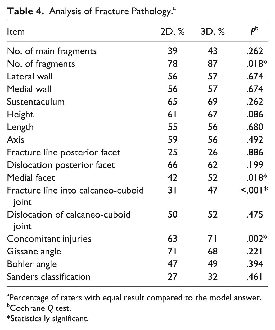
Percentage of raters with equal result compared to the model answer.
Cochrane Q test.
Statistically significant.
A correction of the ratings after presentation of 3D images of 20% or more was regarded clinically relevant. Table 5 shows the results after stratification per item of the questionnaire. Figure 1 summarizes the results after stratification to fracture type (Figure 1A) and experience (Figure 1B). Complex fractures demonstrated more benefit than fractures with lower complexity from 3D CT (Friedman test; P < .01). All groups, except that of surgeons with more than 20 years of experience, benefited from 3D CTs (Friedman test; P < .01). Figure 2 shows a typical case.
Changes of Rating After Presentations of the 3D Computed Tomographs. a
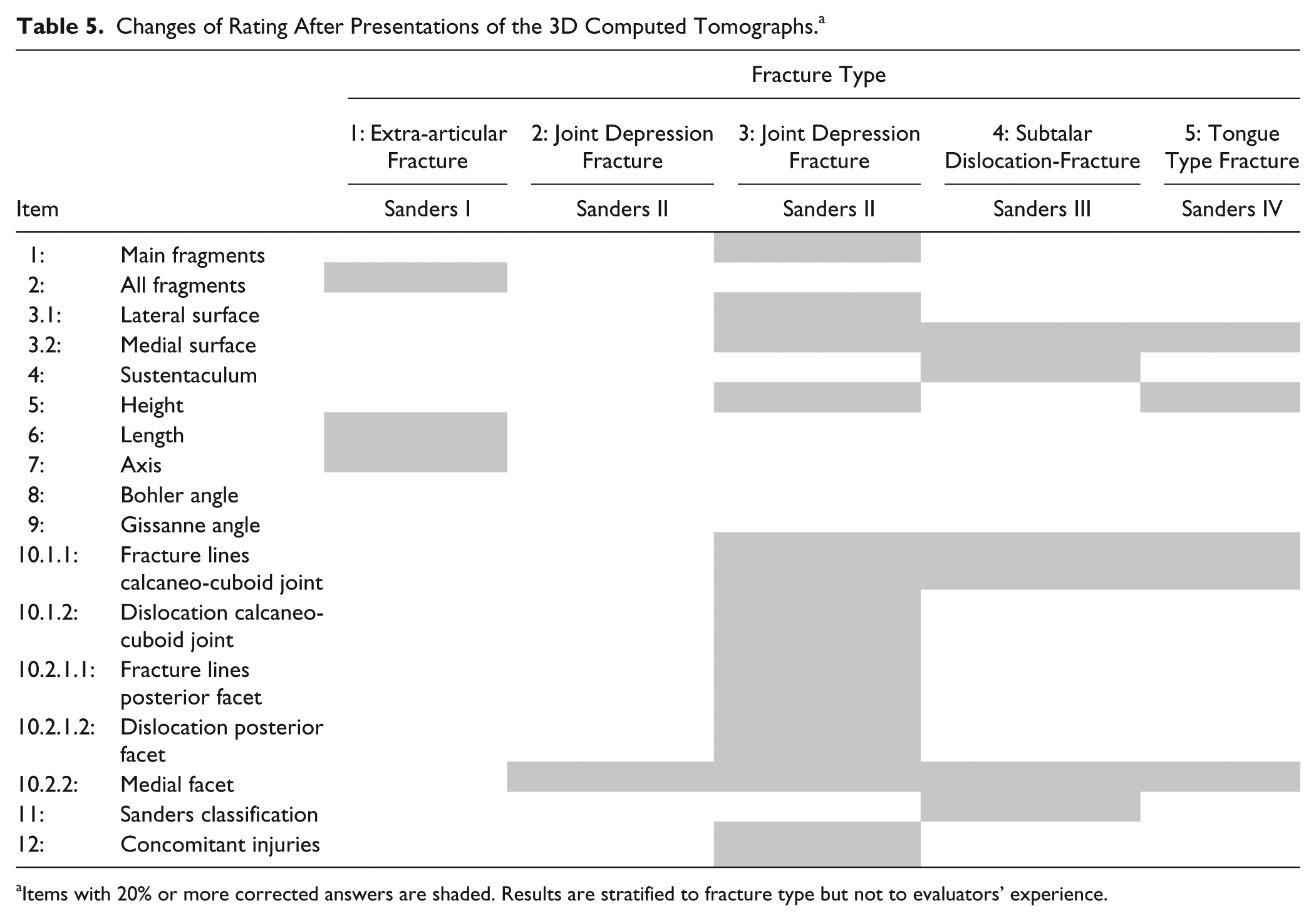
Items with 20% or more corrected answers are shaded. Results are stratified to fracture type but not to evaluators’ experience.
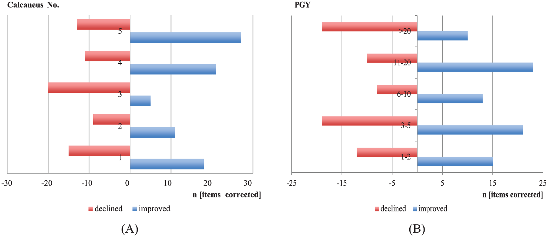
Bar chart of the number of clinically relevant improved and declined items compared to the model answer. Clinically relevant was defined as an improvement or decline of 20% in each group. (A) Rating stratified to fracture type. Note the higher rate of improvements for complex fractures (calcaneus 4 and 5) (Friedman test, P < .01). (B) Rating stratified to experience of the raters (postgraduate years [PGYs]). All groups improved, except that of surgeons with more than 20 years of experience (Friedman test, P < .01).
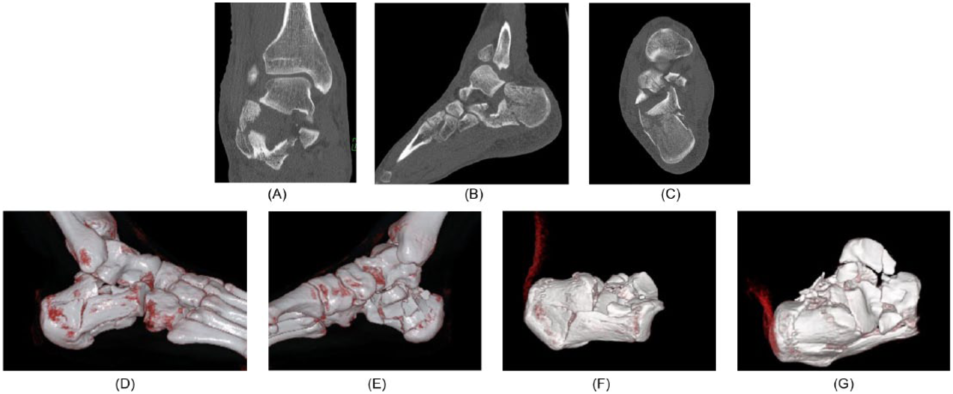
Example of 21-year-old patient with calcaneal fracture. (A) Coronal, (B) sagittal, and (C) axial view of a CT scan. (D) Lateral and (E) medial aspect of the foot using VRT. (F) Lateral and (G) top view of the “exarticulated” calcaneus.
Changes of Treatment Plan
The last 2 items of the questionnaire asked for the preferred approach and implants. After presentation of the 3D images, 49% of the evaluators changed their plan in regard to the approach and 29% in regard to the implants.
Discussion
Analysis of fracture pathology of irregularly shaped bones is demanding, even when using CT scans with multiplanar reformation. 17 Distinct information has to be extracted from the CT scans, and it affects our therapeutic decisions. Recently we showed that interobserver agreement of the interpretation of 2-dimensional CT scans of calcaneal fractures is fair among not only inexperienced but also experienced surgeons. Therefore, our hypothesis was that additional 3D reconstruction of the CT scans would be beneficial for interpretation. Several studies have proposed that this might also be the case in calcaneal fractures. Böhmer et al 2 described 3D CT as a useful instrument in evaluating calcaneal fractures and for preoperative planning because the topographic relationship of the fragments and the surrounding structures can be assessed more easily as the fracture can be seen from unusual perspectives. Likewise, Choplin et al4,5 proposed that 3D images are helpful in the diagnosis of all kinds of foot deformities because they improve the comprehension of the anatomy. Pretorius et al 15 and Cotten et al6,15 recommended the use of 3D CT especially for complex fractures. Allon and Mears 1 compared plain radiography, 2D CT, and 3D CT of 30 fractured calcanei and concluded that 3D CT improves preoperative planning and the choice of an adequate approach. Pate et al 13 evaluated 202 patients with musculoskeletal problems and found that 3D CT was helpful for the analysis of fractures of bones with complex anatomy. Others came to similar results. 12
However, recently the benefit of 3D CT was disputed. Veltman et al 22 concluded that the addition of 3D CT imaging does not increase inter- and intraobserver reliability for the classification of calcaneal fracture and should therefore not be a part of the routine workup of displaced intra-articular calcaneal fractures. Others negated the beneficial effect of 3D CTs earlier.20,21 However, one must consider that at the time these studies were conducted, 3D reconstructions (volume rendering) was affected by artifact and strongly depended on the choice of threshold.
Intra- and interobserver reliability of classification systems of calcaneal fracture has repeatedly been shown to be fair or low in 2D CTs.8,9,19 This is supported by our own studies.10,17 As expected, 3D imaging was not able to make a relevant difference in this respect in our study either. This is explained by the nature of the scoring systems; that is, Sanders classification is based on the coronal view of the 2D CT images. Three-dimensional CT cannot be helpful, especially if the talus is not “exarticulated” virtually since the subtalar joint is not visualized, as in this and other studies. 22
Yet the fate of 3D CT should not be reduced to inter- and intraobserver reliability of classification systems. Brunner et al 3 concluded: “3D reconstructions may have other benefits not evaluated in the presented study and may give useful information not captured by current classification systems.” Therefore, this study aimed to analyze the value of 3D reconstructions in a more holistic approach. Hence, we used a different methodological approach compared to other recent studies, namely, that of Veltmann et al. 22 Besides analyzing inter- or intrarater reliability, our main focus in this study was on the validity of 2D and 3D CTs. Therefore, the ratings obtained were matched to a model answer created by the treating surgeon who reevaluated the images in light of the intraoperative findings and after discussing his results with 2 additional foot and ankle surgeons. The model answer was created based on the consensus of the 3 foot and ankle surgeons. A second difference was the high number of evaluators. Prior investigations included 3 to 4 raters, whereas this study included 57 raters with various levels of experience in 4 different trauma departments.
One item asked for the subjective impression whether the 3D imaging helped when analyzing the fracture. Predominantly inexperienced surgeons felt that they gained additional information with the 3D CT. Comparing the 2D and 3D results, we could demonstrate that the number of correct answers increased for most of the items, in some cases statistically significantly (Table 4). After stratification of the results to experience and fracture type, we found that the benefit of 3D CTs was higher for inexperienced surgeons and complex fracture types. Similar to others, we found that some information was better extracted from conventional image reformations than from the 3D images, especially concerning fracture classification. Therefore, in our daily practice we always use both 2D and 3D images to extract as much information from the CT scan as possible. 22
Recent image-processing technology allows for easily subtracting adjacent bones or fragments overlapping the pathology of interest (Figure 2). Freud et al emphasized that 3D imaging without “exarticulation” is useless for the diagnosis of calcaneal and talar fractures. 7 According to our experience, exarticulation is not routinely necessary; however, it may be helpful in special situations.
Prasartritha et al analyzed the diagnostic quality of 3D CT of 51 fractured calcanei. Important findings best seen on 3D CT images were the number and configuration of displaced posterior facet fragments, fracture lines separating the anterior process and the middle facet, and the extension of fracture lines into the calcaneo-cuboidal facet. 14 These results compare well to those of this study.
One other point to be discussed is the possible costs associated with 3D CT. Almost all modern CTs provide the necessary software tools for volume rendering technology. Therefore, no additional direct costs would be incurred. Many hospitals also provide their users with special image-processing software, which enables to process the images in less than 10 minutes. Hence, the additional costs for 3D rendering are negligible. Moreover, in other fields, an overall cost reduction has been attributed to 3D volume rendering because of the superior information gained. 11
Conclusion
The evaluation of CT scans of calcaneal fractures was improved by the additional use of 3D images (VRT). Inexperienced surgeons benefited more than experienced surgeons and complex fractures more than simple fractures. Specifically, regions of interest such as the middle facet and fractures extending into the calcaneo-cuboid joint were evaluated more precisely. However, the overall rate of “correct” ratings was still low. Thus, 3D scans do not replace our teaching efforts emphasizing not only operative skills and novel implants but also the quality of interpretation of CT scans.
Footnotes
Declaration of Conflicting Interests
The author(s) declared no potential conflicts of interest with respect to the research, authorship, and/or publication of this article.
Funding
The author(s) received no financial support for the research, authorship, and/or publication of this article.