





Abstract
Background:
The combination of neurologic deformity of the ankle and foot with osteomyelitis is a therapeutic challenge. We investigated the role of Ilizarov with regard to eradication of infection, correction of deformities and improvement of patient function.
Methods:
We retrospectively reviewed 77 patients (77 feet) with neurologic deformity of the ankle and foot with osteomyelitis treated sequentially in 3 stages by Ilizarov method. The 3 stages were (1) active debridement and Ilizarov stabilization, (2) further strengthening of Ilizarov fixator and active osteosynthesis consisting of strategies like arthrodesis, osteotomy distraction, bone transport, or combination thereof, (3) consolidation of fusion/regeneration achieved in second stage. Patients with Charcot arthropathy were not included in the study. There were 43 males and 34 females with mean age of 34.4 (range, 18 to 70) years. All patients had a minimum of 1 year follow-up and 49 patients had 5 years follow-up.
Results:
Mean duration of external fixation was 179.9 (range, 128 to 413) days. The success of infection eradication was 90.9% and 91.9% at the end of 1 and 5 years, respectively. We achieved plantigrade feet in all cases with no recurrence of deformity. The American Orthopaedic Foot & Ankle Score (AOFAS) improved from a median preoperative value of 74 (interquartile range, 65 to 75.25) to 81 (interquartile range, 77 to 88) (P < .0001). The improvement of the score was by 7 points only considering the painless nature of the pathology, and the fact that pain contributed to 40% of total score. A total of 39 complications occurred in 30 patients which were managed successfully by alteration of rehabilitation and carefully chosen interventions.
Conclusion:
Ilizarov external fixation was an effective technique to address problems associated with neurologic foot and ankle deformity with infection. It allowed eradication of infection and achievement of a stable plantigrade foot. It had some disadvantages like lengthy duration of treatment, pin-tract infections, need for operative expertise, and supervised rehabilitation, but we feel these were definitely outweighed by its benefits.
Level of Evidence:
Level IV, retrospective case series.
Upper and lower motor neurological diseases can result in stiff deformities of the foot and ankle. Most often they are resistant to correction and relapses are common after conventional modalities of deformity correction. 8 Instability and progressive deformities predispose to callosities, soft tissue breakdown, trophic ulcers, and infections that can lead to osteomyelitis. This combination is considered a formidable therapeutic challenge. 11
Conventional management consists of multiple surgeries along with systemic antibiotics aimed at eradication of infection and achievement of a stable, plantigrade foot. Deformity correction involves extensive soft tissue releases, osteotomies and small joint arthrodeses with extensive removal of bony wedges, and even radical procedures like talectomy and amputations in recalcitrant cases. The problems of this approach include skin breakdown, relapse of infection, reduction of foot height with low-lying malleoli; thus a search for alternative options is warranted. 9
The Ilizarov fixator has emerged as an effective alternative to extensive soft tissue and bony surgery for patients with rigid ankle and foot deformities, especially with complex multiplanar deformities (combination of ankle equinus/calcaneus, heel varus/valgus, forefoot abduction/adduction, cavus and toe contractures), previous operative intervention, scarring, poor skin condition and deep ulceration.7,8 It has also been shown that tissues under tension stress of the Ilizarov apparatus exhibit bactericidal effect due to activation of biosynthetic processes, playing a major role in osteomyelitis management with reduced requirement for systemic antibiotics. 3 We hypothesized that Ilizarov external fixation would be an effective modality addressing all treatment goals in this complex situation. The pathology was secondary to neurologic causes like myelomeningocele and posttraumatic myelopathy (Table 1) and patients with Charcot arthropathy were not included in the analysis. The goals of the study were to assess the effectiveness of Ilizarov method in treatment of neurologic ankle and foot deformity with concurrent osteomyelitis in terms of eradication of infection, correction of deformities and improvement in patient function. We also tried to provide an algorithmic approach to this problem based on our experience.
Clinical Characteristics of Patient Cohort (N = 77).

Methods
We retrospectively reviewed patients who underwent treatment for neurologic ankle and foot deformity with concurrent osteomyelitis (Figures 1A-1D, 2A-2B) by Ilizarov method between 1995 and 2016. We identified 127 patients who were treated for ankle and foot deformity with concurrent osteomyelitis. From this we excluded patients without neurologic etiology (35 patients), with diabetic neuroarthropathy (12 patients) and patients with incomplete records (2 patients). Thus 77 feet in 77 patients (infection was unilateral in cases) were eligible for analysis. The enrollment was independently reviewed by 2 authors. All surgeries were done by or under the direct supervision of the senior author. All available medical records, radiographs and clinical photographs were reviewed. Minimum follow-up of 1 year from removal of the Ilizarov fixator was available for all patients. Informed consent was available for all patients and the study was approved by the Institutional Review Board.

Preoperative clinical photographs of a 39 year old male with partially recovered posttraumatic paraparesis with bilateral equinocavovarus and osteomyelitis of forefoot and midfoot (past history of 2 surgeries including excision of part of fifth metatarsal).

Preoperative Anteroposterior (A) and Oblique (B) radiographs of right ankle and foot of the same patient.
Apart from thorough history and meticulous clinical examination, the patients were evaluated with the following investigations—plain radiography, sinogram, CT scan, hemogram and inflammatory markers, microbiological cultures from sinus swabs, Doppler evaluation of limb vascularity, measurement of pain and temperature sensitivity of the foot and ankle dermatomes, electromyography of muscles around the foot and ankle, and dynamic pedobarography.
Our protocol of management was divided into 3 phases—(1) debridement and stabilization, (2) active osteosynthesis, and (3) consolidation. In the first phase, under spinal anesthesia, sinuses were demarcated by injecting brilliant cresyl green, radical excision of all unhealthy and nonviable tissues including sequestrectomy was performed and skin closure over drains and stabilization with Ilizarov apparatus (Figure 3A). We achieved skin closure by primary suturing or local flaps in 54 patients; the other 23 wounds healed by secondary intention (Figure 3B).

Clinical photographs of the same patient after debridement and stabilization.
Low frequency ultrasonic debridement was employed in all cases with ulcers. This technology worked by delivering low frequency ultrasound through a constant flow of saline. Sound waves were transmitted to tissue, via a liquid medium, through a treatment applicator. The nonthermal effects of ultrasound have been shown to cause 2 phenomena at the ulcer surface—acoustic streaming (a steady mechanical force delivered in a fluid medium, ie, sterile saline) and cavitation (formation of gas bubbles in the fluid creating micro-shockwaves). The combined effects of acoustic streaming and cavitation are thought to alter cell membrane activity and increase the activity of each cell. Subsequently this is thought to have 3 clinical effects: debridement, bactericidal effect and an ulcer healing stimulatory effect. 10 Soft tissue and bone materials collected from the operative wounds were sent for microbiological and histopathological examination. Ilizarov apparatus consisted of 2 tibial rings (attached with 3 tensioned wires on the proximal ring, 2 opposing olive wires on the distal ring and to each other with 4 threaded rods) and 1 or 2 metatarsal half rings (attached to the foot by opposing olive wires passing through the first and fifth metatarsals, along with an oblique non-olive wire). The tibial module was attached to the metatarsal half ring with 2 rods and hinges in maximum possible correction of deformity. (Figures 4, 5A-5B)

Schematic Ilizarov fixator design after first surgery.

Immediate postoperative anteroposterior (A) and lateral (B) radiographs after debridement and stabilization.
The first phase typically lasted for 25 to 30 days or until complete resolution of infection, whichever was later. Intravenous antibiotics were administered according to culture and sensitivity reports for the first 10 to 20 days. The patients were mobilized with full knee range of motion and nonweightbearing and covered with oral chemoprophylaxis agents against deep vein thrombosis. Sutures were removed by 2 to 3 weeks.
The second phase consisted of adding calcaneal rings to this assembly after dismantling the forefoot ring and connecting the 3 modules together with hinges in maximum correction (Figures 6, 7A-7B, 8A-8B). If the infection was confined only to the forefoot, the calcaneal half ring was applied at the first stage itself (Figures 3A-3B). The ring designs differed from case to case based on requirements. In addition, active osteosynthesis was achieved by the following strategies—(1) attempt operative fusion of unstable or painful joints by removal of intervening articular cartilage, (2) multilevel osteotomy (involving calcaneus, talus, or midfoot depending on deformity) and acute deformity correction or gradual correction of deformity by controlled distraction, (3) transport of a segment of calcaneus into an area of bone defect by osteotomy and distraction, and (4) combination of the above.

Schematic design of Ilizarov fixator after second surgery to facilitate residual deformity correction.

Clinical photographs of the same patients after arthrodesis of subtalar and midtarsal joints. Note complete deformity correction and addition of extra calcaneal wires, talar wire, connecting rods, hinges and toe wires.

Immediate postoperative anteroposterior (A) and lateral (B) radiographs after arthrodesis of subtalar and midtarsal joints.
Small joints for fusion were accessed through anterolateral curvilinear incision and osteotomies were done percutaneously. After a latent period of 7 to 8 days, controlled manipulation of the Ilizarov apparatus was commenced by distraction (for deformity correction and transport of bone fragment) or compression (for arthrodesis) or both until slight over-correction of deformity to 5 to 10 degrees was achieved. This phase lasted for 2 to 4 weeks depending on the amount of correction required and patients were allowed to weightbear gradually with support on the operated limb. Intravenous antibiotics were administered for the first 7 days of this period.
The third phase was aimed at consolidating the correction already achieved and lasted for 2 to 4 months. The patients were allowed to weightbear gradually until full weightbearing was achieved. The decision to remove the fixator was based on clinical absence of tenderness (could be elicited in 7 patients only as they had painful foot), satisfactory stress tests after removal of connecting rods and radiological evidence of consolidation of regenerate or complete fusion of the joints. After removal, the limb was protected in a below knee walking cast for 3 to 4 weeks and allowed to progressively increase weightbearing. After removal of the cast, supervised gait training and strengthening exercises continued to achieve best results. Ankle foot orthoses were used for brief periods after fixator removal in selected cases.
After removal of the fixator, the patients were followed up once every 3 months for 2 years and yearly thereafter. At every visit, patients were assessed for any evidence of relapse of infection and maintenance of deformity correction (Figures 9A-9D), radiographic assessment was done (Figures 10A-10B), and functional scoring was done by the American Orthopaedic Foot & Ankle Score (AOFAS). 6

Clinical photographs of the same patient at 2-year follow-up.

Anteroposterior (A) and lateral (B) radiographs of the foot and ankle of the same patient at 2-year follow-up.
All continuous variables with normal distribution (assessed by D’Agostino-Pearson test) are presented as mean (range) whereas those with skewed distribution are presented as median (interquartile range), while all proportions are expressed as numbers (%). Wilcoxon match pair rank sum test was used to compare improvement of AOFAS score from preoperative values to those at final follow-up. Two-tailed alpha < .05 was set as significant beforehand and all calculations were done using MedCalc V15.8 (Ostend, Belgium).
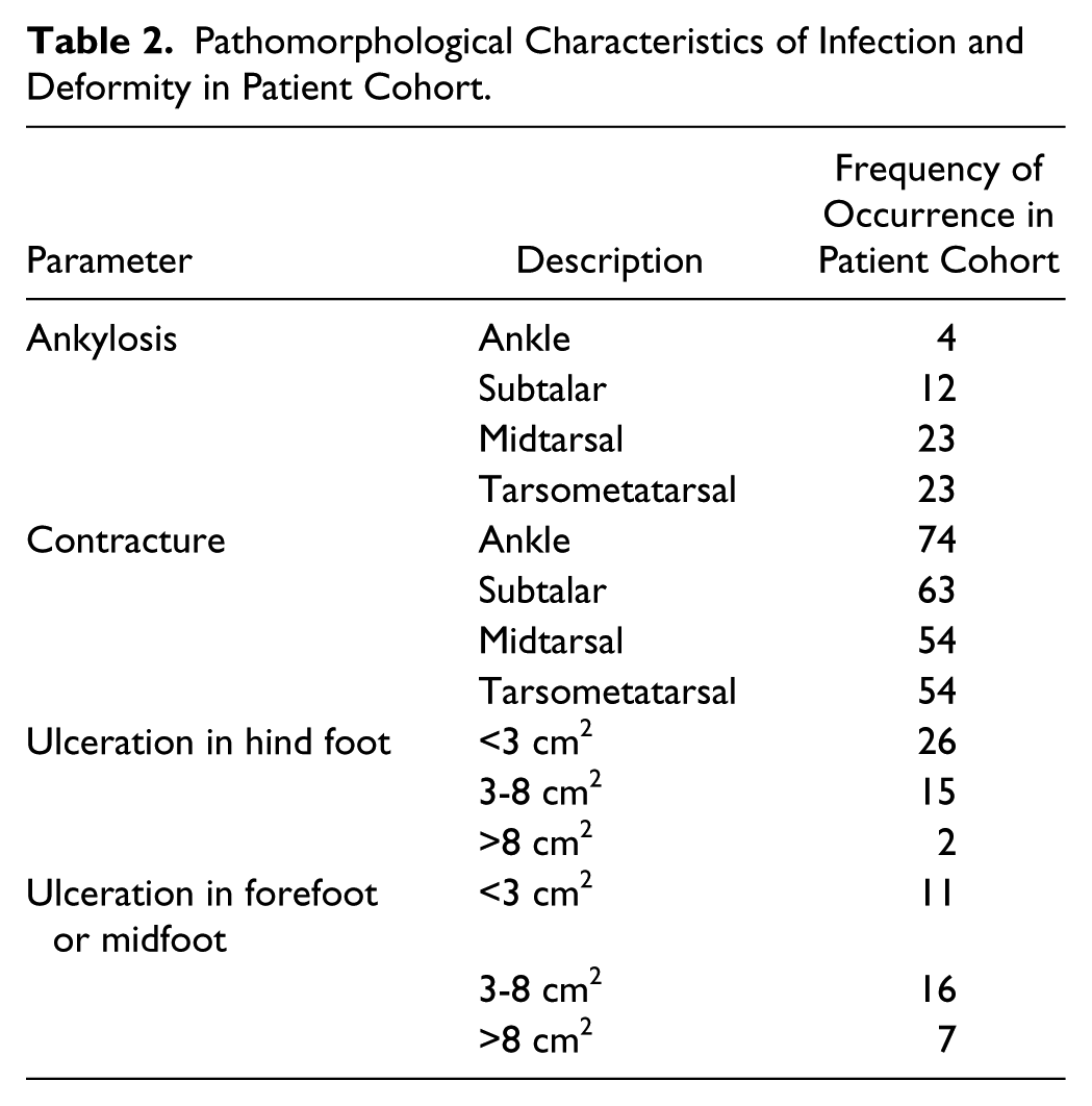
There were 43 males and 34 females with mean age of 34.4 years (range, 18 to 70 years). The clinical characteristics of the patient cohort are summarized in Table 1. The pathomorphological characteristics of infection and deformity are summarized in Table 2. The different components of the deformity when present were as follows: sagittal plane deformity at the ankle (equinus/calcaneus) had an average value of 45.1 degrees (range, 25 to 55 degrees), coronal plane deformity of the heel (varus/valgus) had an average value of 35.5 degrees (range, 5 to 55 degrees) and forefoot deformity (adduction) had an average value of 15.1 degrees(range, 5 to 25 degrees).
Pathomorphological Characteristics of Infection and Deformity in Patient Cohort.
All patients had dorsalis pedis and tibialis posterior pulses though they were weak in some cases. On Doppler evaluation, tibialis anterior (ATA), dorsalis pedis (DPA), tibialis posterior (PTA), and medial plantar (MPA) arteries showed relatively increased diameter, possibly due to inflammation associated vasodilatation. The mean and peak blood flow rates were increased in PTA and MPA and these values were moderately reduced in ATA and DPA. Temperature and pain sensitivity showed significant impairment in deformed and contralateral sides in all cases, especially over L5 and S1. Electromyography was performed in rectus femoris, tibialis anterior, and gastrosoleus and showed significant reduction of M-responses in all 3 muscles bilaterally.
Results
All 77 patients had 1 year of follow-up and during this period, 7 out of 77 patients showed evidence of relapse of infection. These patients had repeat interventions. At the end of 5 years, 49 patients were available for follow-up and 4 of them had relapse of infection between 2 and 5 years. Thus the success rate of eradication of infection (failure defined as relapse of infection after removal of fixator) was 90.9% and 91.9% at the end of 1 and 5 years, respectively. We achieved plantigrade feet in all cases and none of the patients developed relapse of the deformity at any stage. Even in the patients with relapse of infection, deformity correction was maintained. The AOFAS score improved from a median preoperative value of 74 (interquartile range, 65 to 75.25) to 81 (interquartile range, 77 to 88) (P < .0001) (Figure 11).

Boxplot depicting significant improvement of AOFAS score from a median preoperative value of 74 (65-75.25) to 81 (77-88), P value < .0001. The center line inside the box represents median, box represents interquartile range, whiskers represent 1.5 times the interquartile range, and outliers beyond this range are marked separately as dots.
When we assessed the individual components of the AOFAS score separately, only 7 patients had pain preoperatively, considering the neuropathic nature of the problem. The pain was occasional, related to exertion. The pain disappeared with treatment in all but 1 patient. Before treatment, 46 and 21 patients each had obvious and marked gait disturbance, respectively. At final follow-up, gait was normal in 3 patients, 67 had mild gait disturbance, and only 7 patients had obvious gait disturbance. None of the patients had marked limping. In spite of severe deformities preoperatively, 73 patients showed excellent ankle and foot alignment at final follow-up and the remaining 4 patients had good alignment.
The average duration of stabilization phase was 26.1 days (range, 15 to 37 days), of active osteosynthesis phase was 26.3 days (range, 12 to 91 days), and of consolidation phase was 127.5 days (range, 81 to 302 days). Overall, the duration of external fixation was 179.9 days (range, 128 to 413 days). The mean duration of admission was 46.7 days (range, 29-63 days) in patients with static frames and 110 days (range, 101-120 days) in patients with dynamic frames.
In our study group, osteotomy-distraction was used as the sole strategy in 25 out of 77 (32.5%) feet. Some of these patients had fused painless joints that needed deformity correction alone. Arthrodesis was used as the strategy in 39 out of 77 (50.7%) feet. Among them fusion was done in foot joints alone in 23 feet and in 16 cases the ankle also had to be fused. The remaining 13 patients needed a combination of the above strategies and 6 among them had transport of a segment of calcaneus to an area of bone defect by osteotomy and gradual distraction. This strategy is considered separately as osteotomy-distraction and was not intended to correct deformity, but to compensate for a bone defect. A plantigrade foot was achieved in 37 out of 77 (48.1%) cases at the end of second surgery and had static Ilizarov frames placed with only minor adjustments postoperatively. The remaining 40 feet (51.9%) had residual deformity at the end of surgery and were corrected over a period of time with well-constructed dynamic frames.
Microbiological analysis isolated multiple strains of bacteria. Complete agreement between preoperative and intraoperative culture reports was seen only in 38% of cases. The infection (as per intraoperative cultures) was due to gram positive bacteria in 24 patients (31%), gram negative bacteria in another 24 (31%) and combined gram positive and negative in 29 patients (38%). The frequency of occurrence of the various bacteria are summarized in Table 3. Osteomyelitis was confirmed in all cases by histopathological examination.
Microbiology of Infection in the Study Cohort.

All patients had arterial Doppler evaluation after completion of treatment. There was decrease of inner diameter of all 4 vessels, most significantly in the MPA. The peak and the mean blood flow rates increased in the ATA, DPA, and peroneal artery and reduced in PTA and DPA, compared to preoperative values. Thus there was evidence of redistribution of circulation from posterior to anterior vessels following treatment. The clinical significance of this remains unknown.
Comparison of dynamic pedobarography before and after treatment (Figures 12A-12B) showed increase in the weightbearing area of the operated foot by an average of 30% to 40%, both in standing and walking. Also the preoperative areas of local overloading of the feet due to infection and deformity shifted toward normal after surgery. Visual gait analysis revealed improvement of temporal and spatial parameters in all patients.

Comparison of preoperative (A) and postoperative (B) pedobarographs of both feet.
In this series, 39 postoperative complications occurred in 30 patients (Table 4). Early relapse of infection was seen in 5 patients. Two of them had the second phase of surgery inadvertently at 15 and 19 days from first surgery, which could have contributed to the relapse on the fifth and seventh day from second surgery postoperatively. These infections settled with redebridement and continuation of the same type of treatment. Seven patients developed edema of the ankle and foot while in the phase of active osteosynthesis. All of them settled with reducing the dose of distraction or compression and limiting the duration of weightbearing for a brief period. Two of the patients who had edema and 5 others developed marginal necrosis of the operative wound and they also resolved with bed rest, limb elevation, and antiedema measures without the need for any secondary procedures. The most common complication was pin-tract infection that was observed in 21 patients. All of them were detected and managed in a timely fashion with a combination of daily dressings, oral antibiotics, or reduction of dose of distraction as per the individual case. In 16 patients where infection did not settle with these conservative measures, the affected wire had to be removed or replaced.
Occurrence of Complications in the Current Series.

Discussion
Osteomyelitis of the foot and ankle is considered an “expensive and morbid disease” and a significant study of a nationwide sample of foot osteomyelitis concluded that it is essentially an operative disease, leading to failure if treated with antibiotics alone. 5 To our knowledge, this is the only report dealing exclusively with neurologic deformity with coexistent bone infection. We found the Ilizarov method as an effective solution to manage all components of the problem simultaneously. Conservative treatment using a total contact cast may result in a poor outcome because of inadequate stabilization, persistence of infection, compromised blood supply, persistent deformity, instability, and nonunion. Open correction with internal fixation is associated with complications and failure because of infection, bone softening, resorption, fragmentation and breakage of the implant. The main advantages of using an Ilizarov fixator are the ability to gradually correct deformity (without undue stress on soft tissue) and maintain this stable correction while weightbearing, even in the presence of soft bone. Achievement of a stable plantigrade foot prevents further skin breakdown and relapse of infection. Correction of deformity at the level of bone or joint (by osteotomy-distraction or arthrodesis) ensures nonrecurrence of deformity, as happens when deformity correction is achieved by soft tissue releases or distraction.
These outcomes must be interpreted in the light of the limitations of the study. Ours was a retrospective study from hospital records. There were several confounding variables: etiology and severity of the deformity, age of the patients, differences in the duration, severity and microbiology of infection, and so on. A minimum follow-up of 1 year from removal of fixator may be insufficient in this situation where both infection and deformity can relapse over a long term, although a majority of our patients had a follow-up of at least 5 years. Assessment bias may have occurred as surgeons themselves recorded the outcome. We could not apply a prospective protocol considering the rarity of such cases. Also our approach and techniques evolved over time even though the decision making process had not substantially changed. This study was performed in a center dedicated to the Ilizarov technique and our results may not be replicable by surgeons with less experience in the Ilizarov method.
Most of the available reports on Ilizarov management include cases with diabetes and Charcot arthropathy.1,2,4,11-13 We have not included patients with diabetes mellitus in our study because the pathology is different and our approach to treatment is quite different. The pathology of our cases is “neuropathic arthropathy” as opposed to “neuro-ischemic arthropathy” of diabetes mellitus. Based on our experience, we recommend our treatment algorithm consisting of 3 stages—debridement and stabilization, active osteosynthesis and consolidation. The first stage is meant for clearance of infection, reestablishment of hemodynamics and improvement of bone quality. The second stage must be commenced only after complete clinical evidence of clearance of infection. Our attempts to shorten the duration of the first stage in cases with early control of infection led to relapse of infection; we recommend a gap of at least 3 to 4 weeks between the first 2 stages for complete achievement of the treatment objectives. In the second stage of active osteosynthesis, we recommend selection of deformity correction modalities (osteotomy-distraction/arthrodesis) based on clinical findings that include congruity and stability of joints, bone quality, severity of deformity, and so on. Since we are not correcting the underlying neuromuscular imbalance, we strongly recommend the correction of deformity at the level of bone or joint (and not soft tissue). This obviates the requirement of bracing after complete consolidation and reduces the chances of relapse of the deformity. Also, the deformity must be corrected acutely only to the extent that soft tissues permit, the rest must be corrected gradually to prevent soft tissue complications. All of the complications we faced could be overcome with conservative measures, modification of the rehabilitation plan and well-chosen interventions. The surgeon must anticipate and monitor for these complications for their timely diagnosis and management.
In conclusion, Ilizarov external fixation was an effective technique to address problems associated with neurologic foot and ankle deformity with infection. It allowed eradication of infection and achievement of a stable plantigrade foot. It had some disadvantages like lengthy duration of treatment, pin-tract infections, need for operative expertise, and supervised rehabilitation, which we believe were definitely outweighed by its benefits.
Footnotes
Authors’ Note
This work was performed at the Russian Ilizarov Scientific Centre for Restorative Traumatology and Orthopaedics, M.Ulianova, 6, Kurgan, Russia 640005.
Declaration of Conflicting Interests

Funding
The author(s) received no financial support for the research, authorship, and/or publication of this article.
References
Supplementary Material
Please find the following supplemental material available below.
For Open Access articles published under a Creative Commons License, all supplemental material carries the same license as the article it is associated with.
For non-Open Access articles published, all supplemental material carries a non-exclusive license, and permission requests for re-use of supplemental material or any part of supplemental material shall be sent directly to the copyright owner as specified in the copyright notice associated with the article.
