






Abstract
Background:
Hindfoot alignment view (HAV) radiographs are widely utilized for 2-dimensional (2D) radiographic assessment of hindfoot alignment; however, the development of weightbearing computed tomography (WBCT) may provide more accurate methods of quantifying 3-dimensional (3D) hindfoot alignment. The aim of this study was to compare the 2D calcaneal moment arm measurements on HAV radiographs with WBCT.
Methods:
This retrospective cohort study included 375 consecutive patients with both HAV radiographs and WBCT imaging. Measurement of the 2D hindfoot alignment moment arm was compared between both imaging modalities. The potential confounding influence of valgus/varus/neutral alignment, presence of hardware, and motion artifact were further analyzed.
Results:
The intraclass correlation coefficients (ICCs) of interobserver and intraobserver reliability for measurements with both imaging modalities were excellent. Both modalities were highly correlated (Spearman coefficient, 0.930; P < .001). HAV radiographs exhibited a mean calcaneal moment arm difference of 3.9 mm in the varus direction compared with WBCT (95% CI, –4.9 to 12.8). The difference of hindfoot alignment between both modalities was comparable in subgroups with neutral/valgus/varus alignment, presence of hardware, and motion artifact.
Conclusion:
Both HAV radiographs and WBCT are highly reliable and highly correlated imaging methods for assessing hindfoot alignment. Measurements were not influenced by severe malalignment, the presence of hardware, or motion artifact on WBCT. On average, HAV radiographs overestimated 3.9 mm of varus alignment as compared with WBCT.
Level of Evidence:
Level III, retrospective comparative study.
Introduction
Hindfoot alignment is correlated with many disorders commonly encountered by foot and ankle surgeons. The hindfoot alignment view (HAV) radiograph, originally described by Saltzman and El-Khoury, 41 has been commonly used as a standard method for quantitative radiographic assessment of inframalleolar hindfoot alignment.1,3,12,14,15,26,27,46 Accurate assessment of hindfoot alignment is crucial for preoperative planning of surgical deformity correction, hindfoot arthrodesis, and ankle arthroplasty procedures. HAV radiographs, however, may be susceptible to sources of error due to radiographic magnification, variability in patient positioning, anatomic variability, and overlap of bony prominences, leading to a false representation of the true hindfoot moment arm.1,3,40,44
Recent developments in advanced imaging such as weightbearing computed tomography (WBCT) have helped to eliminate sources of error present in depicting 2-dimensional (2D) representations of 3-dimensional (3D) anatomic structures. Additionally, WBCT provides more clinically useful information concerning alignment as it is performed under natural standing conditions.2,17-20,22,23,28,33,34,47 Multiple parameters have been previously used to assess the hindfoot alignment on WBCT, including the foot ankle offset,5,6,17,19,23,29,33-35,47 calcaneal offset,5,6,19,29,35 and hindfoot alignment angle.5,6,10,30 Although WBCT offers the theoretical benefits of increased accuracy present in cross-sectional imaging coupled with physiologic alignment under weightbearing conditions, this technology is currently less prevalent in orthopedic clinics across the community where HAV radiographs are generally utilized.
The aim of this study was to compare the 2D calcaneal moment arm measurements on HAV radiographs with those on WBCT. We hypothesized that these 2 imaging modalities would be reliable but have substantial differences in representation of the calcaneal moment arm.
Methods
Data Collection
This study was conducted in accordance with the Declaration of Helsinki and the Guidelines for Good Clinical Practice. 38 The institutional review board of the University of Utah approved this retrospective study (IRB no. 71733). Informed consent was waived, as all data in the analyses came from chart review. Patient records and radiographs were retrospectively reviewed from January 2016 to December 2018. A total of 375 consecutive patients met the inclusion criteria of having weightbearing HAV radiographs and WBCT scans performed within 3 months of each study. HAV radiographs were obtained as a part of the patients’ standard radiographic foot and ankle assessment performed routinely in our clinical practice. WBCT was indicated by the provider on a case-by-case basis to improve the accuracy of clinical diagnoses. Patients who had injury and/or surgery between imaging modalities were excluded from this study. Measurements were divided between 4 qualified practitioners including (1) a senior orthopedic foot and ankle surgeon, (2) a foot and ankle surgery clinical fellow, (3) a foot and ankle research fellow, and (4) a musculoskeletal radiologist. Twenty-five patients were analyzed by all 4 observers to assess the interobserver reliability. Two of the observers (foot and ankle surgery clinical fellow and musculoskeletal radiologist) evaluated 25 patients twice at an interval of 4 weeks between measurements to determine the intraobserver reliability.
Measurement Methods
A novel method for measuring the hindfoot alignment calcaneal moment arm with WBCT was performed as depicted in Figure 1 and described as follows:
1) The coronal cut is selected based on the widest tibial diaphyseal distance at the most proximal edge of the image.
2) The proximal circle is placed contacting the most proximal portion of the tibial diaphysis, medial tibial diaphyseal cortical edge, and lateral tibial diaphyseal cortical edge.
3) The distal circle is placed contacting the distal tibial plafond, medial tibial metaphyseal cortical edge, and lateral tibial metaphyseal cortical edge.
4) A line is used to measure the distance from the distal tibial plafond to the center of the radius of the proximal circle bisecting the center of the radius of the distal circle. This measure is recorded and rounded to the nearest whole number in millimeters, distance A.
5) The distal aspect of this line is extended to the most inferior portion of the image representing the extension of the tibial axis.
6) Next, the axial cut is visualized in the plane that depicts the most inferior aspect of the calcaneus; the center is marked and transferred to the previously used coronal cut using the “localizer mode” function.
7) A line parallel to the ground is used to measure the distance from the tibial axis to the mark representing the most inferior portion of the calcaneus on the coronal view. Varus alignment is represented by a negative value, while valgus alignment is represented by a positive value in millimeters, distance B.

Novel method of determining hindfoot alignment using weightbearing computed tomography. Details of measurement steps are delineated in the corresponding section of the paper.
Hindfoot alignment was then measured using weightbearing HAV radiographs as depicted in Figure 2 and described by Saltzman and El-Khoury 41 with a 40% magnification correction factor to compare directly to WBCT:
1) Distance A measured on WBCT was increased by 40% (multiplied by 1.4) to become distance C.
2) A circle is placed contacting the medial and lateral aspect of the tibial cortex with the center of radius (distance C) from the tibial plafond.
3) A second circle is placed with 3 edges contacting the distal tibial plafond, medial distal tibial cortex, and lateral distal tibial cortex.
4) A line is drawn from the center of the radius of the proximal circle, passing through the distal circle, to the inferior aspect of the image representing the tibial axis.
5) A line parallel to the ground is used to measure the distance from the inferior aspect of the calcaneus to the tibial axis (distance D). Varus alignment is represented by a negative value, while valgus alignment is represented by a positive value in millimeters. This value is reduced by 40% (divided by 1.4) and compared with distance B obtained on WBCT.

Method of determining hindfoot alignment utilizing hindfoot alignment view radiographs described by Saltzman and El-Khoury. 41 Details of measurement and correction for magnification error are delineated in the corresponding section of the paper.
Varus Versus Valgus Alignment Classification
Neutral inframalleolar alignment was defined as the calcaneal moment arm falling within 1 standard deviation (SD) of the mean for measurements using HAV radiographs and WBCT. Moderate varus or valgus alignment was defined as the calcaneal moment arm greater than 1 but less than 1.96 SDs. Values greater than 1.96 SDs were defined as substantial varus or valgus alignment. 45
Statistical Analysis
The intraclass correlation coefficients (ICCs) and associated 95% confidence intervals (CIs) of the ICCs quantified inter- and intraobserver reliability. ICC values were interpreted as follows: 1, perfect agreement; 0.81 to 0.99, excellent agreement; and 0.61 to 0.80, substantial agreement. 25 The Shapiro-Wilk test and Smirnov-Kolmogorov test were performed to verify whether calcaneal moment arm measurements were normally distributed. A Wilcoxon signed-rank test was used for comparison of paired nonnormally distributed data (calcaneal moment arm assessed by HAV radiographs vs WBCT). A Mann-Whitney rank-sum test was used for comparison of nonpaired normally distributed data (difference of calcaneal moment arm assessment using 2 different imaging modalities in groups with vs without hardware in place and with vs without motion artifact). The chi-square test was used for comparison of binomial data (presence of underlying deformity, yes vs no; presence of hardware, yes vs no; presence of motion artifact, yes vs no). The Kruskal-Wallis test was used for comparison of noncontinuous variables between 3 groups (difference in calcaneal moment arm between neutral vs moderate varus/valgus vs varus/valgus alignment). The Spearman’s rank correlation coefficient was used to assess the linear dependence between data that were not normally distributed (calcaneal moment arm assessed by HAV radiographs vs WBCT). Bland-Altman plots were generated to quantify agreement between the HAV radiographs and WBCT to measure the calcaneal moment arm.7,8 The Bland-Altman plots included the bias (mean of the differences in calcaneal moment arm measurement between both imaging modalities) as well as the upper and lower 95% limits of agreement (estimated as the SD of the differences multiplied by 1.96). A univariate Cox’s regression test was performed to identify factors associated with substantial differences of calcaneal moment arm between both imaging modalities: underlying hindfoot deformity, retained hardware in the hindfoot, and motion artifacts. All data were analyzed using IBM SPSS Statistics version 26.0 (IBM Corp, Armonk, NY).
Results
The ICCs representing interobserver and intraobserver reliability of hindfoot alignment measurements were excellent for both imaging modalities (Supplemental Table 1).
Both imaging modalities were highly correlated (Spearman coefficient, 0.930; P < .001) (Table 1, Figure 3). HAV radiographs exhibited a mean calcaneal moment arm difference of 3.9 mm in the varus direction compared with WBCT (95% CI, –4.9 to 12.8). The difference of hindfoot alignment between both modalities was comparable in subgroups with neutral/valgus/varus alignment, presence of hardware, and motion artifact.
Measurement of Calcaneal Moment Arm Using HAV Radiographs and WBCT: Difference and Correlation Between Both Imaging Modalities.
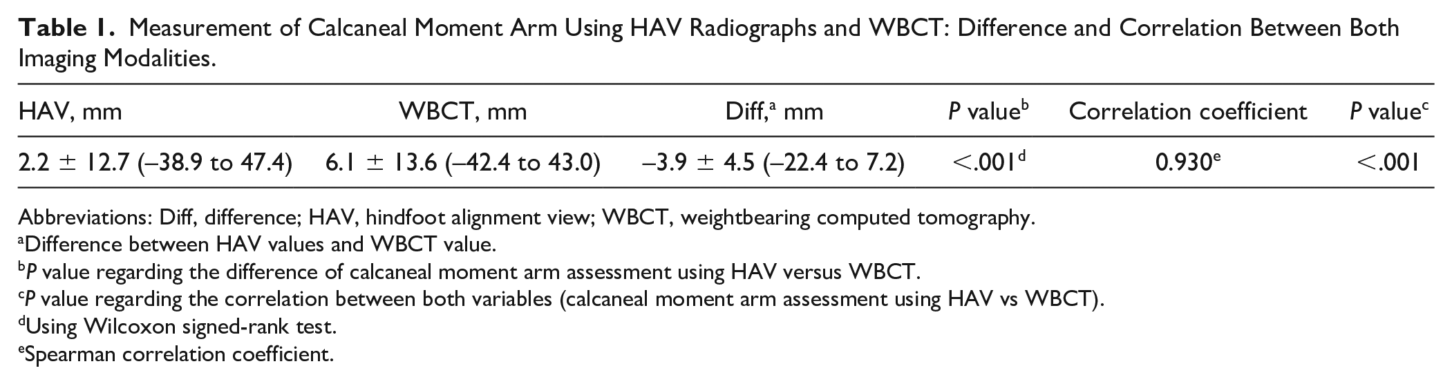
Abbreviations: Diff, difference; HAV, hindfoot alignment view; WBCT, weightbearing computed tomography.
Difference between HAV values and WBCT value.
P value regarding the difference of calcaneal moment arm assessment using HAV versus WBCT.
P value regarding the correlation between both variables (calcaneal moment arm assessment using HAV vs WBCT).
Using Wilcoxon signed-rank test.
Spearman correlation coefficient.

Correlation plot of calcaneal moment arm measurements (375 ankles) using hindfoot alignment view (HAV) radiographs versus weightbearing computed tomography (WBCT; strong and significant correlation with Spearman coefficient, 0.930; P < .001).
The Bland-Altman plot indicated no agreement between hindfoot alignment measurements on the HAV radiographs and those from WBCT (Figure 4). Specifically, the calcaneal moment arm could be measured with an average difference of −3.9 mm. The Bland-Altman plot (Figure 4) indicated that the hindfoot alignment would be measured to within approximately ±8.8 mm in 95% of all observed cases. Twenty-two ankles were defined as outliers outside 1.96 SDs.

Bland-Altman plots showing level of agreement between calcaneal moment arm measurements acquired from hindfoot alignment view (HAV) and those from weightbearing computed tomography (WBCT). The HAV radiographs overestimated the calcaneal moment arm measured on WBCT by −3.9 mm (solid line). The limits of agreement indicated that HAV measurements would be measured to within ±8.8 mm of those from the WBCT in 95% of the cases (2 dashed lines).
Radiographic agreement regarding varus/valgus alignment was observed in 322 of 375 ankles (Table 2). The calcaneal moment difference between both imaging modalities was comparable in patients with versus those without substantial underlying deformity (Supplemental Table 2).
Radiographic Agreement Between Measurements Using Weightbearing Computed Tomography Versus Hindfoot Alignment View in 375 Ankles. a
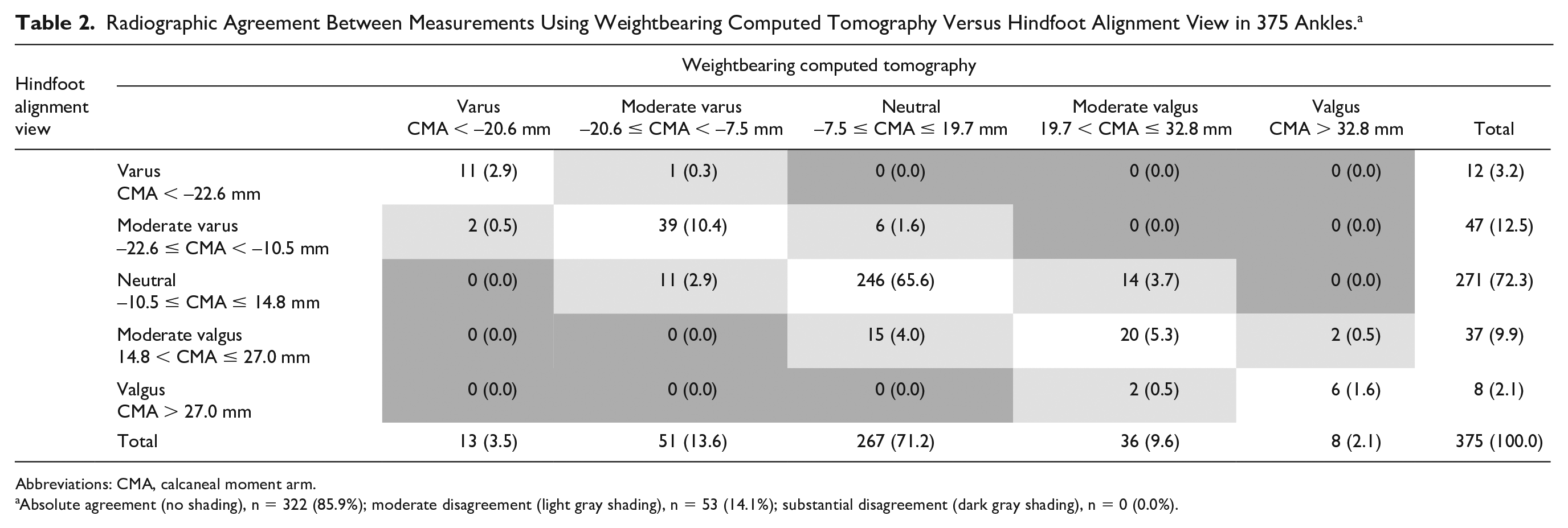
Abbreviations: CMA, calcaneal moment arm.
Absolute agreement (no shading), n = 322 (85.9%); moderate disagreement (light gray shading), n = 53 (14.1%); substantial disagreement (dark gray shading), n = 0 (0.0%).
In 119 cases, hardware from a previous surgery was present in the hindfoot; however, the difference between both types of imaging was comparable in both subgroups, this is with and without hardware (Supplemental Table 3). Motion artifact was observed in 30 WBCT scans; however, the presence of a motion artifact did not negatively influence the measurement of the calcaneal moment arm (Supplemental Table 3).
Univariate analysis did not reveal any significant predictors for substantial differences in calcaneal moment arm between these 2 imaging modalities (Supplemental Table 4, Table 3).
Univariate Analysis of Potential Risk Factors Giving the Odds Ratios With 95% CIs for Substantial Difference of Calcaneal Moment Arm Between Both Imaging Modalities (Outside 1.96 Timed SD).
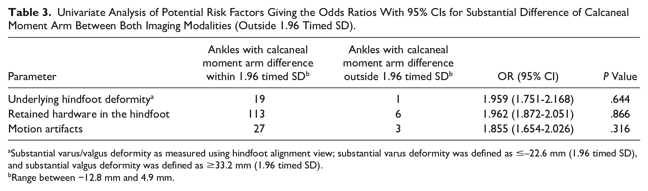
Substantial varus/valgus deformity as measured using hindfoot alignment view; substantial varus deformity was defined as ≤–22.6 mm (1.96 timed SD), and substantial valgus deformity was defined as ≥33.2 mm (1.96 timed SD).
Range between −12.8 mm and 4.9 mm.
Discussion
We found that both HAV radiographs and WBCT could be used to make reliable measurements to quantify inframalleolar hindfoot alignment. With minimal training, observers from varying degrees of orthopedic background were able to measure the calcaneal moment arm using both imaging modalities with both high intraobserver and interobserver reliability. This high degree of integrity of hindfoot alignment measurement is consistent with that observed in previous studies.9-11,16,21,32,37 In addition to high reliability, the calcaneal moment arm measurements using these 2 imaging modalities were found to be highly correlated. WBCT offers a more detailed representation of patient anatomy and provides more precision in determining the true heel point of contact in quiet standing. This makes WBCT less susceptible to the error seen in HAV radiographs that can occur as a result of bony overlap or patient positioning with respect to the orientation of the radiograph capture system. Calcaneal moment arm measurements made from HAV radiographs were therefore treated as estimations of true standing calcaneal moment arm distance as measured with WBCT. Our findings suggest that HAV radiographs overestimate a mean calcaneal moment arm difference of 3.9 mm of varus when compared with WBCT, with 95% of cases falling within ±8.8 mm range. Since WBCT technology is not currently available to many orthopedic surgeons, those with access to only HAV radiographs should understand the implications that the calcaneal moment arm may be overestimated by an average of 3.9 mm of varus and should consider adjusting their operative plan accordingly.
The exact and reliable assessment of some hindfoot measurements can be challenging. A detailed study by Kvarda et al 32 demonstrated that numerous hindfoot parameters, including the tibial articular surface angle, tibiotalar surface angle, talar tilt, and lateral and axial talocalcaneal angles, have substantially lower intra- and interobserver reliability and found that those measurements could be significantly improved by using an autogenerated 3D measurement method. The interobserver reliability of hindfoot alignment angle measurement was high for both methods, including manual 2D and autogenerated 3D methods, with ICCs of 0.98 (95% CI, 0.94-0.99) and 0.98 (95% CI, 0.99-1.00), respectively. 32 Angular measurements between the longitudinal axis of the tibia and the subtalar joint axis have also been described, 40 as well as angular measurements between the longitudinal axis of the tibia and the medial/lateral contours of the calcaneus 24 in assessing hindfoot alignment. In our practice, however, we assess inframalleolar hindfoot alignment using the distance of the calcaneal moment arm from the mechanical axis of the tibia as described by Saltzman and El-Khoury 41 because of the imprecision encountered utilizing angular measurement methods. The complex anatomy of the hindfoot with the nonlinear morphology of the relevant bones and often overlapping structures, in our experience, can prove difficult in identifying reproducible axes. The consistently high intra- and interobserver reliability of assessment of the calcaneal moment arm can be partially explained by the exact definition of anatomic landmarks and the simplicity of the measurement method. 41
The previous study by Chan et al 13 analyzed the calcaneal moment arm in 30 patients who underwent medial displacement calcaneal osteotomy due to progressive collapsing flatfoot deformity. They found a strong linear correlation between the amount of calcaneal shift and postoperative improvement of inframalleolar alignment. The final regression model indicated that each millimeter of medial shifting may result in a 1.52-mm medial displacement of the calcaneal moment arm on the HAV. 13 The difference between clinical and radiographic measurements of the calcaneal shift can be partially explained by magnification distortion, which is similar to the 40% magnification confirmed in our pilot work assessing HAV radiographs (Supplemental File). Utilizing angular measurements to represent hindfoot alignment as opposed to calcaneal moment arm length can also be affected by magnification error. Angular measurements each reference points in different planes with different corresponding distances to the image receiver and are not immune to the errors of magnification we describe in HAV radiographs.
Potential risk factors for observed discrepancies in hindfoot alignment measurements between these 2 imaging modalities were investigated. These included alignment (valgus, varus, neutral), the presence of hindfoot or ankle hardware, and the presence of motion artifact on WBCT. These suspected risk factors did not significantly affect differences in hindfoot alignment measurement by WBCT versus HAV radiographs. The methods described for measuring the calcaneal moment arm in assessment of hindfoot alignment can be recommended in patients regardless of alignment, presence of hindfoot/ankle hardware, or motion artifact present in WBCT.
The observed differences between both imaging modalities may be partially explained by variability in the position of the lower extremities between studies. The assessment of hindfoot alignment using conventional weightbearing radiographs is susceptible to foot/ankle positioning, especially in rotation.1,4,9,40 Variations in bony anatomy may also contribute to misidentification of the “true heel contact point” on HAV radiographs. The human calcaneus may also demonstrate substantial shape variations, as shown in several studies, and thus be misrepresented by a simplified 2D radiographic representation.31,36,39,42
Several limitations of our study exist. The first relates to the retrospective design with no prespecified exclusion factors; however, since this study included a large consecutive patient cohort with contemporaneously performed HAV and WBCT imaging, the results of the study are generalizable to patient populations. Second, the position of the foot/ankle was not quantified in this study. The authors recommend a “position of comfort” for both the limb of evaluation and the contralateral limb while weightbearing for the studies to help minimize error. Variation in patient positioning on HAV acquisition may have occurred; however, in our clinics this is highly regulated with trained technicians who perform these images daily with specific markings on the stand to ensure consistency in positioning from patient to patient. Our pilot study demonstrating magnification distortion on HAV radiographs was performed with the same techniques and standards as those used for the patients in this study. Additionally, the relatively large number of studies evaluated likely reduces the significance of error from malpositioned outliers. The use of all available studies, including potential positional outliers, makes these findings more clinically relevant as they more closely represent what can be expected in clinical practice outside of our own. Third, the true shape of the plantar aspect of the calcaneus was not analyzed in the present study. Further investigations analyzing shape modeling of the calcaneus31,43 may provide more insight into these anatomic variations and the effects they have on hindfoot alignment. Fourth, our study introduces a novel measurement method utilizing WBCT to assess hindfoot alignment, which may add clarity or even confusion to existing measurement techniques. Prior studies have described hindfoot alignment measurements utilizing WBCT, such as foot ankle offset, calcaneus offset, hindfoot alignment angle, 6 talar shift, and talocalcaneal alignment. 10 Although the authors find these parameters useful and well defined, they are not directly comparable to the radiographic measurement of calcaneal moment arm in HAV radiographs. 41 Our novel technique utilizes the lowest weightbearing portion of the calcaneus and tibial anatomic axis as references in both imaging modalities. Lastly, we did not record the time required for each measuring modality and technique. WBCT did require more steps and subsequently took longer to measure. The lowest point of the calcaneus is first identified on axial cross section followed by measurements in the coronal plane. Although these extra steps took longer to measure on WBCT, subjectively, we noticed increased efficiency with more experience and suspect this added time would not be logistically disruptive in the clinical setting.
Conclusion
Assessing hindfoot alignment is a critical component of evaluating many disorders treated by foot and ankle surgeons. In comparing these 2 imaging approaches, we have found them to be highly reliable and highly correlated in measuring the calcaneal moment arm. Measurements were not significantly affected by the presence of hardware in the ankle or hindfoot, motion artifact on WBCT, or inframalleolar valgus/varus alignment. HAV radiographs on average overestimated approximately 4 mm of varus alignment when compared with WBCT. HAV radiographs remain a good standard for the general assessment of inframalleolar alignment due to its ease of use, reliable results, and access throughout the community. Foot and ankle surgeons, however, should consider the potential that varus alignment may be overestimated with this approach and correlate HAV findings with physical examination, clinical alignment, and the results from surgical treatment.
Supplemental Material
sj-pdf-1-fai-10.1177_10711007211014171 – Supplemental material for Assessment of Hindfoot Alignment Comparing Weightbearing Radiography to Weightbearing Computed Tomography
Supplemental material, sj-pdf-1-fai-10.1177_10711007211014171 for Assessment of Hindfoot Alignment Comparing Weightbearing Radiography to Weightbearing Computed Tomography by Christopher B. Arena, Yantarat Sripanich, Richard Leake, Charles L. Saltzman and Alexej Barg in Foot & Ankle International
Footnotes
Declaration of Conflicting Interests
The author(s) declared the following potential conflicts of interest with respect to the research, authorship, and/or publication of this article: Charles L. Saltzman, MD, reports being the Editor-In-Chief of Foot & Ankle International. ICMJE forms for all authors are available online.
Funding
The author(s) received no financial support for the research, authorship, and/or publication of this article.

Supplemental Material
Supplementary material is available online with this article.
References
Supplementary Material
Please find the following supplemental material available below.
For Open Access articles published under a Creative Commons License, all supplemental material carries the same license as the article it is associated with.
For non-Open Access articles published, all supplemental material carries a non-exclusive license, and permission requests for re-use of supplemental material or any part of supplemental material shall be sent directly to the copyright owner as specified in the copyright notice associated with the article.
