
Abstract
Purpose:
To evaluate the intrasession repeatability and validity of posterior corneal curvature and astigmatism measurements provided by a color light-emitting diode reflection topography system in healthy eyes.
Methods:
A total of 40 healthy eyes of 40 patients (age, 16–66 years) were enrolled. A complete eye examination was performed in all cases including posterior topographic analysis with two systems: the Scheimpflug-based system (Pentacam; Oculus Optikgeräte GmbH, Wetzlar, Germany) and the Cassini system (i-Optics; Ophthec, The Hague, The Netherlands). With this last system, three consecutive measurements were taken to assess the level of intrasession repeatability (within-subject standard deviation, Sw; intraclass correlation coefficient). The Bland & Altman analysis was used to evaluate the interchangeability of both devices.
Results:
The Sw was ⩽0.06 mm for all posterior corneal radius measurements, with intraclass correlation coefficient of ⩾0.960. The Sw for the magnitude of astigmatism, J0, and J45 were 0.15, 0.04, and 0.04 D, respectively, with intraclass correlation coefficient values of 0.876, 0.897, and 0.840, respectively. Statistically significant differences between devices were found in all parameters evaluated (p ⩽ 0.025). The interchangeability analysis revealed the presence of clinically relevant limits of agreement for the flattest (0.03 to 0.50 mm) and steepest posterior corneal radii (–0.01 to 0.39 mm). In contrast, limits of agreements were not clinically relevant for the magnitude of posterior astigmatism (–0.17 to 0.27 D) and their power vector components (–0.11 to 0.15 D).
Conclusion:
The Cassini system provides consistent measures of posterior corneal curvature and astigmatism in healthy eyes, but only measures of posterior astigmatism can be considered as interchangeable with those provided by the Pentacam.
Keywords
Introduction
Several devices based on different optical principles have been developed and commercially released in the last years for the analysis of the shape of both anterior and posterior corneal surfaces. 1 One of these technologies is the use of Placido disk systems that are based on specular reflection. 1 Although these systems can be very useful in clinical practice, 1 they have certain restrictions, such as the limited ability to identify contour topographic changes, 2 or the inability of measuring the curvature of the posterior corneal surface. 1 In 1997, a new corneal topography technology based on specular reflection was developed to overcome the limitations of Placido disk systems: point-source color light-emitting diode (LED) topography. 3 The Cassini system (i-Optics; Ophthec, The Hague, The Netherlands) is a relatively new topographer based on this concept. Specular reflection of 679 colored LEDs is used by this topography system to generate anterior corneal topographic maps. Similarly, the curvature of the posterior corneal surface is measured by analyzing the specular reflections of seven additional infrared LEDs. 4
Several studies have been conducted to evaluate the precision and validity of anterior corneal curvature5–9 and total corneal astigmatism measurements4,10,11 provided by the Cassini system. Similarly, preliminary evidence has been reported about the usefulness of this system in the clinical management of keratoconus 12 and scarred cornea. 13 However, to this date, there are no studies reporting the precision and validity of posterior corneal curvature measurements provided by the Cassini system. As the manufacturer of this device states that this type of measurements can be obtained with Cassini system, it is crucial to test the consistency of such measurements and their interchangeability with those provided by a gold standard. The aim of the current study was to evaluate the intrasession repeatability and validity of posterior corneal curvature and astigmatism measurements provided by this color LED reflection topography system in healthy eyes.
Material and methods
Patients
A total of 40 healthy eyes of 40 patients ranging in age from 16 to 66 years were included in this study evaluating the Cassini technology. All participants were selected from the Optometry Clinic of the University of Alicante (Alicante, Spain), where this investigation was developed. The performance of this study was conducted in accordance with the ethical standards laid down in the Declaration of Helsinki (Seventh Revision, October 2013, Fortaleza, Brazil), being approved by the ethics committee of the University of Alicante. All subjects provided written informed consent, being free to withdraw their participation at any time without reason.
Inclusion criteria for the study were signed informed consent, no contact lens wear and presence of a healthy eye according to the ocular examination performed. Exclusion criteria were previous ocular surgery, corneal opacities or scars, contact lens wear, previous diagnosis of dry eye, and presence of any active ocular or systemic disease. To avoid potential interference in the outcomes of the correlation that often exists between the two eyes of the same person, only one eye from each patient was chosen for the study randomly.
Measurement protocol
A very complete eye examination was performed in all cases comprising uncorrected and best-corrected visual acuity, manifest refraction, Goldmann tonometry, slit-lamp biomicroscopy examination, optical biometry, and keratometry with the IOL-Master 500 system (Carl Zeiss Meditec AG, Jena, Germany), and posterior corneal curvature and astigmatism analysis with the Scheimpflug imaging–based system (Pentacam software version 1.14r01; Oculus Optikgeräte GmbH, Wetzlar, Germany) as well as with the color LED reflection topography system Cassini (i-Optics; Ophthec). The measurements were performed by the same single experienced examiner (D.P.P.) following the specific sequence Cassini–Pentacam. Data analysis extraction and analysis were performed by another independent examiner (V.C.S.).
Measurement devices
The Cassini system is a topographer employing multi-spot (up to 700), multi-color (red, yellow, and green) LED tear film-reflection imaging. The arrangement pattern of LEDs is designed to ensure that there is a one-to-one correspondence in the projection between source and image points. This potentially decreases source-image mismatch and artifacts caused by shadow. 9 Points in the LED tear film-reflection image are processed by an image algorithm that accounts for smearing and deformation in irregular corneas. 12 Specifically, a ray-tracing processing is done by every three spots identified by the software, defining the relevant local elevation. This process is theoretically unbiased by media opacity. 13 This technology is combined with second Purkinje imaging technology (reflection of seven infrared LEDs) to obtain measures of the posterior corneal surface. 4 The system has received US Food and Drug Administration (FDA) approval for clinical use in corneal topography. 12
The Pentacam system is a rotating Scheimpflug camera system with the ability of obtaining 100 images with 500 measurement points on the anterior and posterior corneal surfaces over a 180° rotation. 14 A combination of the elevation data obtained from all these images is combined to generate a three-dimensional reconstruction of the corneal structure. After all the information is processed, the internal software provides many different calculations. In this study, the Pentacam software version 1.14r01 was used (Oculus Optikgeräte GmbH).
The measurement procedure of corneal topography used was the same in all cases. The patient was asked to blink twice and to maintain the fixation before initiating the measurement with each device once the patient was positioned adequately in front of the device. The examiner adjusted the joystick until the perfect alignment was obtained. Then, the system automatically took the measurements within a short time period. With both topographers, scans not meeting acceptable criteria (blinks during the scan or other artifacts) according to the Pentacam and Cassini software indications were repeated. Only data provided for posterior corneal surface were extracted and analyzed in this study. Calibration of both topographers was performed according to the manufacturer’s instructions before initiating the study.
Statistical analysis
Sample size estimation
The Dupont–Plummer approach was used for sample size estimation. 15 For unpaired t-tests, we estimated the number of pairs of patients needed to detect a true difference in population means δ with Type I error probability α given a standard deviation (SD) σ. Our sample size of 40 eyes was found to provide a statistical power of 99%, with a difference between means in posterior keratometry of 0.23 D, SD of the difference in the response of matched pairs of 0.23 D, and an alpha error of 0.05.
Data analysis: intrasession repeatability and agreement between devices
The statistical data analysis was performed using the software SPSS version 15.0 for Windows (SPSS, Chicago, IL, USA). Normality of all data distributions was confirmed by means of the Kolmogorov–Smirnov test. Then, parametric statistics was always applied. The intrasession repeatability for posterior corneal curvature and astigmatism data provided by the Cassini system was assessed by calculating the within-subject SD (Sw) of the three consecutive measurements, the intrasubject precision (1.96 × Sw), and the intraclass correlation coefficient (ICC).
After intrasession repeatability analysis, interchangeability of posterior corneal topographic measurements provided by the Cassini system with those provided by the Pentacam system was evaluated. Differences between devices were evaluated using the paired Student t-test. After this, an evaluation of the interchangeability of corneal topographic measurements obtained with both measurement devices evaluated was performed using the Bland–Altman method. The limits of agreement (LoA) were defined as the mean ± 1.96 SD of the differences. Finally, the Pearson correlation coefficients were used to assess the correlation between the magnitude of posterior corneal topographic variables measured with the Cassini system and differences between the Cassini and Pentacam systems in such variables. All statistical tests were two-tailed, and p-values less than 0.05 were considered statistically significant.
Analysis of astigmatism
Posterior corneal astigmatic measurements were expressed and analyzed as power vectors. It should be considered that power vectors are more helpful for detecting complex changes in astigmatism, because with this methodology trajectories are traced in a uniform dioptric space. 16 The vector components J0 and J45 were calculated for each posterior astigmatic measurement using the standard procedure defined for such purpose. 16
Results
The study involved 40 eyes (21 right eyes and 19 left eyes) of 40 subjects (22 males and 18 females) with a mean age of 32.2 years old (ranging from 15 to 53 years). Mean axial length in the analyzed sample was 23.71 mm (SD: 0.92; median: 23.73; range: 21.90–25.57 mm) and mean anterior chamber depth was 3.40 mm (SD: 0.33; median: 3.40; range: 2.47–4.05 mm). Mean white-to-white corneal diameter was 12.44 mm (SD: 0.43; median: 12.40; range: 11.70–13.20 mm). Mean anterior keratometric reading and astigmatism were 43.44 D (SD: 1.27; median: 43.60; range: 41.21–45.79 D) and 1.33 D (SD: 0.95; median: 1.00; range: 0.30–3.91 D), respectively.
Intrasession repeatability analysis (precision)
Table 1 summarizes the outcomes of the intrasession repeatability analysis for posterior corneal topographic parameters provided by the Cassini system. The Sw was 0.06 mm or below for all corneal radius measurements, with ICC of 0.960 or higher. The Sw for the magnitude of astigmatism was 0.15 D, with ICC value of 0.876. The Sw value for the power vector components of corneal astigmatism was 0.04 D, with ICC of 0.897 and 0.840 for J0 and J45, respectively. No statistically significant correlations of the magnitude of each variable evaluated with its corresponding Sw were found (‒0.127 ⩽ r ⩽ 0.355, p ⩾ 0.064).
Intrasession repeatability analysis for posterior corneal topographic parameters provided by the Cassini system.
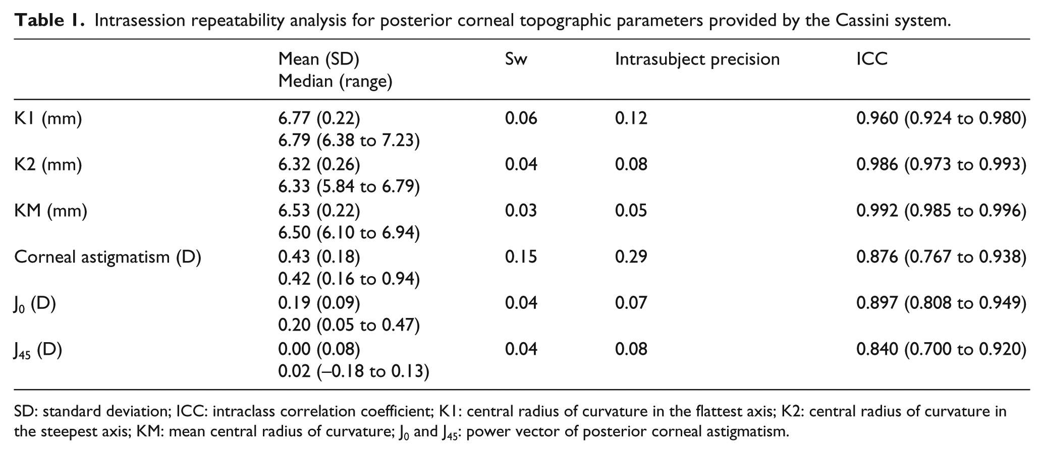
SD: standard deviation; ICC: intraclass correlation coefficient; K1: central radius of curvature in the flattest axis; K2: central radius of curvature in the steepest axis; KM: mean central radius of curvature; J0 and J45: power vector of posterior corneal astigmatism.
Interchangeability of corneal topographic measurements (validity)
Table 2 summarizes the posterior corneal topographic data obtained with the Pentacam and Cassini systems. As shown, there were statistically significant differences between devices in all parameters evaluated, providing the Cassini system steeper curvature measurements (p < 0.001) and higher values of astigmatism (p = 0.025) than the Pentacam system. However, no significant differences between devices were found in power vector components of astigmatism, J0 (p = 0.178) and J45 (p = 0.280).
Posterior corneal topographic data obtained with the Cassini and Pentacam systems.
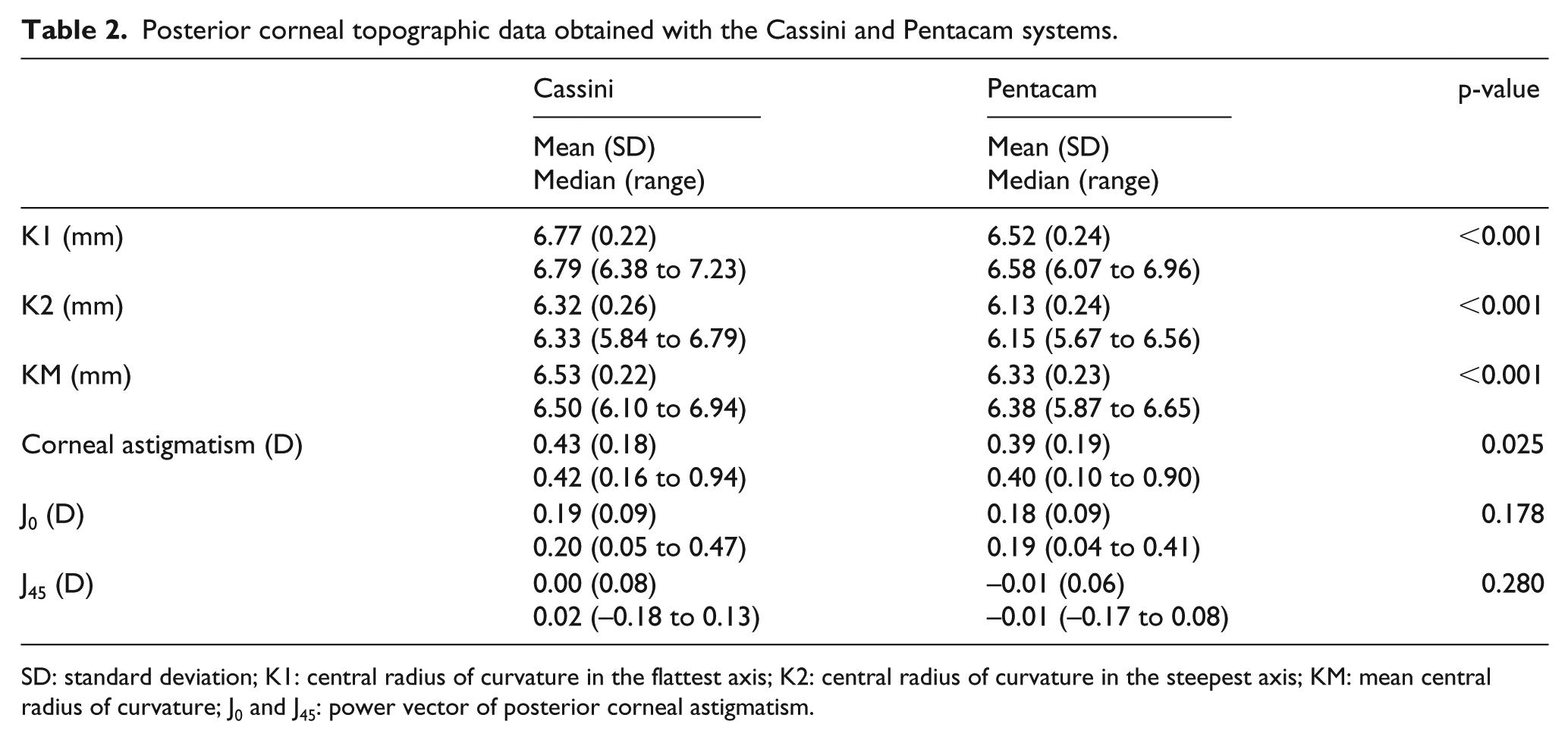
SD: standard deviation; K1: central radius of curvature in the flattest axis; K2: central radius of curvature in the steepest axis; KM: mean central radius of curvature; J0 and J45: power vector of posterior corneal astigmatism.
Table 3 summarizes the results of the interchangeability analysis of the posterior corneal measurements obtained with the two topographic devices used. As shown, a clinically relevant trend to obtain flatter curvatures (especially for the radius of curvature in the flattest corneal meridian) (Figure 1) with the Cassini system was observed (Figure 2). In contrast, differences between devices in the measurement of the magnitude of posterior corneal astigmatism were not clinically relevant, with inter-device variability of less than 0.50 D (Figure 3). Concerning the variability in the power vector components of astigmatism between devices, it was within a range of ±0.15 D, which can be considered as acceptable clinically (Table 3). No significant correlations were found between the magnitude of the topographic variables evaluated and the difference between devices in such variables (–0.115 ⩽ r ⩽ 0.318, p ⩾ 0.106).
Bland and Altman analysis outcomes of the comparison of posterior corneal topographic measurements obtained with the Cassini and Pentacam systems.
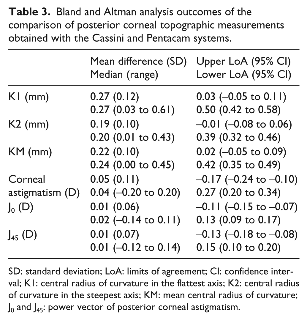
SD: standard deviation; LoA: limits of agreement; CI: confidence interval; K1: central radius of curvature in the flattest axis; K2: central radius of curvature in the steepest axis; KM: mean central radius of curvature; J0 and J45: power vector of posterior corneal astigmatism.
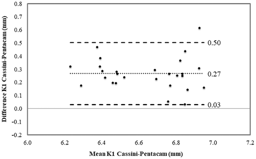
Bland–Altman plots for the comparison of the values of the posterior radius of curvature in the flattest meridian (K1) obtained with the Cassini and Pentacam systems. The dotted lines show the limits of agreement (±1.96 SD).
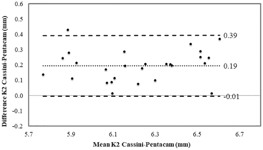
Bland–Altman plots for the comparison of the values of the posterior radius of curvature in the steepest meridian (K2) obtained with the Cassini and Pentacam systems. The dotted lines show the limits of agreement (±1.96 SD).
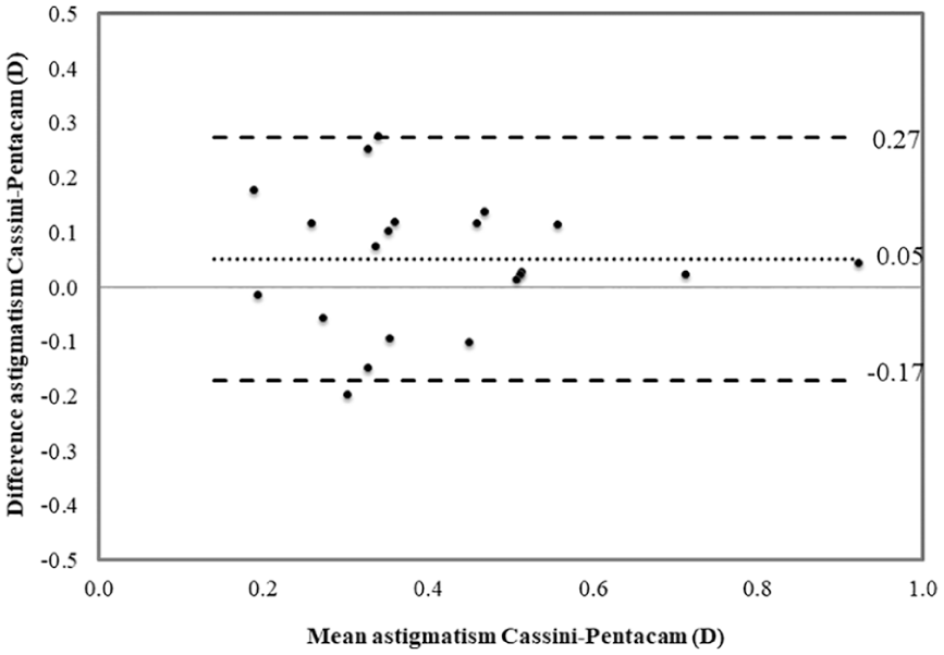
Bland–Altman plots for the comparison of the values of the magnitude of posterior corneal astigmatism obtained with the Cassini and Pentacam systems. The dotted lines show the limits of agreement (±1.96 SD).
Discussion
The current study has shown that the color LED reflection topography system evaluated is able to provide consistent central curvature and astigmatism measurements of the posterior corneal surface in healthy eyes. To our knowledge, this is the first study reporting a validation of the posterior corneal topographic measurements provided by this topography system. First, the intrasession repeatability of central posterior corneal curvature measurements was evaluated, obtaining Sw values of 0.06 mm or lower, and ICC values of 0.960 or higher. This level of intrasession consistency of posterior corneal curvature measurements is in the same magnitude or somewhat worse than that reported by different authors for different Scheimpflug imaging–based devices.17–26 Montalbán et al. 25 reported Sw values of 0.04 mm or lower and ICC of more than 0.990 for measurements of the posterior corneal radius of curvature obtained with a Scheimpflug camera topography system (Sirius; CSO, Costruzione Strumenti Oftalmici, Florence, Italy) in healthy eyes. Recently, Savini et al. 17 have found a Sw value of 0.03 D for posterior keratometric power measured with an optical coherence tomography–based system. Similarly, Kim et al. 19 reported a Sw value of 0.03 D for posterior keratometry in a sample of healthy eyes measured with a dual Scheimpflug analyzer (Galilei G4, Ziemer, Port, Switzerland). Concerning the Scheimpflug imaging–based system (Pentacam; Oculus Optikgeräte GmbH), mean Sw values for flattest and steepest posterior keratometric powers of 0.029 and 0.038 mm, respectively, were reported in a validation study. 26
Besides the excellent level of intrasession repeatability obtained for posterior corneal curvature measurements with the Cassini system, good repeatability was found for the posterior astigmatism measurements. Specifically, Sw for the magnitude of posterior corneal astigmatism was 0.15 D, with ICC value of 0.876. This outcome is consistent with the results of previous studies evaluating the consistency of posterior corneal astigmatic measurements provided by different types of Scheimpflug imaging devices.17–26 Bao et al. 18 reported Sw values of less than 0.12 D for posterior corneal astigmatism measured with two Scheimpflug-based devices (Sirius; CSO and TMS-5; Tomey Corp., Nagoya, Japan). In another study, good intrasession precision values (1.96 × Sw) were obtained for keratometric readings (<0.09 D) obtained with the Scheimpflug-based system Pentacam. 26 Concerning the variability observed in the power vector components of corneal astigmatism obtained with the Cassini system, it was found to be acceptable in clinical terms, with a range of variability not exceeding 0.15 D. The ICC values obtained for J0 and J45 were like those reported by Cerviño et al. 22 for the total cornea (a combination of anterior and posterior corneal surface contributions) using a dual-camera rotating Scheimpflug–Placido imaging system (Galilei G4; Ziemer). Montalbán et al. 25 obtained Sw of 0.04 mm or lower and ICC higher than 0.990 for power vector components of posterior corneal astigmatism using the Scheimpflug-based system Sirius; CSO. It should be additionally mentioned that in the current study, no statistically significant correlations were found between the magnitude of each variable evaluated and its corresponding Sw. This confirms that the level of consistency of the posterior corneal measurements obtained with the Cassini system did not vary with the magnitude of the variable measured. Therefore, no more variability was present in the measurements obtained in healthy eyes with the Cassini system in corneas with steep or flat mean curvatures or more or less astigmatism.
Besides the evaluation of precision of posterior corneal curvature measurements obtained with the Cassini system, the agreement of such measurements with that provided by another “gold standard” device was assessed (Scheimpflug-based system Pentacam). This type of analysis is called with the term validity and is commonly performed when evaluating new diagnostic technology as the true value of any ocular measurement cannot be known with complete certainty. 27 The comparative analysis between the two devices, Cassini versus Pentacam, revealed that there were statistically significant and clinically relevant differences between them in terms of posterior corneal curvature data. Specifically, the Cassini system provided flatter posterior curvature measurements than the Pentacam system. According to this, it seems that the Cassini system has a trend to underestimate such measurements if we used as reference or gold standard the Pentacam system. However, the range of posterior corneal curvature measured in our sample with the Cassini system is equivalent than that reported with other Scheimpflug-based devices in large samples of healthy eyes.17,18,28 There are studies demonstrating that the Pentacam posterior corneal measurements cannot be used interchangeably with those provided by scanning-slit and other Scheimpflug imaging–based devices.23,24,29,30 In some of these studies, a similar trend to obtain steeper measurements of curvature with the Pentacam system compared to other devices has been reported.24,30 More studies are necessary to confirm these findings, but at present this trend to obtain flatter posterior corneal curvature measurements with the Cassini system should be considered when comparing or using this system and the Pentacam system in clinical practice.
Finally, the level of agreement in terms of the magnitude of posterior corneal astigmatism was good, with the presence of statistically significant differences in the measurement of this parameter with both devices, but not reaching clinical relevance considering the potential impact on clinical decisions such as corneal ectasia diagnosis or toric IOL power calculation (LoA, ‒0.17 to 0.27 D). Similarly, differences in the power vector components of posterior corneal astigmatism between Pentacam and Cassini systems were not statistically significant and not clinically relevant, with ranges of agreement not exceeding 0.15 D. This should be confirmed in future studies evaluating the differences between Cassini and Pentacam devices in eyes with high levels of corneal astigmatism, including irregular corneas.
In conclusion, the color LED reflection topography system Cassini provides consistent measures of posterior corneal curvature and astigmatism in healthy eyes. Therefore, this system is a useful instrument to assess the posterior corneal geometry in clinical practice. Concerning the agreement with the Scheimpflug imaging–based system Pentacam, only the measurement of the magnitude and power vector components of astigmatism could be considered as interchangeable with both devices. In contrast, the agreement between devices was poor in terms of measurements of posterior corneal curvature and axis of astigmatism and therefore, such measurements cannot be considered as interchangeable, with a clear trend of Cassini system to provide flatter curvature measurements compared to the Pentacam system. Future studies should be conducted to provide a full comparison of posterior corneal measurements of Cassini system with other devices providing information about the posterior corneal shape. Similarly, more studies must be conducted to evaluate the interchangeability of posterior topographic data provided by Cassini in eyes with altered posterior corneal shape (i.e. keratoconus) compared to other topography systems.
Footnotes
Declaration of conflicting interests
The author(s) declared no potential conflicts of interest with respect to the research, authorship, and/or publication of this article.
Funding
The author(s) disclosed receipt of the following financial support for the research, authorship, and/or publication of this article: This study was supported by the Ministry of Economy, Industry and Competitiveness of Spain within the program Ramón y Cajal, RYC-2016-20471 (to D.P.P.).