
Abstract
This study aims to evaluate the correlation between fungi causing paint deterioration and air contamination in Assiut University hospital to give a complete picture for the fungal quantity and spectrum. Seventeen fungal species were isolated from 15 samples of deteriorated water-based paint collected from the hospitals. Chaetomium globosum was the most common paint-deteriorating fungal species, followed by Alternaria alternata, Aspergillus parasiticus, Penicillium oxalicum and Setosphaeria rostrata. Direct examination confirmed the ability of these fungi to colonize the paint samples producing mycelia, conidia and fruiting bodies. In vitro, these fungi exhibited high potential to utilize the thin layer of polyacrylic paint and significant enzymatic activities of cellulase, lipase and urease that may play a main role in paint degradation and as virulence factor of human diseases. Moreover, 27 fungal species were isolated as air contaminating mycobiota. Aspergillus spp., Cladosporium cladosporioides, P. oxalicum, A. alternata and C. globosum caused a considerable amount of indoor air contamination. The results indicated that there is a clear correlation between fungi causing paint deterioration and air contamination, whereas certain fungi were responsible for wall paint deterioration and frequently indoor air contamination. The current study suggests that improvement of antimicrobial additives of paints may be a promising approach to reduce paint biodeterioration and subsequently air contamination of indoor environments.
Introduction
There is a persistent interest in the use of biocidal approaches or irradiation to disinfect indoor environments for the control of infectious diseases in hospitals, other health care facilities and the public sector.1,2 Various studies were conducted in the fungal contamination of hospital environments to demonstrate the spectrum and levels of airborne pathogenic fungi, air quality and effectiveness of air handling systems. 1 Paint deterioration caused by microbial attack is one of the main sources of air contamination in the indoor environments. 3 Water-based wall paints have mainly acrylic-structure; these are vulnerable target materials to fungi and bacteria due to the fact that they contain cellulosic compounds as thickeners.
These compounds may be utilized by microorganisms as carbon source producing different miens of paint deterioration such as colour changes, chalking and adhesion loosening.4,5 Under sufficient moisture, opportunistic fungi can attack and colonize wide varieties of binders producing large amount of propagules, hyphal fragments, allergens, mycotoxins and volatile organic compounds (VOCs) into indoor environments.6–8 The increases of these contaminating agents in the indoor environment are potential human health hazards. Moreover, these microbial contaminants play an essential role in respiratory symptoms, allergies, asthma and immunological reactions causing 20% of the infected cases with sanitary assistance.9–11 Therefore, fungi causing paint deterioration and subsequently air contamination should be controlled using integrated strategies.12,13
Antimicrobial agents added during the paint manufacture are the main key for controlling and suppressing microbiological attack and colonization.5,14 There are many efforts for using effective and less environmental impact biocides replacing the traditional organochlorides and organometallic compounds.5,15 Recently, new trends have been employed for discovery and application of operative antimicrobial coatings that significantly inhibit the infections propagation in health care centres and public places.5,16,17 In indoor environments containing deteriorated wall paints, fungal species are able to grow, producing several types of VOCs and toxins that are suspended in the air or adsorbed on dust particles. 18 Therefore, the determination of indoor air quality is needed to understand the potential risk for the human health of workers and visitors as well as to suggest fast remediation avoiding sick building syndrome. 19 Several fungi were reported for their ability to aggressively infest, colonize paint films, produce different compounds including mycotoxins and negative impacts on human health. 20 In this respect, Alternaria, Aspergillus, Penicillium, Chaetomium and Cladosporium were frequently found to deface acrylic painted surfaces depending on their enzymatic activities to degrade the cellulosic thickeners employed in paint formulation.5,21,22 Many investigations confirmed that during the building renovation in health care centres, immunosuppressed patients face the risk of developing invasive pulmonary aspergillosis and other opportunistic infections.23,24 To reduce the risk of invasive fungal hospital-acquired infections, monitoring of the microbial contamination of the environment (air, surfaces, water) as well as determination of the contamination sources represent an effective remedial procedure.25,26
The objective of the present study aimed to evaluate the correlation between fungi causing paint deterioration and air contamination in Assiut University hospital to give a complete picture about the quantity and spectrum of fungal propagules. The role of fungal enzymatic activity involved in the paint degradation was also addressed.
Materials and methods
Studied site and sample collection
Fifteen samples of deteriorated water-based paints were collected from 11 medical units belonging to the Assiut University hospital, Assiut Governorate, Egypt. Moreover, details of the studied sites as well as the description of paint deterioration were illustratively presented in the supplementary data. Samples of deteriorated paints were scrapped off using sterilized scalpel and collected in sterilized plastic airtight vials. The samples were transferred to the microbiological Laboratory of Botany and Microbiology Department, Assiut University for isolation and identification of fungi.
Isolation and identification of fungi causing paint deterioration
Sabouraud’s dextrose agar (SDA) medium supplemented with 250 mg.dm−3 chloramphenicol was used for fungal isolation. SDA medium (g.dm−3) was composed of mycological peptone, 10; glucose, 40; agar, 15 and the pH has been adjusted to 5.5. 27
The dilution plate method was applied for fungal isolation. 28 Five grams of the flakes of the deteriorated paints were put in sterilized 100 mL Erlenmeyer flasks containing 50 mL sterilized distilled water and then the flasks were shaken for 20 min. Ten millilitres of the suspension were transferred into another sterilized flask containing 90 mL sterilized distilled water and then the flasks were shaken by hand for 5 min. One millilitre of the final dilution (1/100) was transferred into sterilized Petri plate, and then about 15 mL of melted isolation medium was poured. Five replicates were applied for each sample and then the plates were incubated at 28°C. The developing colonies were isolated and purified using hyphal tip techniques suggested by Leslie. 29 Then, the purified fungi were identified according to their morphological and microscopical characteristics as described by Domsch et al., 30 Samson and Pitt 31 and Harman and Kubicek. 32
Preliminary detection of fungi causing paint deterioration
The collected deteriorated paint flakes were washed by immersing in sterilized distilled water to remove the spores deposited from environmental air. Then, they were transferred into moist-chamber designed according to Tranchida et al. 33 and incubated at room temperature for 30 days. Paint flakes were examined using research zoom trinocular stereomicroscope (Model SZ61, Olympus, Germany) supplemented with a digital camera (model SC100, Olympus, Germany) and light microscope (model CX31-P, Olympus, German) connected with a digital camera (SC30, Olympus, Japan). Specimens were exposed to lactophenol, examined for observation of spores, mycelia, sometimes fruiting bodies as well as other characteristics, photographed and then the captured images analysed using analySIS getIT program.
Assay of paint utilization by fungi
The physico-chemical characteristics of the tested paints consisted of an acrylic based emulsion paint (acrylic polymers/hydroxyethyl cellulose) with low VOCs content and free of alkyl phenol ethoxylates (APEO). Also, the allowed dilution was prepared in 10% water. Thin layer of the tested water-based paint was placed on the surface of solid minimal medium (MM) poured on Petri plates. The composition of the minimal medium was 0.7% K2HPO4, 0.2% KH2PO4, 0.05% sodium citrate, 0.01% MgSO4.7H2O and 0.1% NH4SO4. 34 Subsequently, the tested fungal isolates were inoculated and incubated at 25°C for 10 days. The ability of the tested fungi to utilize the paint was expressed by the rate of fungal growth (mm) on the thin layer of the paint. 35 Also, the fungal sporulation was measured by recovering 5 mm of the fungal culture using cork borer, suspended in 10 mL sterilized distilled water, agitated and then the fungal spores were counted using haemocytometer. 36
FT-IR analysis
The FT-IR analysis of the paints was carried out at the Chemistry Department of the Assiut University, using a Thermo Scientific Nicolet (FT-IR; Madison, WI, USA) 710 FT-IR. The infrared spectra were scanned at a resolution of 2 cm−1, over a frequency ranged from 4000 to 400 cm−1. The samples were well mixed with dried KBr powder and pressed to form disc tablets and used for measurements. 37
Assay of paint-degrading enzymes
Cellulase activity
Five-day-old potato dextrose agar (PDA) cultures of the tested fungal isolates were inoculated into 250 mL Erlenmeyer flasks containing 100 mL basal medium broth (BMB) amended with 1% cellulose. The flasks were incubated at 28°C under an agitation (120 r/min) for seven days. The medium of BMB composited of: 1.4 g dm−3 (NH4)2SO4; 2.0 g dm−3 KH2PO4; 6.9 g dm−3 NaH2PO4; and 0.3 g dm−3 MgSO4.7H2O. The pH of the medium was adjusted to 6.0. After incubation, the contents of the flasks were passed through Whatman filter paper No.1 to separate mycelial pellets from culture filtrates. Cellulase activity was determined using 3,5-dinitrosalicylic acid (DNS) method 38 and the intensity of the colour was measured by a spectrophotometer at 540 nm. 39 One unit of cellulase activity was defined as μg glucose min−1 from 1 mL of the fungal filtrate under the standard assay conditions. A standard curve was estimated by preparation of serial dilution of glucose.
Lipase activity
Erlenmeyer conical flasks containing 100 mL of medium of lipase production were autoclaved at 121°C for 20 min. and then each conical flask was amended with sterilized 100 µL Tween 80. The medium of lipase production composed of 1 g.dm−3 NH4NO3; 1.5 g.dm−3 K2HPO4; 0.025 g.dm−3 MgSO4; 0.025 g.dm−3 CaCl2; 0.015 g.dm−3 FeSO4; and 0.005 g.dm−3 ZnSO4. The pH of the medium was adjusted to 5. The flasks were inoculated with the tested fungal isolates, whereas three replicates were applied for each fungal isolate. The flasks were incubated at 30°C on a rotary shaker at 120 r/min for seven days. The biomass produced was recovered by filtration through a dried and weighed Whatman filter paper. Culture filtrate was centrifuged under cooling at 8000 r/min for 30 min at 4°C. Lipase activity of supernatants (extracellular crude enzyme) was determined with p-nitrophenylpalmitate (pNPP) as described by Rehman et al. 40 The reaction mixture included 1 mL of the substrate solution, 0.5 mL of buffer (Tris buffer, pH 7, 100 mM), 1 mL of supernatants (crude enzyme) and the volume was completed into 3 mL using distilled water. The reaction mixture was incubated for 45 min at 30°C and then the reaction was stopped by adding 0.2 mL of isopropanol. Absorbance was measured spectrophotometrically at 410 nm. 41 One unit of enzyme activity was defined as μg p-pNPP min−1 using 1 mL of the fungal filtrate under the standard assay conditions.
Urease activity
The growth medium used in urease production contained (w/v) 0.2% glucose, 0.5% citrate, 1% Na2HPO4, 0.0032% NiSO4. 6H2O and the pH of the medium was adjusted to 5.0 with tartaric acid. Erlenmeyer conical flasks (250 mL) containing 100 mL of this medium were sterilized by autoclaving and then supplemented with 0.02% sterilized urea (by filtration). The flasks were inoculated with the tested fungal isolates and then incubated at 30°C on a rotary shaker at 120 r/min for seven days. Urease activity was determined using phenol-hypochlorite method. 42 Eppendorf tubes containing 1 mL 100 mM-potassium phosphate buffer (PH 8.0), 1 mL 50 mM-urea and 1 mL crude enzyme were incubated at 37°C for 30 min. The reaction was stopped by adding 1 mL 5.0% (w/v) phenol-nitroprusside solution. After adding 500 μL of 0.2% alkaline hypochlorite solution, the mixture was shaken, incubated for 10 min at room temperature and the ammonia concentration was determined at 600 nm. 43 One unit of urease activity was calculated as μg NH4 min−1 using 1 mL of the fungal culture filtrate, under the standard assay conditions.
Determination of extracellular protein
The concentration of extracellular protein in the crude enzyme was measured as described by Lowry et al. 44 The protein concentration in the tested samples was calculated as mg/mL. A standard curve was prepared using bovine serum albumin.
Airborne fungi
Airborne fungi of the studied sites containing the deteriorated paints were investigated using the sedimentation method according to Polish Norm (PN-89/Z-04008/08).
45
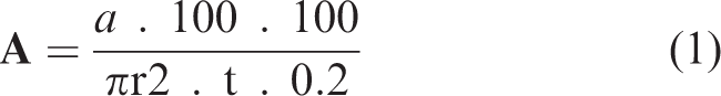
The airborne fungi were recovered, during assemblage of the deteriorated paints, four times per month for each studied site. Five Petri plates containing SDA media were exposed to the air of the selected sites for 30 min, at 150 cm above the floor level. The plates were transferred to the microbiological laboratory for incubation at 28°C. The growing colonies of fungi were counted and calculated using equation (1), as colony forming unit per m3 of the air (CFU/m3), based on Omeliański’s formula
46
Statistical analysis
The obtained results were statistically analysed via one-way ANOVA using the SPSS 10.0 (SPSS, Chicago, IL, USA) software program. The mean as well as standard deviation were computed for three replicates. Means were compared by the Duncan’s multiple tests and then the statistical significance was determined at 5% level.
Results
Fungi causing paint deterioration
In this study, 17 species belonging to eight genera were isolated from 15 samples of deteriorated water-based paint that collected from the Assiut University hospital. Generally, the deterioration included shifting in the colour (from white to pale and dark brown), gloss reduction, poor adhesion, off-colour and uneven colour, porous film and agglomeration (supplementary data).
The mycological analysis showed that Aspergillus and Penicillium were represented by seven and four species, respectively. The remained fungal genera (six genera) were constituted by only one species. The most common fungal species causing paint deterioration was Chaetomium globosum contributing 53.33% of the collected samples. Alternaria alternata, A. parasiticus, P. oxalicum and Setosphaeria rostrata came in the second rank with frequencies up to 13.67% for each. Moreover, all remained fungal species were isolated as one case comprising 6.33% of total collected samples. Remarkably, investigation of the status of fungal isolation showed that C. globosum, Mucor hiemalis and P. oxalicum individually caused the paint deterioration in their samples, while other fungi have a mixed association with samples of the deteriorated paint (Table 1).
Paints deteriorating fungi isolated from water-based samples collected from different medical units in Assiut University hospital (15 samples).
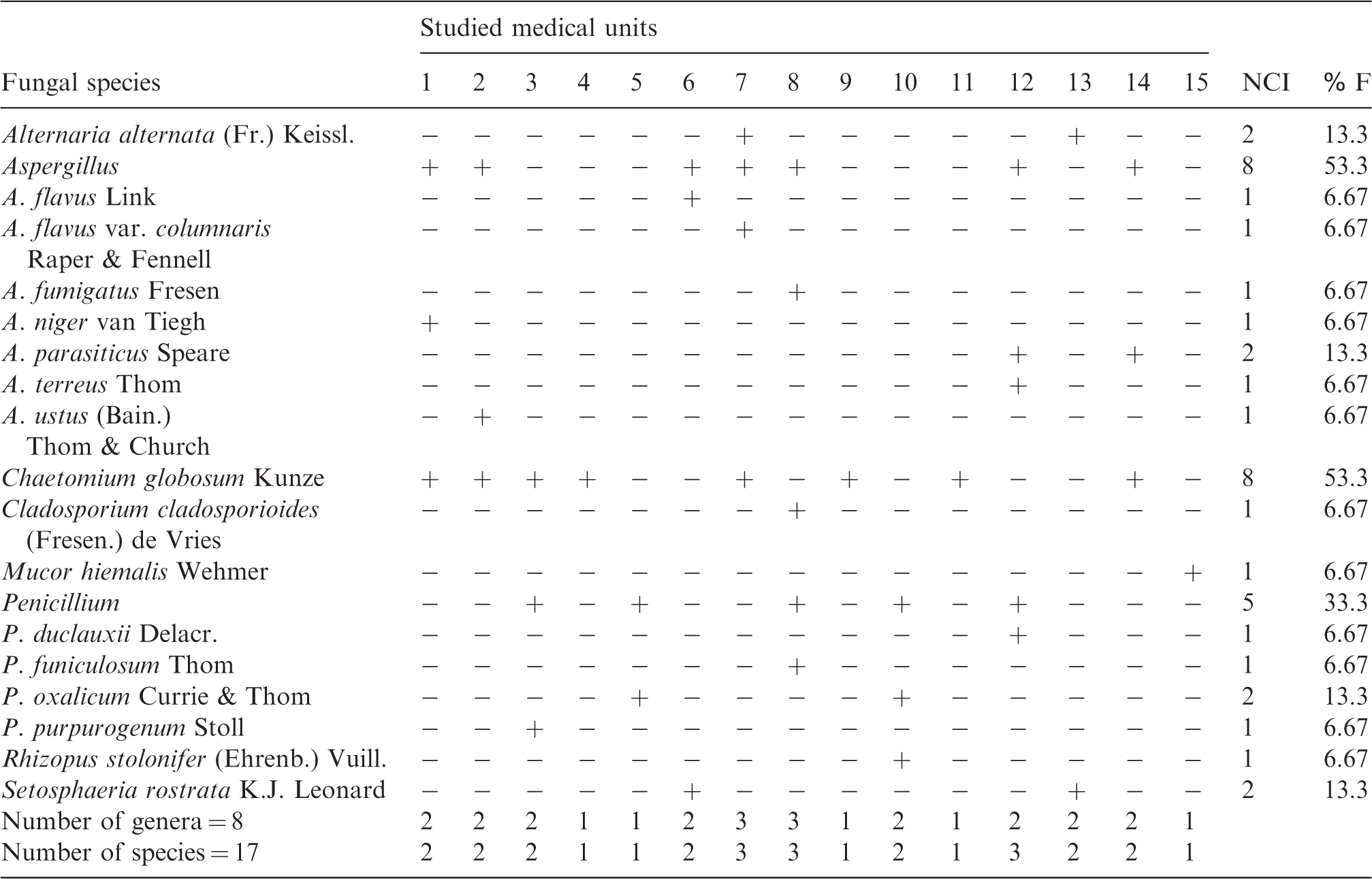
The number of case of isolation (NCI) and % frequency (%F) were recorded. Studied medical units: 1: Receiving Care Injuries; 2–4: Reception Injuries; 5: Echocardiography Clinics; 6: Digestive system Diseases Clinics; 7: Respiratory Intensive Care Unit (RICU); 8: Orthopedic Surgery Department; 9: Emergency Pharmacy; 10: Nervous Clinics; 11–12: Ophthalmology Department; 13–14: Special care unit (the 5th Department); 15: South Egypt Cancer Institute.
Preliminary detection of the fungi causing paint deterioration
Direct examination using stereomicroscope and light microscope confirmed that the isolated fungi have the ability to grow, invade and colonize the collected paint samples forming mycelia, conidia and sometimes fruiting bodies. C. globosum was individually responsible for the deterioration of paint samples collected from Reception Injuries, Emergency Pharmacy and Ophthalmology departments producing their own distinct fruiting bodies on these paint samples (Figure 1). Likewise, P. oxalicum was solely the causative agent of the paint deteriorates on Echocardiography Clinics and the examination showed the presence of its distinguished penicillus structure (Figure 1). Also, the examination exhibited that Aspergillus niger colonized the paint sample of Receiving Care Injuries generating the distinct structure as the conidial head of this fungal species (Figure 1).

Direct examination via stereomicroscope showing the colonization of the deteriorated paint samples by fungi. (a,b) Deteriorated paint flacks containing ascoma of C. globosum; (c,d) Deteriorated paint flacks containing conidial head of A. niger; (e,f) Deteriorated paint flacks containing penicillus structure of P. oxalicum.
Assay of paint utilization by fungi
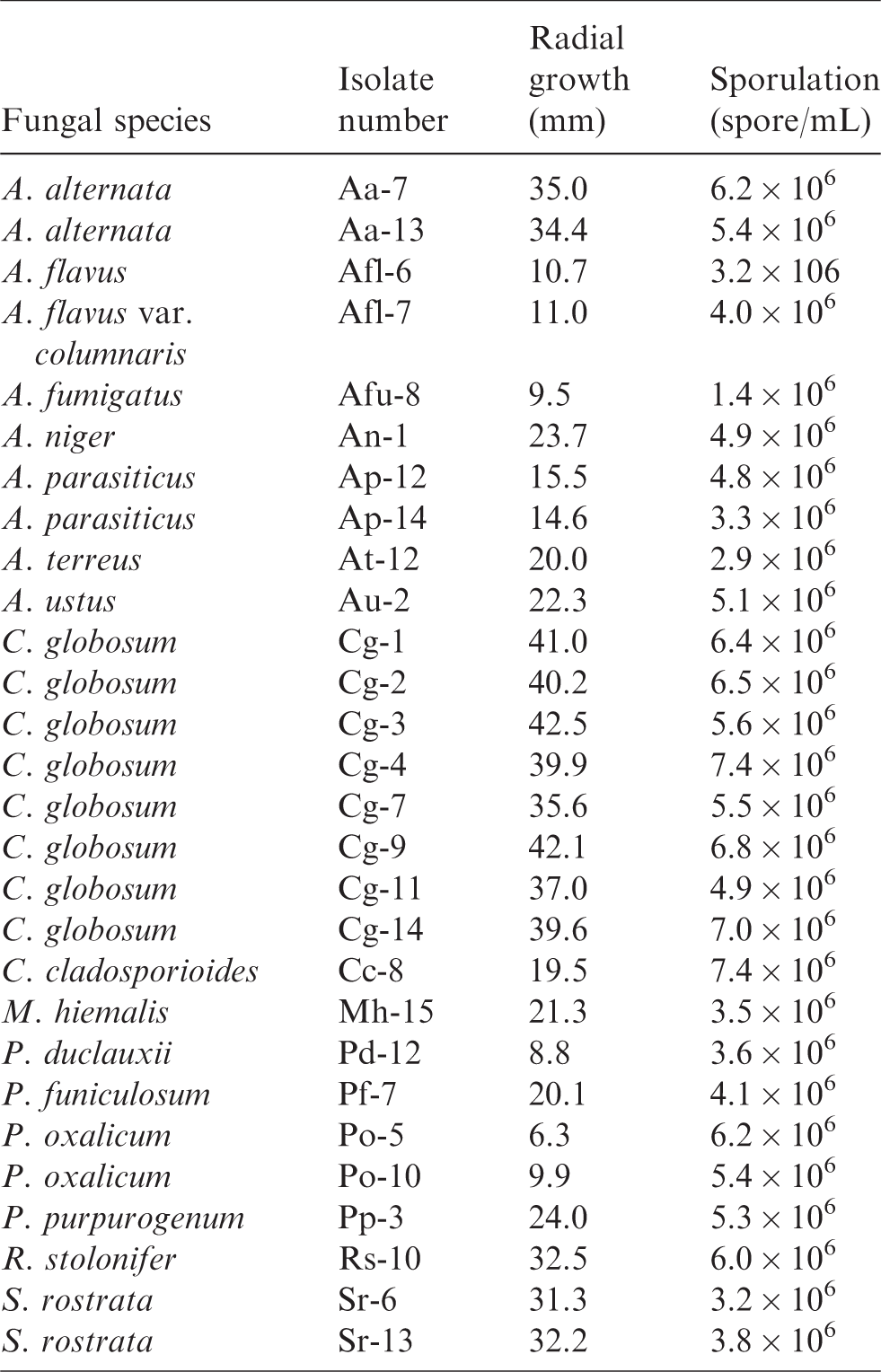
The results presented in Figure 2 show that there were considerable differences between the tested fungal isolates in their growth rate on the paint layer. The eight isolates of C. globosum showed the highest growth rate on the paint ranging 35.6–42.5 mm and sporulation of 4.9 × 10⁶ to 7.4 × 10⁶ spore/mL. The two isolates of A. alternata grew on the paint layer producing 34.4 and 35.0 mm with sporulation rate of 5.4 × 10⁶ and 6.2 × 10⁶ spore/mL. The radial growth of the two isolates of S. rostrata were 31.3 and 32.2 mm with sporulation rate of 3.2 × 10⁶ and 3.8 × 10⁶ spore/mL. Rhizopus stolonifer grew on the paint layer of 32.5 mm and 6.0 × 10⁶ spore/mL. On the other hand, 15 fungal isolates exhibited growth on the paint layer less than 25 mm (Table 2).
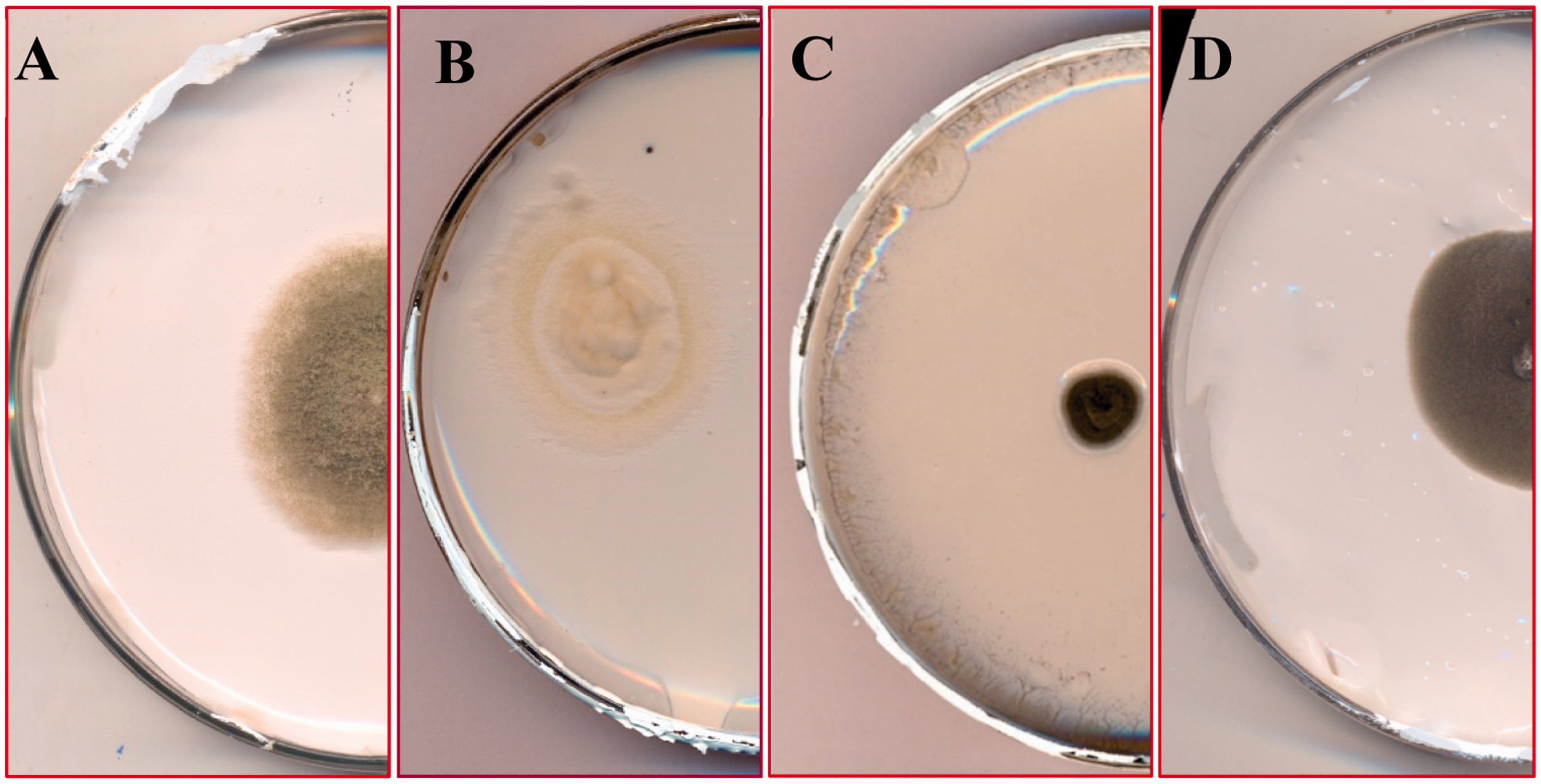
Assay of utilization of water-based paint by fungi. (a) C. globosum; (b) A. parasiticus; (c) Cladosporium cladosporioides (CC16); (d) A. alternata.
Determination of the growth rate of 28 fungal isolates on thin layer of water-based paint.
FT-IR analysis
The FT-IR spectrum of the tested water-based paint showed the absorption peak at 1798 cm−1 corresponding to the ester carbonyl group (C = O). Moreover, the peak of the C–O–C groups appeared in the spectrum at 1100 cm−1 (Figure 3). There was a peak detected at 2925 cm−1 corresponding to stretching vibration of C–H bonds of methylene groups (–CH2– stretch, aliphatic). On the other hand, other peaks were detected at 712 cm−1 (C–H, bend), 874 cm−1 (C–H, bend), 1426 cm−1 (C–H bend, methylene groups), 2513 cm−1 (O–H stretch, acid), 2850 cm−1 (C–H stretch, aldehyde) and 3410 cm−1 (N–H stretch, amine).
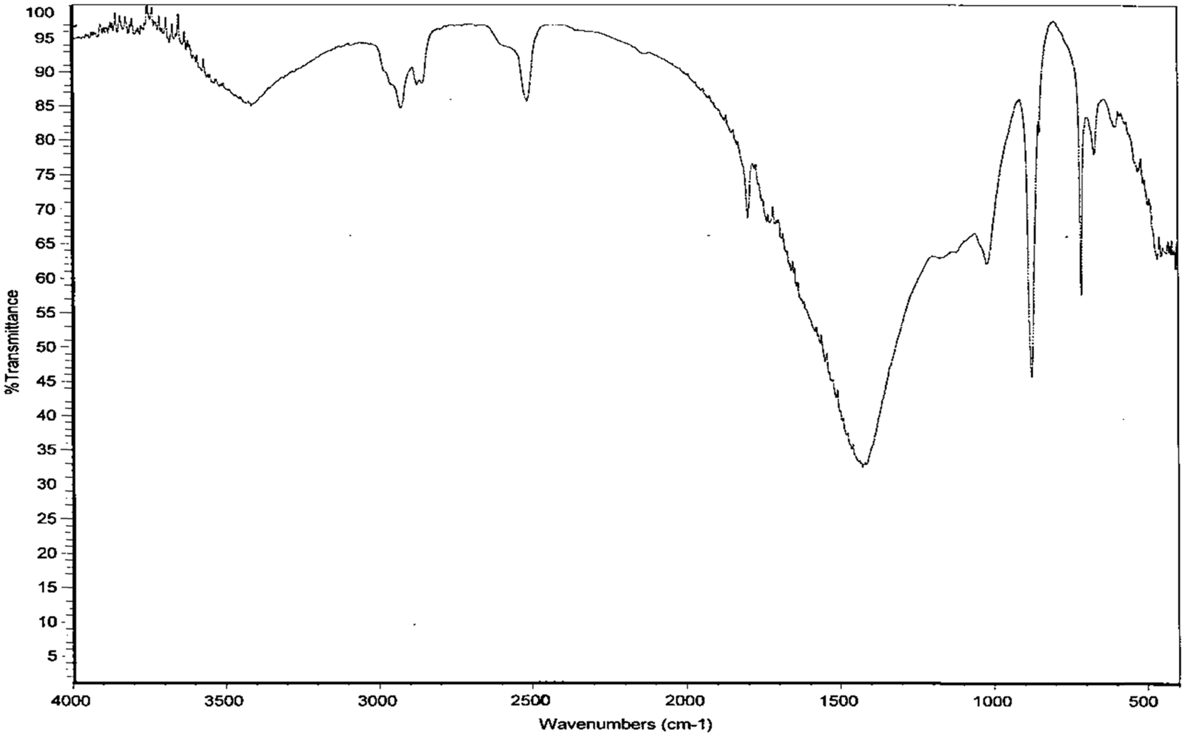
FT-IR analysis of the water-based paint showing the main functional chemical groups.
Assay of paint-degrading enzymes
According to the functional groups detected by FT-IR analysis as well as the chemical composition of the tested paint that included acrylic co-polymers and hydroxyethyl cellulose (HEC), we assayed the activities of cellulase, lipase and urease enzymes that may have the ability to utilize and degrade the paint components. Twenty-eight fungal isolates belonging to 17 species were tested for production of paint-degrading enzymes.
Cellulase activity
The results presented in Table 3 show that there were significant differences between the tested fungal isolates in their extracellular cellulase activity ranging from 4.32 to 0.01 μg glucose min−1. The isolate C. globosum (Cg-9) exhibited the highest cellulase activity of 4.32 U/mL with specific activity up to 6.66 U/mg protein. Also, there were three fungal isolates namely Aspergillus ustus (Au-2), C. globosum (Cg-7) and R. stolonifer (Rs-10) demonstrated cellulase activity 3.57, 3.52 and 3.02 U/mL corresponding specific activities 4.19, 3.47 and 2.49 U/mg protein. While the cellulase activity of C. globosum (Cg-4), Aspergillus terreus (At-12) and P. oxalicum (Po-10) were 2.50, 2.27 and 2.01 U/mL equivalent specific activities 4.05, 7.47 and 4.60 U/mg protein. On the other hand, 21 fungal isolates weakly showed cellulase activity less than 1.40 U/mL (Table 3).
Assay of cellulase activity of fungi causing water-based paint deterioration.
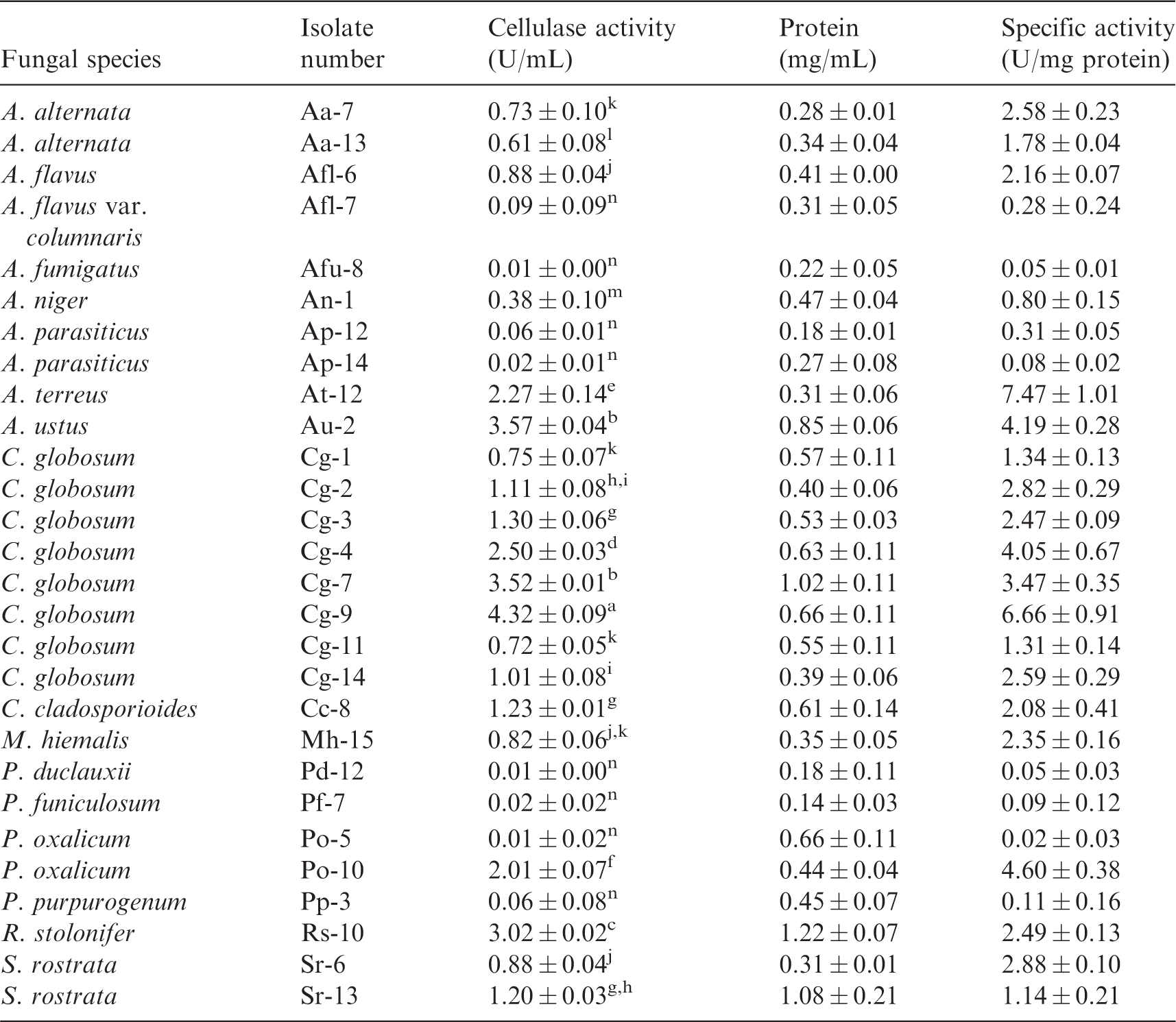
Numbers given in the table are mean of three replicates ± standard deviation. Values of activity associated with same letters indicate no significant differences (P ≤0.05) based on one-way ANOVA.
Lipase activity
Interestingly, all tested fungal isolates showed significant differences in lipase activity contributing 0.71–15.31 U/mL. The maximum lipase activity was obtained by the two isolates of P. oxalicum (Po-5 and Po-10) comprising 15.31 and 13.11 U/mL with specific activities 62.84 and 85.84 U/mg protein, respectively. In the second rank, C. globosum (Cg-2), M. hiemalis (Mh-15) and Penicillium purpurogenum (Pp-3) showed high lipase activity (12.53, 12.09 and 12.06 U/mL) corresponding specific activities up to 89.20, 56.48 and 83.19 U/mg protein, respectively. Moreover, seven fungal isolates namely A. alternata (Aa-7) A. ustus (Au-2), A. parasiticus (Ap-12 and Ap-14), A. terreus (At-12), Aspergillus flavus var. columnaris (Afl-7) and C. globosum (Cg-1) displayed lipase activities ranging 10.27–11.93 U/mL. On the other hand, 16 isolates had lipase activity less than 10 U/mL with specific activities varying between 3.24 and 51.14 U/mg protein (Table 4).
Assay of lipase activity of fungi causing water-based paint deterioration.
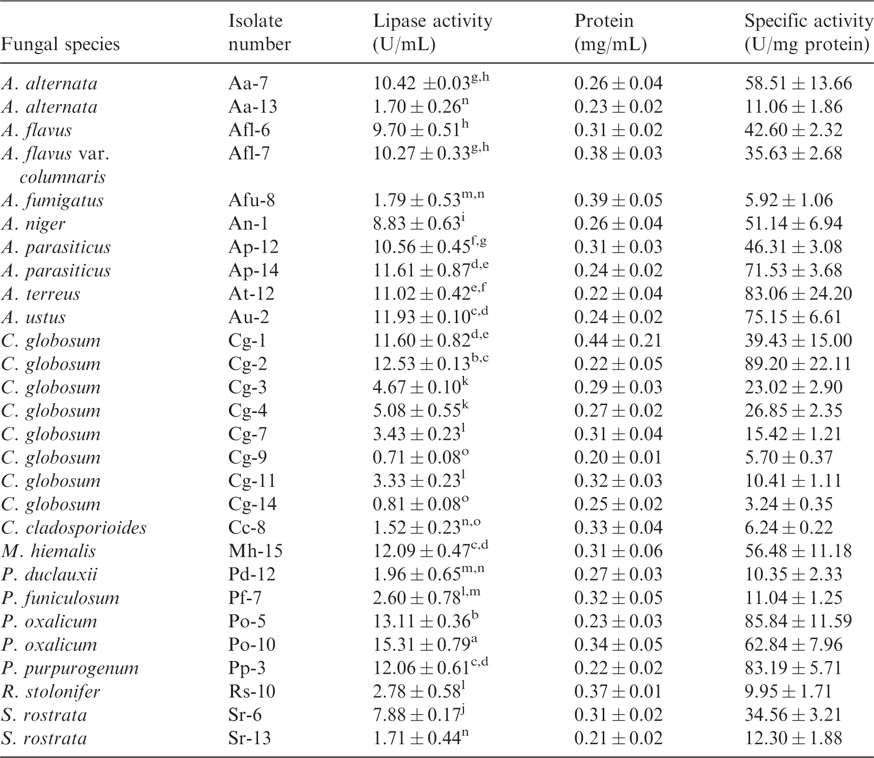
Numbers given in the table are mean of three replicates ± standard deviation. Values of activity associated with same letters indicate no significant differences (P ≤0.05) based on one-way ANOVA.
Urease activity
The results in Table 5 reveal that the urease activity was significantly varied between the tested fungal isolates ranging 98.44–692.11 μg NH4 min−1 mL−1. C. globosum (Cg-3) was the most active urease producer with activity value 692.11 U/mL including specific activity value 1570.64 U/mg protein. In that manner, A. niger (An-1), C. globosum (Cg-9), S. rostrata (Sr-13) and A. alternata (Aa-13) exhibited high urease activity comprising 557.67, 546.56, 545.00 and 527.22 U/mL, respectively. Also, elevated urease activities were produced by A. terreus (At-12), A. ustus (Au-2), C. globosum (Cg-4 and Cg-7), P. oxalicum (Po-10) and A. alternata (Aa-7) ranging 405.44–492.67 U/mL. On the other hand, 17 isolates displayed urease activity less than 400 U/mL with specific activities varying between 230.57 and 885.39 U/mg protein (Table 5).
Assay of urease activity of fungi causing water-based paint deterioration.
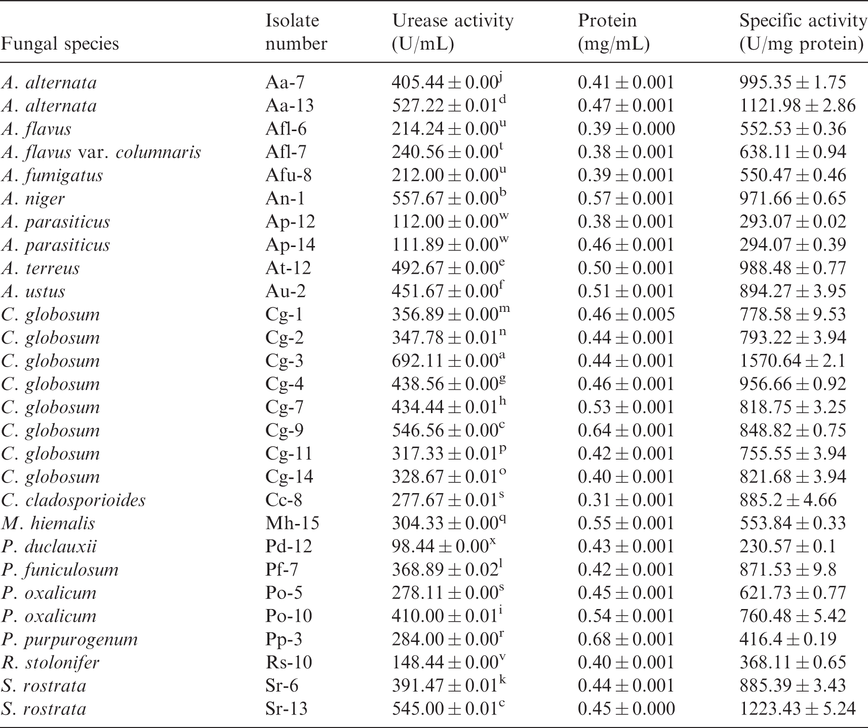
Numbers given in the table are mean of three replicates ± standard deviation. Values of activity associated with same letters indicate no significant differences (P ≤0.05) based on one-way ANOVA.
Airborne fungi
Twenty-seven species belonging to 15 genera were recovered, as air contaminating fungi, from 11 medical units in the Assiut University hospital. These mycobiota are related to three main fungal groups as Zygomycetes, Ascomycetes and Hyphomycetes. The total count of fungi recovered from all studied medical units was 969 CFU/m3. Aspergillus and Cladosporium were the most prevalent fungal genera (100% of total samples) comprising 42.3% and 44.0% of total fungi, respectively. Aspegillus was represented by nine species, while Cladosporium included only one species namely Cladosporium cladosporioides. Out of Aspergillus species, A. niger, A. flavus and A. fumigatus were recovered in high frequencies contributing 100%, 82% and 55% of total samples, respectively. On the contrary, the other remained Aspergillus species were isolated in frequencies less than 20% of total samples. Other than Aspergillus and Cladosporium, Penicillium (five species) and A. alternata were categorized in the second rank (82% and 73% of total samples) comprising 5.8% and 4.3% of total samples. C. globosum was isolated from three medical units comprising 27% of total samples. On the other hand, the remained fungal genera were recovered in frequencies less than 20% of total samples (Table 6).
Total fungal count (CFU/m3), percentage of total fungal count (%TC) and frequency (F%) of air-contaminating fungi collected during 2015–2016 from 11 medical units in the Assiut university hospital.
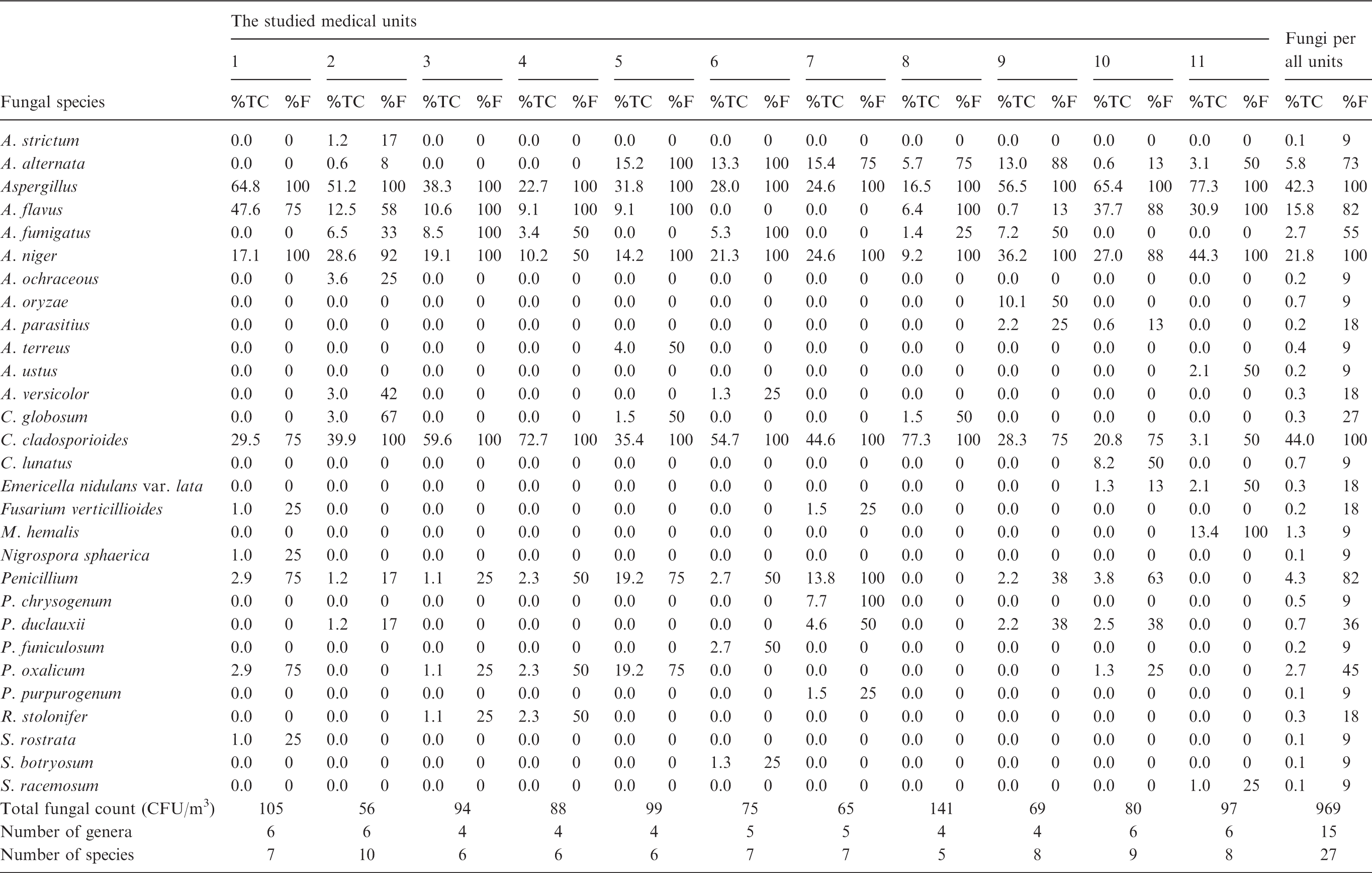
1: Receiving Care Injuries; 2: Reception Injuries; 3: Echocardiography Clinics; 4: Digestive system Diseases Clinics; 5: Respiratory Intensive Care Unit (RICU); 6: Orthopedic Surgery Department; 7: Emergency Pharmacy; 8: Nervous Clinics; 9: Ophthalmology Department; 10: Special care unit (the 5th Department); 11: South Egypt Cancer Institute.
Out of the studied medical units, the highest total fungal count was recovered from the unit of Nervous Clinics (141 CFU/m3), while the lowest fungal population was recovered from the unit of Reception Injuries (56 CFU/m3). On another side, there were no considerable differences between the studied medical units in the spectrum of airborne mycobiota (number of fungal genera and specie) as presented in Table 6.
Acremonium strictum, Aspergillus ochraceous, A. oryzae, A. terreus, A. ustus, Cochiobolus lunatus, Mucor hemalis, Penicillium chrysogenum, Penicillium funiculosum, P. purpurogenum, S. rostrata, Stemphylium botryosum and Syncephalastrum racemosum were recovered from only one medical unit comprising 9% of total samples (Table 6).
Our study of the correlation between fungi causing paint deterioration and air contamination showed that A. niger was responsible for paint deterioration in Receiving Care Injuries and was isolated in high frequency (100%) from the indoor air up to 17.1% of total fungi. Also, A. flavus attacked the paint in Digestive System Diseases Clinics and frequently was isolated from air of 9.1% of total fungi. C. globosum caused paint deterioration and air contamination (67% of total sample) in Reception Injuries of 3.0% of total fungi. Regarding the Respiratory Intensive Care unit, A. alternata and A. flavus produced paint deterioration as well as air contamination (frequency 100%) of 15.2% and 9.1% of total fungi, respectively. C. cladosporioides and A. fumigatus could cause occasional paint erosion and high frequently air contamination (frequency 100%) in the Orthopedic Surgery Department accounting for 54.7% and 5.3% of total fungi, respectively. Moreover, M. hemalis produced individual wall paint degradation in South Egypt Cancer Institute causing high indoor contamination (100% frequency) of 13.4% of total fungi (Table 6).
Discussion
Indoor environments could have direct impacts on the human health. The hazardous to health pollutants posed by contaminated indoor environments include non-biological and biotic agents. Fungi are ubiquitous in distribution and are a serious threat to public health in indoor environments. 1 Improper maintenance, poor building design or occupant activities often result in a condition called ‘Sick Building Syndrome’ in indoor environments, where visitors or inhabitants undergo adverse health effects which related to the time spent in a building. 47
Many reports exhibited that fungi can attack painted surfaces and subsequently caused air contamination. Different Aspergillus species are able to grow on almost all natural and synthetic materials, especially if they are hygroscopic or wet inorganic materials. They frequently become colonized as they are absorbed on dusts, which serve as good growth substrates. 48 Painted wooden furniture is highly vulnerable to fungal attack such as Cladosporium and Penicillium. 49 Acrylic painted surfaces are attacked by Alternaria, Chaetomium, Cladosporium and Aspergillus. 50
In this investigation, 17 species belonging to eight genera were isolated from 15 samples of deteriorated water-based paint that were collected from the Assiut University hospital. The most common fungal species causing paint deterioration was C. globosum, followed by A. alternata, A. parasiticus, P. oxalicum, M. hiemalis and S. rostrata.
Indoor wall paints and paintings are widely recognized as a good substrate for microbial biofilms in general and for fungi in particular. 51 Under favourable environmental conditions as high temperature and humidity, violent changes in semblance, colour and structure take place on the indoor wall paint which has a large surface exposure to fungal development. The microbial decay process of paints and original wall painting has already received much attention. 52
Previous studies reported that C. globosum, A. alternata, Penicillium citrinum and Aspergillus spp. have an aggressive ability to hydrolyse the cellulosic thickeners, grow on the acrylic paint films and could negatively impact on human health.5,35,53 Ugbogu et al. 54 found that fungal deterioration of painted wall surfaces, caused by Aspergillus, Penicillium and Mucor, is a great crisis in buildings of Wukari (Taraba State). Moreover, Arreche et al. 55 reported that the fungal growth of C. globosum and A. alternata on surfaces of buildings is an increasing severe problem around the world, which has an adverse effect on both people's health and buildings.
The current investigation assayed the enzymatic activities of the fungi causing paint deterioration to understand and evaluate their biodegradative and biodeteriorative potential. According to the presence of hydroxyethyl cellulose (HEC) on the water-based paint as well as the results of FT-IR analysis that proved the occurrence of ester and amine groups, the activities of cellulase, lipase and urease were measured.
Our results indicated that C. globosum, A. ustus, A. terreus and P. oxalicum successively showed high enzymatic activities of cellulase, lipase and urease that may individually or collectively play an essential role in the degradation of paint components. A. alternata, A. niger, A. parasiticus, M. hiemalis, P. purpurogenum and S. rostrata exhibited high activities of lipase and urease with moderate cellulase activity. On the other hand, other fungi have weak enzymatic activities, so they may stand together in the hydrolysis of paint components.
Under moist conditions, susceptibility of the paint surface to fungal attack generally depends on enzyme availability and chemical bonds in the polymers for enzyme attack. Fungi have the ability to utilize paint components as a sole carbon and energy source. These fungi developed a particular strategy by excretion of extracellular enzymes which depolymerize the polymer complex outside the cells yielding short chains or small molecules (oligomers, dimers and monomers) that are small enough (water soluble) to pass the cell membranes and then to be utilized as carbon and energy sources.56,57
Several investigators found that the microbial attack on paint components could be through the enzymatic action of hydrolases such as ureases, cellulase, lipase and esterases. 58 Cellulase enzymes have been observed to contain three main structural elements: the hydrolytic domain, a flexible hinge region and a C-terminus tail region involved in substrate binding. 59 A. niger possess one of cellulases β-1,4-endoglucanase which cleaves internal β-1,4-glycosidic bonds of pseudoplastic paint thickener (hydroxyethyl cellulose) causing paint hydrolysis. 35
The mechanism of lipase action in degradation of acrylic paints was previously discussed by Bellucci et al. 60 who reported that the water-based paint is basically a backbone of repeating acrylic and methacrylic acid units, esterified with methyl and ethyl residues, respectively. Lipases are subclass of the esterases and the ideal substrates for lipases appear to be either simple ester (monoesters) or glyceride of long chain fatty acids. Therefore, lipases are capable of hydrolysing some ester groups in the acrylic paints to form free carboxylic acid groups (Figure 4).
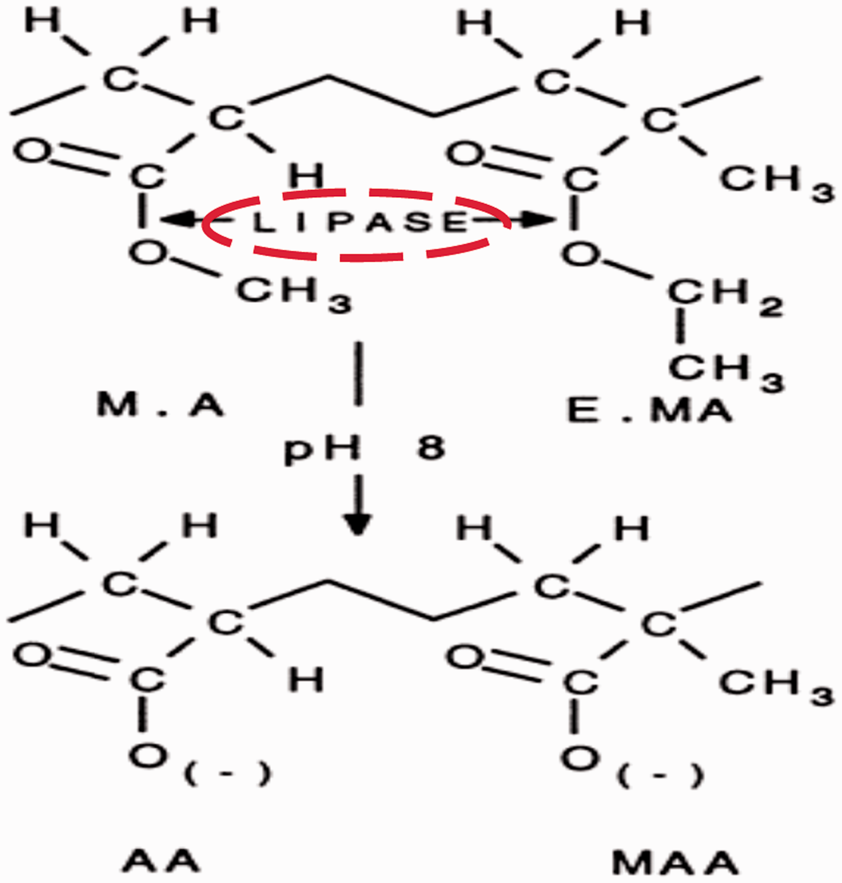
The mode of action for lipase on acrylic paints (Bellucci et al., 1999). 60 AA, MAA, MA and EMA are acrylic acid, methacrylic acid, methylacrylate and ethylmethacrylate, respectively.
On the other hand, FT-IR analysis confirmed the presence of (C-NH2) in the acrylic paint. Therefore, we suggest that urease may degrade the paint through this structure producing NH3. Additionally, several investigations studied urease production, by microorganisms, as a virulence factor of human diseases. Hence, this study gives a clear caution that the fungi causing paint deterioration may be aggressive human pathogens.
Several researchers demonstrated that some enzymatic mechanisms are implicated in the microbial degradation of various paints.10,61 An extracellular and membrane bounded lipase has been characterized for the urethane bond hydrolysis by many studies. 62 Also, esterase and lipase are the two well-known enzymes responsible for the biodegradation of acrylic and polyurethane paints. Hence, the ability of fungus to attack the paint were studied by measuring the esterase and lipase activities in broth culture. 61
In the paint industry, storage stability of paint emulsions is a critical issue. When a complex paint emulsion was exposed to microbial enzymes, the storage stability is of paramount importance and ultimately drives the efficacy of the product. The chemical modifications targeting the amino residues of the microbial enzymes have been shown to be effective in delaying their rapid rate of inactivation in the paint. 63 Fungi produce extracellular enzymes according to the chemical composition of the substrates they consume for growth. Moreover, secretions of hydrolytic enzymes as lipases and urease have been recognized as a main virulence factor of diseases by human pathogenic microorganisms. 64
In the current study, 27 species belonging to 15 fungal genera were isolated, as air contaminating fungi, from 11 medical units in the Assiut University hospital. A. niger, A. flavus, A. fumigatus and C. cladosporioides were the most prevalent fungal genera causing indoor air contamination in the studied hospital. Penicillium, A. alternata and C. globosum were also shown to cause a considerable amount of air contamination on certain studied medical units.
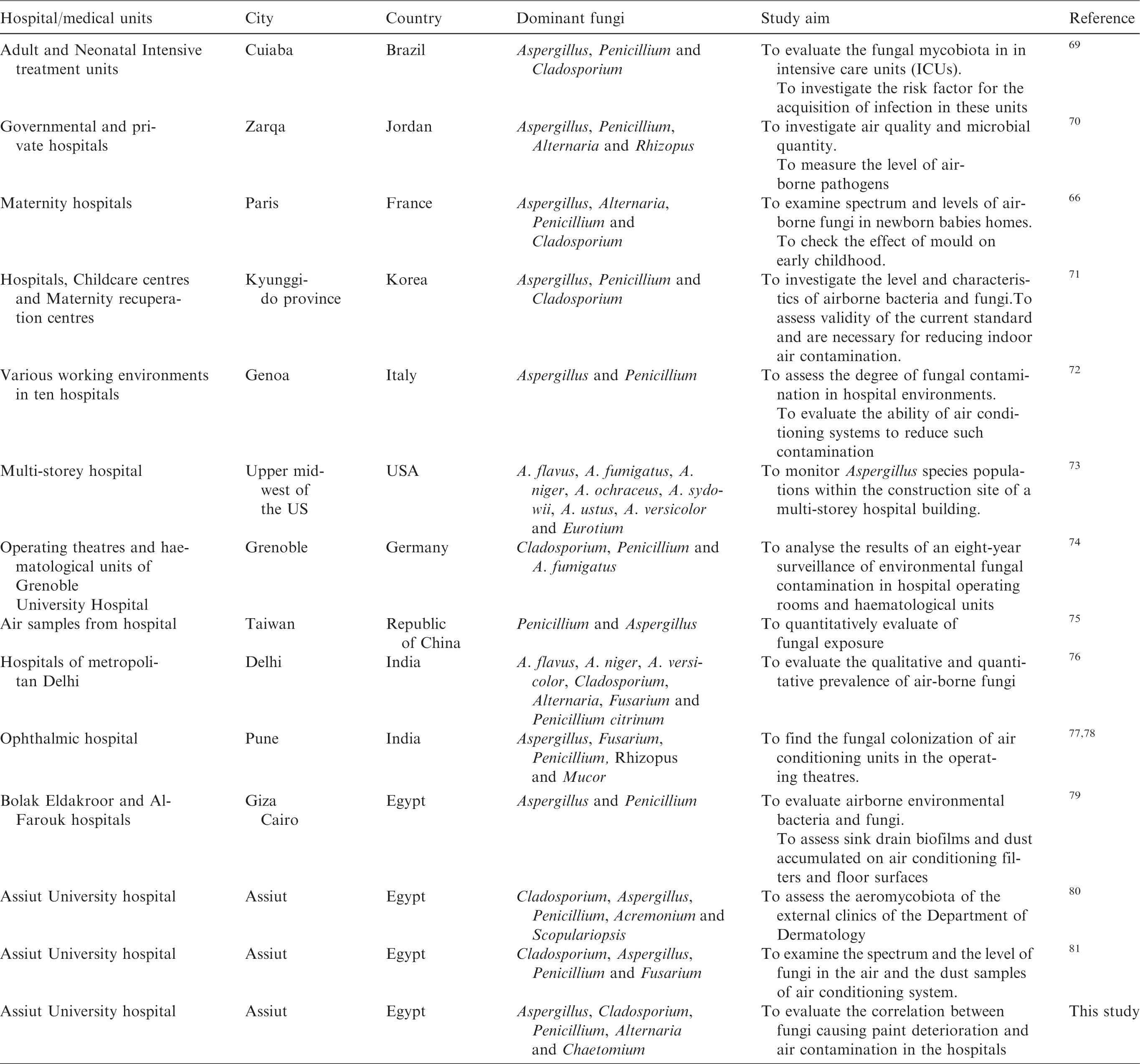
In this respect, data in Table 7 show that there are several investigations focused on fungi isolated from indoor environments of the hospital. Hospital-acquired fungal infections are a big danger to human health. 65 Aspergillus, Penicillium, Cladosporium and Alternaria may assume a menace to vulnerable individuals 66 and Aspergillus can cause invasive aspergillosis 67 even in low concentration. 68
Previous investigations on indoor airborne fungi of hospitals in several countries.
The results of the current investigation illustrated that there is a clear correlation between fungi causing paint deterioration and air contamination. A. alternata, A. niger, A. fumigatus, A. flavus, C. cladosporioides, C. globosum and M. hemalis were responsible for wall paint deterioration as well as frequent air contamination in some medical units in the Assiut University hospital.
In conclusion, airborne fungi in hospitals could be a potential threat to patient and visitors even at low microbial concentrations. Effective attempts should be performed to minimize microbial deterioration of paint and fungal air contamination inside the hospital. Moreover, this study suggests that improvement of antimicrobial additives of water-borne paints may be a promising approach to reduce paint bio-deterioration and subsequently air contamination of indoor environments.
Supplemental Material
Supplementary data -Supplemental material for Fungi-induced paint deterioration and air contamination in the Assiut University hospital, Egypt
Supplemental material, Supplementary data for Fungi-induced paint deterioration and air contamination in the Assiut University hospital, Egypt by Ismail R. Abdel-Rahim, Nivien A. Nafady, Magdy M. K. Bagy, Mohamed H. Abd-Alla and Ahmad M. Abd-Alkader in Indoor and Built Environment
Footnotes
Authors’ contribution
All authors contributed equally in the preparation of this manuscript.
Declaration of conflicting interests
The author(s) declared no potential conflicts of interest with respect to the research, authorship, and/or publication of this article.
References
Supplementary Material
Please find the following supplemental material available below.
For Open Access articles published under a Creative Commons License, all supplemental material carries the same license as the article it is associated with.
For non-Open Access articles published, all supplemental material carries a non-exclusive license, and permission requests for re-use of supplemental material or any part of supplemental material shall be sent directly to the copyright owner as specified in the copyright notice associated with the article.