
Abstract
Spontaneous or fine-needle aspiration (FNA)-induced remission of primary hyperparathyroidism (PHPT) is an extremely rare phenomenon with variable outcomes. We report a 75-year-old Male who initially presented with left ureteric calculi and was found to have PHPT. Imaging studies including ultrasound neck, parathyroid sestamibi scan and computed tomography of thorax, abdomen, and pelvis failed to identify the culprit lesion and exploratory parathyroidectomy was planned. Before surgery, he underwent FNA for cytology of a right cold thyroid nodule which was complicated with a large neck haematoma and dysphagia. The cytology of the aspirated fluid was consistent with a benign cyst. One month after the procedure, serum calcium and phosphate normalised along with resolution of haematoma. He remained in biochemical remission at 1-year follow-up with the latest ultrasound of neck showing resolution of a large colloid nodule that was previously seen occupying the right thyroid lobe.
Keywords
Introduction
Primary hyperparathyroidism (PHPT) is the most common cause of hypercalcemia and it is usually caused by a single adenoma (85%). Surgical excision of the abnormal parathyroid gland remains the only permanent and curative treatment of PHPT. Reports of spontaneous remission of PHPT following ultrasound-guided fine-needle aspiration (FNA) are extremely rare with variable outcomes. We report a case of spontaneous resolution of PHPT following a neck haematoma that developed post-FNA.
Case presentation
A 75-year-old gentleman initially presented to a urologist in 2019 for haematuria secondary to a ureteric stone at the left vesico-ureteric junction. He was later found to have parathyroid hormone-related hypercalcemia and referred to the endocrine unit. He was otherwise asymptomatic of hypercalcemia and had no constitutional symptoms or history of fracture. He has underlying hypertension and benign prostate hypertrophy on telmisartan/hydrochlorothiazide and terazosin. There was no family history of hypercalcemia.
The baseline biochemical analyses showed serum calcium of 2.62 mmol/L (normal range: 2.18–2.60 mmol/L) and parathyroid hormone of 25 pmol/L (normal range: 1.5–7.6 pmol/L). Serum 25-hydroxy vitamin D and phosphate were below the lower limit of the normal range (Table 1). 24-hour urine calcium creatinine ratio was done after vitamin D replacement and withholding hydrochlorothiazide. A ratio of 0.02 favoured PHPT. He had normal thyroid function. There was no renal impairment and repeated ultrasound kidney–ureter–bladder showed no evidence of renal/urinary bladder calculus or obstructive uropathy. The was mild splaying of the right pelvicalyceal system that may suggest a recent passed-out calculus. The bone mineral density showed osteopenia of the lumbar spine and neck of the femur with an FRAX score indicating an 8.7% risk for major osteoporotic fracture and 2.8% risk for hip fracture. The patient decided to wait and watch as it was the COVID-19 era and he was asymptomatic with mildly elevated calcium level. However, in the year 2021, the patient had hypercalcemia (>3.0 mmol/L) requiring repeated doses of pamidronate. He was then counselled and agreed to surgical intervention. Ultrasound neck performed showed a right colloid thyroid nodule measuring 3.6 × 4.6 × 4.8 cm with no sonographic evidence of parathyroid adenoma. Other imaging studies including sestamibi parathyroid scan, computed tomography of thorax, abdomen, and pelvis (CT TAP) and CT parathyroid multi-phase also failed to localise the culprit lesion.
Results of blood investigations before and after FNA of the thyroid nodule.
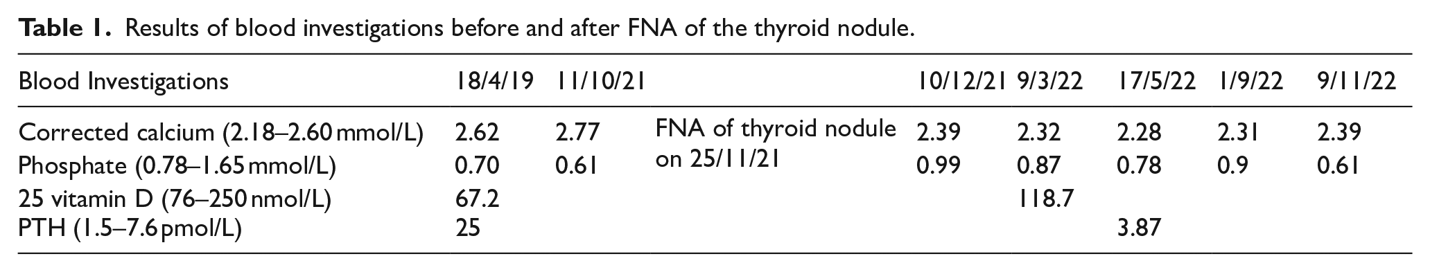
He was then referred to an endocrine surgeon for exploratory parathyroidectomy. Ultrasound-guided FNA for cytology of the right cold thyroid nodule done prior to surgery led to a large neck haematoma that was associated with dysphagia. The cytology of 20 ml of light brownish fluid aspirated was consistent with a benign cyst, Bethesda category 1. Surprisingly, 1 month after the procedure, the serum calcium and phosphate normalised along with the resolution of haematoma. Ultrasound neck done 6 weeks later showed an enlarged right thyroid lobe with retrosternal extension and increasing size of right colloid thyroid nodule (5.3 × 5.3 × 5.2 cm). However, the magnetic resonance imaging (MRI) of the thyroid done 6 months after FNA, in preparation for surgery (right hemithyroidectomy, bilateral neck exploration, total parathyroidectomy with intra-operative parathyroid hormone monitoring), noted a significantly smaller cystic nodule in the right thyroid lobe resulting in the smaller right lobe of thyroid with reduced retrosternal extension. The patient opted not for surgical intervention as he was biochemically in remission and imaging showed reducing size of the lesion.
To date, 12 months following the FNA, the patient remained in biochemical remission and the latest ultrasound which was done 10 months following the procedure showed both thyroid lobes normal in size with no retrosternal extension. The previously mentioned large colloid nodule seen occupying the right thyroid lobe is no longer seen. There is a well-defined oval-shaped hypoechoic nodule in the right thyroid lower pole: 0.8 × 1.0 × 0.6 cm TR4. No sonographic evidence of parathyroid nodules.
Discussion
Sporadic PHPT is most often caused by a single benign parathyroid adenoma (85%), less often by multiple parathyroid gland involvement (hyperplasia and less frequently synchronous or asynchronous adenomas (15%)) and very rarely by parathyroid carcinoma (<1%). Multi-gland disease is more likely to have a genetic or hereditary basis.1,2 In about 80% of all cases, PHPT is asymptomatic.
Parathyroid imaging is not used for diagnostic purposes. 3 It is used to locate the abnormal parathyroid gland(s) and should not be ordered until diagnosis and indications for surgery have been established and the patient has agreed to undergo surgery.3,4 Ultrasound is the preferred initial localisation study 4 and it has a sensitivity of 84% in the hands of an experienced sonologist. 5 Although normal parathyroid glands cannot generally and reliably be visualised with high-resolution ultrasound, enlarged hypercellular glands have a distinct appearance on grey scale imaging. The American Head and Neck Society Endocrine Surgery recommended that planar sestamibi imaging, single photon emission computed tomography (SPECT), SPECT/CT, CT neck/mediastinum with contrast, MRI, and four-dimensional CT may be used in conjunction with high-resolution neck ultrasound to preoperatively localise pathologic parathyroid glands. 4 Parathyroid cytology is not recommended by current guidelines as it has low diagnostic value. It can be easily confused with that of the thyroid and can lead to histological alteration. 6 Parathyroidectomy is the treatment of choice for patients with symptomatic PHPT and for those with asymptomatic PHPT who meet the criteria recommended by the latest guidelines unless medically contraindicated. 3
Reports of spontaneous remission of PHPT are rare and ultrasound-guided FNA biopsy-induced remission of PHPT is even rarer with only a few cases reported. Nylen et al have suggested that autoparathyroidectomy can be divided into three broad aetiologies: auto-infarction, acute extracapsular haemorrhage and acute intracapsular haemorrhage. 7 To better understand this condition, Wotten and Orzeck have reported a case and conducted a meta-analysis of 50 cases of autoparathyroidectomy. 8 The cases were analysed with respect to the classification suggested by Nylen and the investigators found that in acute extracapsular haemorrhage, the prominent clinical signs were neck swelling or pain. Unique to these patients, they can have ecchymosis, haematoma or hoarseness of voice. On the other hand, for both auto-infarction and acute intracapsular haemorrhage, the prominent clinical signs were neck swelling and neck pain.
Based on this meta-analysis, our patient’s clinical scenario was consistent with an acute extracapsular haemorrhage. The extracapsular haemorrhage/haematoma likely has caused compression to the vascular supply leading to tissue ischemia and death. However, the exact aetiology of spontaneous resolution of PHPT in our patient remained unclear. We did not manage to identify any parathyroid lesions on imaging studies and our patient declined surgery. There was no histological confirmation. Besides, these classifications were based on spontaneous, or non-traumatic, remission of PHPT. Long-term normalisation of calcium and parathyroid hormone level following FNA is rare but has been reported by Kara et al. 9 and Falcetta et al. 10 In these two cases, the patients had resolution of hyperparathyroidism following FNA biopsy of parathyroid adenoma. The duration of follow-up was 9 years and 12 months, respectively. Due to the limited number of cases reported, it is unknown how FNA-induced remission affects outcomes in these patients or the likelihood of recurrence. Adequate long-term surveillance is warranted in all of these cases.
Conclusion
Spontaneous resolution of hyperparathyroidism following procedure is rare but has been reported. Our patient likely had parathyroid apoplexy secondary to haematoma post-FNA of thyroid nodule leading to remission of PHPT. As it is unknown how the procedure will affect outcomes, adequate long-term surveillance remains mandatory.
Footnotes
Acknowledgements
We would like to extend our gratitude to the endocrine unit of Hospital Melaka.
Contributors
All authors made individual contribution to authorship. All authors reviewed and approved the final draft.
Declaration of conflicting interests
The author(s) declared no potential conflicts of interest with respect to the research, authorship, and/or publication of this article.
Funding
The author(s) received no financial support for the research, authorship, and/or publication of this article.
Informed patient consent for publication
Signed informed consent was obtained directly from the patient.
Data availability statement
Original data generated and analysed during this study are included in this published article.