






Abstract
Keywords
Introduction
Infrapopliteal arterial disease in patients suffering from critical limb ischemia (CLI) is often characterized by diffusely calcified lesions, frequently involving the tibial bifurcation. 1 According to recent therapeutic protocols and to TASC (TransAtlantic Inter-Society Consensus) recommendations, percutaneous transluminal angioplasty (PTA) is generally considered as a well-proven first-line option in these patients.2,3 Several aggressive techniques have been developed to improve patency of PTA in vessels below the knee; the “kissing dilation technique,” for example, is often used to avoid recurrent problems of plaque shifting observed when the two branches are dilated separately.4,5
Percutaneous transluminal angioplasty achieves a technically successful result (<30% residual stenosis) in most cases, but it is limited by high restenosis rates owing to elastic recoil or flow-limiting dissection requiring stent implantation or, when dealing with late restenosis, to vascular inflammatory reactions in terms of negative remodeling or neointima formation. 6 In the last few years, a new system of balloon angioplasty has emerged; based on a controlled and less traumatic dilation of the lesion, cutting balloon angioplasty (CBA) has the potential to improve the outcome of catheter-based therapy by minimizing elastic recoil and achieving better angiographic results.7–9
To the best of our knowledge, there are no reports about the efficacy of CBA in infrapopliteal lesions involving the arterial bifurcations. Hence, the aim of our pilot study was to evaluate the safety, feasibility, and effectiveness of the cutting balloon in the management of infrapopliteal bifurcation lesions.
Methods
This retrospective single-center pilot study evaluated the safety, feasibility, and effectiveness of the CBA technique for the treatment of short fibrocalcific infrapopliteal bifurcation lesions with a length <3 cm. All patients with CLI (at least Rutherford category 4) and an infrapopliteal bifurcation lesion, either the popliteal bifurcation (distal popliteal/anterior tibial/tibioperoneal arteries) or the tibioperoneal bifurcation (tibioperoneal/posterior tibial/peroneal arteries), treated with the CBA were included.
Patients were selected for CBA by a team composed by vascular diagnostic and interventional radiologists, vascular surgeons, and angiologists based on careful clinical evaluation, with treadmill testing (3.5 km/h; 12% slope), measurement of the ankle-brachial index (ABI), color duplex ultrasonography, and computed tomographic angiography (CTA). Significant clinical disability was evidenced as rest pain, nonhealing ulceration, or gangrene that could require major amputation, plus evidence of diffuse pedal ischemia, resting ankle pressure <60 mm Hg, and toe pressure <30 mm Hg. 7 A stenosis detected by duplex was considered significant if the peak systolic velocity ratio (PSVR) was at least 2.5. CTA assessment of stenosis severity was based on the ratio of the minimal intrastenotic to the prestenotic vessel diameters; a reduction in vessel diameter of at least 50% was considered significant. Each patient provided informed consent prior to endovascular treatment, with specific acceptance of CBA. This study was approved by our department review board.
Lesion Classification
Lesions were classified according to the Medina classification, the most commonly used classification for coronary bifurcations, 10 evaluating the presence of significant stenoses in the three components of the bifurcation (proximal main branch, distal main branch, and the side branch). The distal main branch was defined according to the angiosome concept in patients with Rutherford category 5 or 6 or according to the caliber (the largest artery feeding the foot) in patients with rest pain (Rutherford category 4).11,12
Bifurcation lesions were considered as true or false based on how many and which components of the bifurcation were involved; specifically, true bifurcation lesions were those presenting significant stenosis involving both the distal main branch and the side branch (with or without the involvement of the proximal main branch), while false bifurcation lesions were those with significant stenosis of at least one component and a nonsignificant stenosis of the others.
Study Population
Between November 2010 and March 2013, 23 CLI patients (mean age 69.6±9.01 years, range 56–89; 16 men) with at least 2-vessel runoff to the foot (present or after PTA treatment distal to the related bifurcation) and no aortoiliac disease underwent CBA for the treatment of infrapopliteal artery bifurcation lesions in 25 limbs (16 popliteal bifurcation lesions and 9 tibioperoneal bifurcation lesions). Patients with proximal inflow disease had the proximal lesion treated prior to the infrapopliteal intervention. An endovascular treatment of more distal disease prior to treating the bifurcation was performed in 15 patients (16 limbs). The baseline clinical data of these patients are shown in Table 1.
Baseline Clinical Data. a
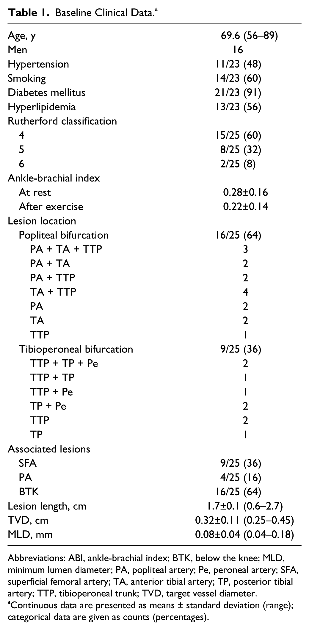
Abbreviations: ABI, ankle-brachial index; BTK, below the knee; MLD, minimum lumen diameter; PA, popliteal artery; Pe, peroneal artery; SFA, superficial femoral artery; TA, anterior tibial artery; TP, posterior tibial artery; TTP, tibioperoneal trunk; TVD, target vessel diameter.
Continuous data are presented as means ± standard deviation (range); categorical data are given as counts (percentages).
Eleven limbs had true bifurcation lesions (7 popliteal, 4 tibioperoneal), while the other 14 limbs had false bifurcation lesions (9 popliteal, 5 tibioperoneal). In detail, the other 28 branches of the diseased bifurcation had no or nonsignificant stenosis (patent components). The median lesion length was 1.7±0.1 cm (range 0.6–2.7), the baseline mean target vessel diameter was 0.32±0.11 cm (range 0.25–0.45), whereas the minimal lumen diameter (MLD) was 0.08±0.04 cm (range 0.04–0.18).
Revascularization Procedures
Before intervention, all patients were administered 325 mg of aspirin and clopidogrel (75 mg/d for 3 days before treatment). All procedures were carried out under local anesthesia via an antegrade approach to the ipsilateral common femoral artery. Patients received 5000 units of standard heparin, and saline infusions containing 5000 U/L of heparin were administrated throughout the procedure (maximum 7000 units).
Through a 5- or a 6-F vascular sheath, two 0.014-inch, 300-cm J-tip guidewires (ChoICE Extra Support Guide Wire; Boston Scientific, Marlborough, MA, USA) were placed across the lesions and into in the main branch and side branch, respectively, with the aid of roadmap guidance. Endovascular treatment was performed using a monorail technique with a cutting balloon (Boston Scientific) ranging from 2 to 4 mm in diameter and 15 mm long, using 1 to 3 inflations at a maximum pressure of 8 atm. The balloon was inflated and deflated 101 kPa (1 atm) every 2 to 3 seconds, holding inflation for <5 seconds, with a dilation time of <1 minute. The diameter of the cutting balloon catheter was selected to approximate a 1.0:1.1 ratio (balloon to the reference vessel diameter).
Angioplasty was performed according to the bifurcation lesion morphology and site using any of 3 techniques:
Single cutting balloon for false bifurcation lesions: the cutting balloon was inflated across the bifurcation with the distal end placed in the dominant artery if only the proximal main branch was stenosed or in the stenosed distal main branch or side branch regardless of whether the proximal main branch was involved;
T-shaped double cutting balloon for true lesions of the tibioperoneal bifurcation involving the tibioperoneal trunk: one cutting balloon was inflated across the bifurcation with the distal end placed in the distal main branch, whereas a second cutting balloon was inflated in the proximal tract of the side branch, across the ostium, with a modified kissing technique;
Double kissing cutting balloon for true lesions involving the popliteal bifurcation or for true lesions not involving the tibioperoneal trunk: both cutting balloons were inflated across the bifurcation, with the proximal end placed in the distal popliteal artery and the distal end placed in the distal main and side branch, respectively.
Postprocedure angiography of the treatment site and runoff vessels was done to evaluate blood flow improvement and potential complications. Stent implantation was limited to suboptimal angioplasty results or early technical failure, such as flow-limiting dissection or elastic recoil.
Follow-up
Clopidogrel was maintained for 3 months after the procedure (aspirin indefinitely). Surveillance included clinical evaluation, Doppler-based ABI measurements, and duplex examination at 1 month and every 3 months thereafter. CTA was carried out only in patients with positive clinical and diagnostic examinations in order to assess the need for further treatments, to decide the type of therapy, and to plan an eventual endovascular/surgical approach.
Definitions and Outcome Measures
The primary outcome measure was technical success; secondary outcomes were 1-year primary and secondary patency rates, clinical improvement, and limb salvage, as well as intra- and postoperative complications and overall and amputation-free survival. Angiographic outcomes also included postprocedure MLD and acute gain, defined as the difference between the postprocedure and baseline MLDs.
Technical success was defined as residual stenosis <30% on digital subtraction angiography (DSA) without flow-limiting dissection. The result of the procedure was scored using a 5-point scale based on the DSA images: score 1 for residual stenosis <20%, score 2 for residual stenosis <20% with non-flow-limiting dissection, score 3 for residual stenosis ranging from 20% and 50%, score 4 for residual stenosis >50% and/or flow-limiting dissection, and score 5 for technical failure. Procedure success was defined as a DSA residual stenosis <20% without flow-limiting dissection (score 1, 2). For procedures scored 3 (residual stenosis from 20% to 50%) resistant to additional dilation using a larger conventional balloon, no further treatment was done, whereas a stent was implanted for flow-limiting dissections and/or significant residual stenosis >50% (score 4).
Patency at follow-up was defined as <50% diameter stenosis determined by either duplex or CTA. Treated lesions were considered for analysis of primary and secondary patency rates according to accepted reporting guidelines. 13 Primary patency referred to persistent patency without any reintervention at the level of the target lesion at the time of the follow-up visit. Secondary patency was defined as patency restored by a catheter-based reintervention for clinically relevant restenosis or reocclusion of the target lesion before the follow-up visit plus all cases of primary patency. The indication for target lesion revascularization was exclusively driven by recurrent symptoms; changes in the ABI or a rise in PSVR detected by duplex only, without concomitant lifestyle-limiting symptoms, did not lead to reintervention. Significant changes in clinical status were based on an upward or downward shift in clinical category combined with a change in the ABI (>0.10).
Perioperative outcomes referred to events occurring during the first 30 days. Minor complications that did not need any treatment were not included in morbidity. Patients who died from causes unrelated to the procedure or had no follow-up were excluded from the follow-up evaluation.
Continuous data are presented as the means ± standard deviation; categorical data are given as the counts (percentage). Patency rates per vessel and considering the bifurcation as a whole were estimated with the Kaplan-Meier method. All hypothesis testing was 2-tailed, and a value of p<0.05 was considered significant. Statistical analyses were carried out by an independent statistical institute using SAS software (release 8.2; SAS Institute, Inc, Cary, NC, USA)
Results
A total of 47 stenoses (9 popliteal, 11 anterior tibial, 16 tibioperoneal trunk, 6 posterior tibial, 5 peroneal) were treated using 36 cutting balloons, predominately in the single balloon configuration (14/25). The other 11 bifurcation lesions were treated using a double cutting balloon technique (9 kissing, 2 T-shaped).
DSA performed at the end of the endovascular treatment showed 100% technical success (Figures 1 and 2), with the majority of lesions (44/47 94%) receiving a procedure success score of 1 or 2. The remaining 3 lesions had a score of 3, and despite additional conventional dilation using a larger balloon, a ~30% residual stenosis persisted (presence of heavy or eccentric calcification). Due to significant clinical status improvement, no further treatment was required in these 3 patients. No flow-limiting dissection requiring stent implantation was observed. The mean postprocedure MLD and acute gain were 0.28±0.04 and 0.20±0.06 cm, respectively, with a residual stenosis of 0.04±0.02 cm.

(A) A 79-year-old man (Rutherford category 4) had a left focal, fibrocalcific, ~80% distal popliteal artery stenosis (arrowhead) just above its bifurcation, with significant stenosis (>70%) of the tibioperoneal trunk and proximal peroneal artery (arrows) and steno-obstruction of the middle and distal tract of the anterior tibial artery (B, C). Owing to the location of the popliteal lesion just above its bifurcation, two 0.014-inch guidewires were placed in the anterior tibial and peroneal arteries, respectively, for treatment using a 2.5-mm cutting balloon (D, arrow). (E) In detail, endovascular treatment of the popliteal and peroneal lesions achieved correct remodeling of the popliteal artery (arrowhead) and tibioperoneal trunk and proximal peroneal artery (arrows) lesions, without procedure-related complications. (F, G) A 0.014-inch guidewire was then advanced until it reached the dorsalis pedis in order to recanalize the anterior tibial artery using a 2.5-×150-mm Coyote over-the-wire balloon.

(A, B) An 82-year-old woman (Rutherford category 4) showed a left focal, fibrocalcific, ~90% distal popliteal artery stenosis below a distal femoropopliteal bypass graft anastomosis and just above the popliteal bifurcation (arrowhead in B), with occlusion of the posterior tibial artery. (C, D) Two 0.014-inch guidewires were placed in the anterior tibial and peroneal arteries, respectively, to treat the stenosis using a 3-×15-mm cutting balloon, achieving remodeling of the lesions (arrowhead in C) without procedure-related complications.
There were no 30-day deaths or adverse events requiring treatment. One puncture site hematoma not requiring any treatment was the only minor complication registered. No arterial damage was observed; in particular, no dissection or arterial tear at the treatment site or damage to uninvolved patent components of the bifurcations.
Mean ABI significantly increased (p<0.05) at rest (from 0.28±0.16 to 0.68±0.13) as well as after exercise (from 0.22±0.14 to 0.65±0.18). The clinical status improved in all patients. An improvement of at least two Rutherford categories was achieved in 21 patients (23 limbs). Preplanned minor amputations were required in 2 patients.
Over a mean 13-month follow-up (range 6–21), 4 of the 23 patients died from unrelated causes at 3, 8, 11, and 12 months, respectively; furthermore, 2 patients were lost at 12-month follow-up. Ten lesions in 6 bifurcations developed recurrent stenosis; 5 lesions in 3 bifurcations were successfully treated with a new endovascular procedure (CBA), with a consequent reintervention rate of 11% (5/47). No revascularization was offered for 3 restenoses in 2 bifurcations due to the absence of lifestyle-limiting symptoms. In the remaining patient (2 restenoses in 1 bifurcation), a major amputation was necessary at 4 months due to poor runoff, intractable wound infection, and gangrene. The 12-month limb salvage rate was 96%. No surgical bypass was needed in follow-up due to target lesion restenosis/occlusion.
Primary and secondary patency rates were 89.3% and 93.5% at 6 months and 77.7% and 88.8% at 12 months, respectively; 1-year primary and secondary patency rates of the treated bifurcation were 74.2% and 87.0%, respectively (Figure 3A and B). The survival rate estimated by Kaplan-Meier analysis was 82.5% at 1 year (Figure 3C).

Kaplan-Meier estimates of (A) primary patency, (B) secondary patency, and (C) patient survival. The standard error did not exceed 10% at 12 months in any curve.
Discussion
Percutaneous transluminal angioplasty is still the gold standard in the treatment of infrapopliteal lesions due to acceptable clinical benefit, limb salvage, and morbidity.14,15 However, it is well known that PTA is characterized by initial technical failure, such as flow-limiting dissection and elastic recoil, and by a reported high late restenosis rate owing to negative remodeling and neointima proliferation.7,16 These limitations are more common in the infrapopliteal district because of the small caliber of the tibial arteries and the prevalence of calcified and diffuse atherosclerotic disease; moreover, the involvement of arterial bifurcations increases lesion complexity and, consequently, the risk of complications. In particular, the treatment of bifurcation lesions with a single balloon dilation increases the risk of plaque shift to the side branch and recurrent stenosis involving the main vessels or, more commonly, the side branch of bifurcated lesions.4,5,17 It is well known that maximizing blood flow to the foot by revascularizing more than one tibial artery speeds wound healing and increases limb salvage rates.18–20 On the other hand, choosing which artery to preserve can be difficult, especially when a dominant vessel cannot be recognized, and both vessels are patent to the distal foot.
Owing to advances in endovascular therapy and technologies, the interest in treatment of infrapopliteal arteries has grown, and the above circumstances are driving the development of specifically designed materials to deal with small-artery disease. For example, the interest in stent-supported below-the-knee (BTK) angioplasty is on the rise thanks to studies that have investigated the safety and efficacy of both bare metal stents or drug-eluting stents, potential alternatives to standard balloon angioplasty.21,22 However, long-term patency of these stents is undermined by the constant foreign body reaction, stress on the vessel wall, and stent fractures; furthermore, stents could also hinder subsequent surgical treatment.
In the past years, several studies have shown the effectiveness of specialty balloon catheters in overcoming the limitations of conventional PTA. In the infrapopliteal district, the use of a scoring balloon is gaining ground in the treatment of patients with CLI, as well as the use of balloon catheters with a controlled-inflation system, such as a nitinol constraining structure. 23
Cutting balloon angioplasty represents another treatment option. The new system of balloon angioplasty combines the features of a conventional noncompliant balloon with advanced microsurgical capabilities provided by 3 to 4 atherotomes mounted on its surface. Based on the concept “to cut first and dilate next,” this device allows controlled dissection, reducing the vascular injury and inflammatory reaction, with less traumatic dilation of the target lesion, potentially improving the outcome of angioplasty. Using less force of dilation, and consequently less barotrauma on the arterial wall, CBA allows optimal remodeling of the treated lesions with a potential significant reduction in procedure-related complications, such as arterial tears, rupture, or flow-limiting dissections requiring stent implantation. Based on this advantage, CBA should be preferred to PTA in the treatment of complex lesions in which the aim is to avoid stent implantation.24,25 CBA is routinely used in other fields of interest,7,8,26,27 with limited experiences in the treatment of infrapopliteal disease; however, to the best of our knowledge there is no published work regarding the use of this device in the treatment of infrapopliteal bifurcation lesions.
It is mandatory to underline that CBA requires specific training in its use and has a learning curve. In order to avoid complications, the balloon has to be correctly sized, trying to approximate a 1.0:1.1 ratio (balloon to the reference vessel diameter); furthermore, dilation and deflation of the device have to be performed slowly to allow safe extrusion of the blades and refolding into their protective sleeves.
In our retrospective pilot study, the use of CBA with different techniques based on lesion morphology and site produced excellent initial success without complications and 1-year primary and secondary patency rates comparable to and perhaps better than those reported in literature for BTK dedicated stents. The 96% limb salvage in this study was also comparable. The clinical status of all patients improved, with only 2 preplanned minor amputations. The treatment proposed in the study is appropriate for all types of bifurcated lesions; the main branch can be treated while preserving and also eventually restoring the side branch. This benefit of treating the entire bifurcation as a single lesion is reflected by the good 1-year primary and secondary patency rates obtained for the 25 treated bifurcations.
Limitations
The main limitations of our study are represented by the small number of patients involved and by the retrospective review of our routine experience, with no control or comparison group data. Another potential limitation is the method of imaging used in follow-up: duplex, although operator-dependent, is our standard postintervention surveillance method to reduce the invasiveness and cost of follow-up. A CTA examination was performed only in patients with positive/suspected clinical and/or diagnostic examinations.
Conclusion
The results of our pilot study indicate that CBA seems to be a safe and effective tool in the routine treatment of short/ostial infrapopliteal bifurcation lesions, avoiding procedure-related complications, overcoming the limitations of conventional angioplasty, and improving the outcome of catheter-based therapy.
Footnotes
Declaration of Conflicting Interests
The author(s) declared no potential conflicts of interest with respect to the research, authorship, and/or publication of this article.
Funding
The author(s) received no financial support for the research, authorship, and/or publication of this article.