






Abstract
Purpose:
To describe the implantation steps of the latest generation of candy-plug device (third CP generation [CP III]) and to illustrate its possible pitfalls by discussing a case in whom this device was employed to occlude the false lumen (FL) of a chronic type B aortic dissection.
Technique:
A 69 year-old male patient who underwent a frozen elephant trunk arch repair due to residual type A aortic dissection developed a FL aneurysmal degeneration limited to the descending thoracic aorta. Two thoracic stent-grafts were deployed into the true lumen up to the celiac trunk origin. Then, the FL was occluded with a self-occluding CP III device (Cook Medical, Bloomington, Indiana), placed at the same level as the distal thoracic stent-graft. The distal un-stented sleeve was pushed upward to allow immediate occlusion of its central lumen, avoiding interference with reno-visceral arteries arising from the FL. Both intraoperative transesophageal echocardiography and follow-up computed tomographic angiography scan demonstrated complete FL thrombosis.
Conclusion:
The introduction of CP III with its self-occluding mechanism helped to shorten and standardize the procedure. However, adjunctive steps may be needed to immediately obtain FL occlusion and avoid hampering the perfusion of vessels arising from the FL.
Keywords
Introduction
The candy-plug (CP) technique was developed to avoid retrograde perfusion of the thoracic false lumen (FL) in chronic type B aortic dissection (TBAD) cases treated by standard thoracic stent-grafting.1–3 Since its first report, in 2013 by Kölbel et al, 1 the CP technique has increasingly improved from homemade vascular plugs and off-label devices to dedicated custom-made FL exclusion devices.1,4–10
The first CP generation (CP I) consisted of a double-tapered configuration with greater proximal and distal diameters and a small diameter in the central midsection, enabling the dilator tip retraction. This version required occlusion of the central channel with a plug increasing the procedure complexity. The second CP generation (CP II) was based on the Cook Alpha nitinol platform and consisted of a nontapered stent-graft with an internal self-occluding un-stented fabric channel that did not require the deployment of the plug (Figure 1A).4,7,9–11 This version was soon abandoned due to regulatory problems related to the use of the Cook nitinol platform in dissections. Therefore, a third CP generation (CP III) was designed with the self-occluding un-stented fabric channel, called sleeve, at the distal end of the stent-graft (Figure 1B). 4
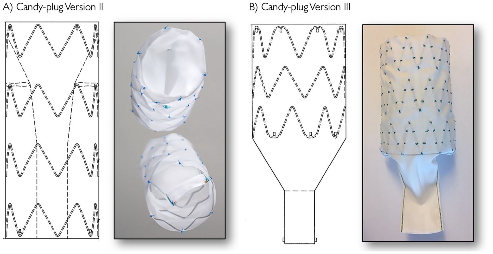
Technical drawings and pictures of the custom-made candy-plug (CP) device with self-occluding central channel: version II (A) and version III (B). The 2 versions differ each other depending on the conical sleeve segment which it is sutured inside the device in CP II (A) and at the distal part of the CP III (B).
The goal of this technical note is to describe the procedural steps of CP III deployment to obtain immediate occlusion of the distal sleeve and avoid possible interference of its distal end protruding in the abdominal FL.
Technique
Case Description
The CP III deployment technique is demonstrated in a 69 year-old male patient with a past medical history of type A aortic dissection and ascending aorta and aortic valve replacement with a 30 mm Hemashield Dacron graft (Maquet, Rastatt), and a 27 mm Medtronic Freestyle bioprosthesis (Medtronic, Minneapolis, Minnesota), respectively. One year after the procedure, the patient underwent a frozen elephant trunk repair with a 30 mm Vascutek Lupiae graft (Vascutek, Inchinnan, Glasgow) and E-vita Open Plus 28 mm (JOTEC GmbH, Hechingen) due to persistent and enlargement of the aortic arch FL. The following year, a large FL aneurysm in the descending thoracic aorta was identified at the annual follow-up computed tomographic angiography. The patient was treated with a thoracic endovascular aortic repair (TEVAR) to the celiac trunk in the TL (Zenith Cook Alpha ZTA-P-30-201 + ZTA-P-30-155; William Cook Europe, Bjaeverskov) and FL occlusion with a CP III (please refer to device description). Immediate FL occlusion was demonstrated at completion angiography and intraoperative transesophageal echocardiography (TEE). The patient was discharged on postoperative day 3, without neurological deficits, after an uneventful recovery. Computed tomographic angiography at 6 months showed complete FL thrombosis proximal to the CP III, without device migration or visceral arteries occlusion (Figure 2).

Preoperative computed tomographic angiography (CTA) 3D reconstruction and axial images showing retrograde false lumen (FL) perfusion after frozen elephant trunk repair and its extension with 2 standard stent-grafts (A). Six-month follow-up CTA imaging demonstrating FL occlusion after candy-plug version III placement (B).
Device Description (Figure 1B)
The CP III (William Cook Europe) is a custom-made thoracic stent-graft with 3 sealing stents of a diameter ranging from 26 to 46 mm and a total length of 97 to 104 mm depending on diameter, including a distal sleeve segment with a 14 mm wide central fabric channel, sutured outside the graft. The 3 independent nitinol Z-stents are covered by a woven polyester graft fabric. The 14 mm wide sleeve, which allows the dilator tip retraction, is tapered down from the nominal diameter of the device. Radiopaque markers are placed 1 mm from the edge of the graft material, placed in the apex of the proximal stent, and further markers in the bottom of the distal stent. The distal part of the sleeve is marked with 2 large lateral gold markers to assist in the deployment accuracy.
The stent-graft is loaded in a 16- to 20-Fr delivery system, depending on the CP III diameter, with a hydrophilic coated sheath of 6.7 mm inner diameter and 7.7 mm outer diameter, with an 85 cm usable length and a flexible atraumatic tip. The CP III device is available in Europe as a custom-made device and is under the Food and Drug Administration as an investigational device exemption protocol in the United States. Deployment technique (Figure 3 and Video 1 of the Supplemental material): After accessing the FL through a distal reentry, an ultra-stiff Lunderquist guide wire (William Cook Europe) is placed in the FL of the proximal descending thoracic aorta to deploy the CP III device above the celiac trunk origin. In case of proximal thrombosis of the FL, a glide wire is advanced through the thrombus as cranial as possible with the help of 0.035″ support catheter and then exchanged for the stiff wire to gain enough support for the CP advancement and length for the dilator tip of the introduction system. The device is introduced and positioned at the same level of the distal end of the previous TL stent-graft. The sheath is withdrawn through a standard pullback maneuver until the device is fully unsheathed. Then, the dilator tip is retracted passing the 14 mm sleeve distally to the CP III, and the stiff wire is left in place. Figure 3 depicts suggested additional procedural steps to manage the distal sleeve. The aims of these additional procedural steps are (1) to avoid interference between the sleeves deployed distally with reno-visceral arteries originating from the FL (Figure 4); (2) to allow closing immediately the central core of CP with thrombosis of the FL (Figure 5 and Video 1 of the Supplemental material).

Candy-plug (CP) version III step-by-step deployment sequence. The CP device is placed at the same level of the distal end of the previous true lumen stent-graft. The proximal and mid gold markers represent the proximal and distal end of the cylindrical stent-graft part of the device (A). The introducer sheath is withdrawn through a standard pullback maneuver until the device is fully unsheathed (white arrows). Note that the proximal edge of the stent-graft is still constrained (black arrow) (B). The trigger wires are removed by rotating the blue handle of the delivery system to free the graft from the delivery system by opening the proximal stent-graft end (black arrow) and allow the removal of the tip dilator and the delivery system (white arrows). The wire is still in place through the lumen of the CP (C). The sleeve end (black arrows) has to be repushed upward (D). The introducer sheath is readvanced without the dilator tip over the wire to voluntarily engage the distal end of the polyester sleeve with the sheath (E). The introducer is pushed upward (white arrows) to drag the sleeve as close as possible to the stented portion of the CP: the 2 distal lateral gold markers help to guide this maneuver (black arrow) (F). The wire is retracted within the sheath (G). The J tip of the stiff wire is readvanced out of the sheath to eventually disengage the sleeve from the sheath (H) and allow its final retraction (white arrow) (H), avoiding pulling the sleeve distally again. Final result showing the retraction of the sheath and the guide wire, keeping the sleeve close to the CP-stented portion (I).

Intraoperative angiography (A) and digital subtraction (B) showing an example of possible interference between a candy-plug (CP) version III distal sleeve (white arrow) deployed without the adjunctive maneuvers described in Figure 3 and the left renal artery originating from the false lumen. Coronal computed tomographic angiography (CTA) reconstruction (C) and axial CTA image (D) showing the CP III sleeve markers (white arrow) in front of the left renal artery ostium.

Example of a persistence false lumen patency after candy-plug (CP) III deployment demonstrated by a 1 month computed tomographic angiography. The cause was the absence of thrombosis of the central lumen of the CP (white arrows) which was persistent at 6 months.
Discussion
Most patients with chronic TBAD treated with TEVAR alone have retrograde FL perfusion through distal reentries, leading to aneurysmal FL degeneration of the distal aortic arch and proximal and mid-descending thoracic aorta. 12 The occlusion of such connections between TL and FL can be achieved by further distal coverage using fenestrated or branched endografts into the iliac arteries, allowing complete FL exclusion and consequent thrombosis. 13 It should be noted that occluding these distal reentries is necessary to successfully occlude the FL irrespectively that the aneurysmal degeneration involves the abdominal FL. However, these extensive thoracoabdominal procedures are plagued with a substantial risk of spinal cord ischemia (SCI), unpredictable FL perfusion due to type II endoleaks, and an increased rate of reintervention.7,10,14
As the FL aneurysmal degeneration in most cases is limited to the thoracic aorta, less invasive FL thoracic occlusion techniques, such as the CP technique, may allow a less extensive repair by limiting stent-graft coverage to the thoracic aorta and thereby deduce the risk for SCI. False lumen occlusion techniques targeting the connections between the 2 lumens directly aim at the occlusion of the thoracic FL, avoiding unnecessary coverage of nondilated dissected abdominal aortas. 8 More recently, this technique has been proposed as a stage of fenestrated or branched repairs to precondition the spinal cord and avoid intersurgical thoracic ruptures of large aneurysms during the waiting time of custom-made devices.6,7
Since the first description of this technique, improvements have been developed to the CP device to allow a better occlusion of the FL, avoiding persistent retrograde reperfusion, and limiting concerns about the possibility of narrowing the TL.4,11 The first device was obtained with a back table modification of a 42 mm Zenith thoracic TX2 Pro-Form stent-graft (Cook Medical, Bloomington, Indiana). 1 After the initial proof of concept, the CP I was developed as a custom-made device, and feasibility studies reported good early outcomes with low morbidity and mortality. 10 However, the need for an adjunctive off-label procedure to occlude the center of the CP I, with increased procedural complexity and the risk of inadequate CP occlusion, pushed the natural evolution of the device toward the current self-occluding generations of CP.
The CP version III differs from the previous generation because the central channel fabric with the above-reported self-occluding mechanism comes outside the graft at the distal part instead of inside the stent-graft (Figure 1). Recent studies reported the feasibility of the CP III technique with low mortality and morbidity and a high rate of aortic remodeling.4,7,15 Despite device improvements that allowed avoiding the need for central plug deployment steps and the increased conformability of the 3 nitinol stents to the false aortic wall with better occlusion rates, some possible technical pitfalls deserve further discussion. After implantation in the FL, the sleeve might remain in front of a FL originating reno-visceral arteries. To overcome this issue, an option might be to deploy the CP more cranially. However, different drawbacks can be anticipated. First, FL CP placement at the same distal edge level of the stent-graft positioned into the TL prevents exerting unilateral force on the lamella. Stent-induced new entry tears may occur placing the CP device without TL stent-graft at the same level. 16 Second, the region close to the diaphragmatic crus is usually the less dilated thoracic FL section and, therefore, the best place to obtain sealing with the CP.
The above-described procedural steps of CP III placement allow a correct distal deployment of the CP with TL opposing stent-graft and, by pushing the sleeve upward, avoid any reno-visceral artery interference. In addition, the described maneuver enhances immediate sleeve occlusion and consequent FL thrombosis, as observed in the reported case by completion angiography and TEE. However, in some cases, a late occlusion of the central core of the self-occluding mechanism was occasionally observed (Figure 5). This potential drawback might be crucial when an immediate thrombosis of the FL is required (eg, impending or free ruptures). The presence of intercostal or lombar arteries and abdominal reentries might create a persistent flow within the sleeve that prevents its immediate occlusion and therefore the use of the abovementioned technique is suggested. We should acknowledge that this occurred in only in the case here described and at 6 month follow-up no FL growth was observed . The persistent backflow through the CP device central channel can be eventually addressed with adjunctive distal FL embolization with a vascular plug or coil, or by complete distal FL exclusion with fenestrated/branched endografts, in case of aneurysmal FL progression.
Conclusion
The new generation of the CP III device facilitates a simplified occlusion of the FL in patients with postdissecting aneurysms. The described adjunctive procedural steps allow an early occlusion of the CP central channel and avoid interference of the sleeve with vessels arising from the FL.
Footnotes
Declaration of Conflicting Interests
The author(s) declared no potential conflicts of interest with respect to the research, authorship, and/or publication of this article.
Funding
The author(s) received no financial support for the research, authorship, and/or publication of this article.

Supplemental Material
Supplemental material for this article is available online.
References
Supplementary Material
Please find the following supplemental material available below.
For Open Access articles published under a Creative Commons License, all supplemental material carries the same license as the article it is associated with.
For non-Open Access articles published, all supplemental material carries a non-exclusive license, and permission requests for re-use of supplemental material or any part of supplemental material shall be sent directly to the copyright owner as specified in the copyright notice associated with the article.
