






Abstract
Purpose:
To report a unique entity and its management that occurred at our facility: combined spontaneous isolated renal and celiac arterial dissection (SIRCAD) with resultant renal and gastrointestinal symptoms.
Case Report:
A 50-year-old man with no past medical history presented with a 4 day history of nausea, intermittent stabbing epigastric pain, right flank pain, and uncontrolled hypertension. After full physical examination and imaging studies, the diagnosis of SIRCAD was established and confirmed. Selective right renal artery catheterization revealed dissection limited to the main trunk, and after careful selective hand-injection and successful cannulation of the distal renal artery branches through the true lumen assisted by intravascular ultrasound, a balloon expandable covered stent (6 mm in diameter and 60 mm in length) was deployed in the main renal artery. The same steps were performed for management of the celiac artery dissection. The patient was treated with clopidogrel 75 mg for 6 weeks and lifetime aspirin. A week after the procedure, his symptoms completely resolved.
Conclusion:
The pathology of SIRCAD in the absence of other vascular dissections is extremely rare, which speaks for the necessity of reporting this case and highlights the great role of evolving imaging modalities in the diagnosis and management of such cases.
Clinical Impact
Symptomatic combined spontaneous isolated renal and celiac arterial dissection (SIRCAD) remain rare despite the increased frequency of reports on asymptomatic dissections. The etiology of SIRCAD is not precisely defined. Moreover, treatment of SIRCAD remains controversial with only a few cases of percutaneous interventional treatment are reported in the literature.
Keywords
Introduction
Symptomatic combined spontaneous isolated renal and celiac arterial dissection (SIRCAD) remains rare despite the increased frequency of reports on asymptomatic dissections with technical advances in multidetector contrast-enhanced computed tomography angiography (CTA). Spontaneous isolated renal and celiac arterial dissection is a rare cause of renovascular hypertension combined with duodenitis, acute pancreatitis, and cholecystitis. The most common symptom in acute dissection is epigastric, abdominal, or flank pain.
The etiology of SIRCAD is not precisely defined. Although fibromuscular dysplasia, severe atherosclerosis, malignant hypertension, Marfan syndrome, and severe trauma are known to be associated with SIRCAD according to some reports, this condition can develop in a healthy person with normal blood pressure.
Treatment of SIRCAD remains controversial. Surgical intervention is usually suggested for cases with medically uncontrolled hypertension, progressive renal dysfunction, ischemic duodenitis, acute pancreatitis, or cholecystitis although some authors have observed that medical treatment alone provides blood pressure control. Only a few cases of percutaneous interventional treatment are reported in the literature.
The purpose of this article is to report a unique entity and its management that occurred at our facility: combined SIRCAD with resultant renal infarcts and gastrointestinal symptoms.
Case Report
A 50-year-old man with no medical history presented to our hospital with a 4-day history of nausea, intermittent stabbing epigastric pain, right flank pain, and uncontrolled hypertension. He denied fevers, chills, acholic stools, or bilirubinuria and was continuing to tolerate intake despite his nausea. He had no prior similar events, no inciting causes, and no worsening or alleviating factors, and he denied use of tobacco or alcohol. His father died of a myocardial infarction, and his brother died of aortic dissection.
On physical examination, he experienced right flank and epigastric tenderness, no jaundice or abdominal distention, and only mild tachycardia (100 beats per second). He displayed a leukocytosis (white blood cell count: 22×103). However, other laboratory investigations were within normal limits. Abdominal ultrasound was insignificant apart from grade I fatty liver.
Initial blood pressure was 170/100 mm Hg. After administration of felodipine 10 mg/day, bisoprolol 5 mg/day, and doxazosin 4 mg/day, his blood pressure decreased to 150/90 mm Hg but pain did not resolve.
Computed tomography angiography of the aorta and splanchnic and renal arteries showed a linear dissecting flap involving the celiac artery reaching the ostia of the common hepatic and splenic arteries with no evidence of occlusion (Figure 1).
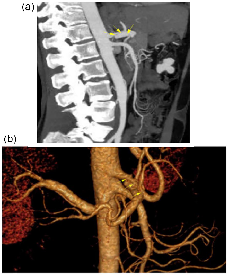
Computed tomography angiography (CTA) images illustrating spontaneous isolated celiac artery dissection and its extent to the origin of the common hepatic artery in a sagittal view (a) and 3D reconstructed image (b).
Another linear dissecting flap is seen within the right renal artery with no evidence of occlusion. (Figure 2) There was no history of catheter angiography that might have caused iatrogenic injury to the celiac or renal artery.

Computed tomography angiography (CTA) images illustrating spontaneous isolated right renal artery dissection with no evidence of occlusion or extension to distal renal branches in a sagittal view (a) and 3D reconstructed image (b).
Selective catheterization was performed very carefully because there is no real arterial wall surrounding the false lumen, and perforation could lead to catastrophic complications. Selective right renal artery catheterization was performed, which confirmed that the focal lesion was limited to the main trunk of the renal artery and did not extend to the division branches, and then selective hand-injection was performed carefully. The tip of the catheter in the false lumen revealed the extent of the dissection (Figure 3).

Diagnostic angiography via a pig tail catheter placed in the suprarenal aorta illustrating spontaneous isolated right renal artery dissection and its extent before distal branches with healthy aorta and a proximal right renal artery segment.
An intravascular ultrasound (IVUS) catheter was then advanced over a wire passing through the caudal channel. The live images demonstrated that the caudal channel did not have an intimal echogenic reflection, suggesting that this lesion was a dissection rather than a fenestration. In addition, the thickened intimal flap with a single layer of intimal echogenic reflection suggested that the IVUS catheter was passing through the false lumen of subacute-chronic focal dissection (Figure 4).

Intravascular ultrasound image obtained by the ultrasound catheter within the caudal channel (FL, false lumen) showing the right renal artery intimal flap with bright intimal reflection (arrow) at the side of the cranial lumen (TL, true lumen).
Pressure measurements were then obtained, revealing a 35 mm Hg systolic pressure gradient across the dissection. Following a successful cannulation of the distal renal artery branches through the true lumen, a balloon expandable covered stent measuring 6 mm in diameter and 60 mm in length was deployed across the dissected truncal segment of the main renal artery. Care was taken to avoid extending the stent into the branches. Control arteriogram revealed a significantly enlarged true lumen with a widely patent stent in excellent position. The false lumen became compressed by the stent with faint filling of contrast medium (Figure 5).

Completion angiography after a covered stent measuring 6 mm in diameter and 60 mm in length was deployed across the dissected truncal segment of the main right renal artery revealed a significantly enlarged true lumen with a widely patent stent in excellent position.
Next, selective celiac artery catheterization was then performed, which confirmed that the focal lesion was limited to the main trunk of the celiac artery and did not extend to the common hepatic or splenic arteries. Selective hand-injection was performed carefully. The tip of the catheter in the false lumen revealed the extent of the dissection (Figure 6).

Diagnostic angiography via a pig tail catheter placed in the supra-celiac aorta illustrating spontaneous isolated celiac artery dissection (arrow) and its extent before the common hepatic artery origin with healthy aorta and a proximal celiac artery segment; notice the previously inserted right renal artery stent (*).
Following a successful cannulation of the distal hepatic artery branches through the celiac artery true lumen confirmed by IVUS imaging, a balloon expandable covered stent measuring 6 mm in diameter and 60 mm in long was deployed across the dissected truncal segment of the main celiac artery, with distal landing zone extended to the common hepatic artery. Control arteriogram revealed a significantly enlarged true lumen with a widely patent stent in excellent position. The false lumen became compressed by the stent with faint filling of contrast medium (Figure 7).

Completion angiography after a covered stent measuring 6 mm in diameter and 60 mm in length was deployed across the dissected truncal segment of the celiac artery revealed a significantly enlarged true lumen with a widely patent stent; notice the previously inserted right renal artery stent (*).
The patient was treated with clopidogrel 75 mg for 6 weeks and lifetime aspirin. A week after the procedure, his symptoms were completely resolved, his blood pressure was 120/70 mm Hg, and it remained within normal limits for the remainder of the follow-up period. Although the CTA obtained at 2 month follow-up revealed very slight residual filling of the right renal artery false lumen, CTA at 6 month follow-up revealed a widely patent renal artery stent with no residual filling of the false lumen.
Discussion
Arterial dissection has been defined as the cleavage of the arterial wall by blood flow resulting in intramural hematoma between 2 elastic layers. Isolated arterial dissection occurring in the absence of aortic dissection has been a reported entity in the carotid and renal arteries. However, it is rarely reported in the visceral arteries. 1
Despite the clinical significance of aortic dissection, there is an ongoing debate with regard to the pathophysiology of the initial damage to the intima. The trigger in aortic dissection is believed to be a tear or hemorrhage in the aortic wall that leads to a separation of the media with potential end-organ damage from propagation of the dissection and obstruction of the origins of vital vessels such as the celiac, mesenteric, renal, or iliac arteries. 2
Suspected causes of arterial dissection are numerous and include atherosclerosis, trauma, iatrogenic causes, pregnancy, fibromuscular dysplasia, infectious disease, hypertension, Marfan syndrome, and Ehlers-Danlos syndrome. 3
Clinical presentation of isolated dissection of the renal and splanchnic arteries may be classified as acute or chronic. Acute dissections are classified as spontaneous and iatrogenic, whereas chronic dissections are classified as functional and silent. Spontaneous arterial dissection is more common in men, a reported 5:1 occurrence ratio with an average incidence age of 55 years. Iatrogenic dissections may be caused by guidewires, catheters, and angioplasty balloons. Because there was no history of catheter angiography in this patient, the diagnosis in this case merits comment.4,5
Dissections of chronic renal and splanchnic arteries may be entirely asymptomatic or result in renovascular hypertension, an episode of flank pain, abdominal pain, weight loss, hemorrhage, and more atypical symptoms like malabsorption, jaundice, or pancreatitis. Chronic dissections are most frequently associated with fibromuscular dysplasia of the renal artery, which accounts for 5% to 10% of renal artery stenosis.6,7
Renal and visceral arteries have been shown to have a decrease in dissection rates compared to the aorta and carotid arteries, with dissection of the celiac artery being very rare. 8
Spontaneous isolated renal and celiac arterial dissection seems to be a complication of primary hypertension, and it is the most likely etiology in our case; however, the patient also had a brother who died recently of aortic dissection, thus raising the possibility of a hereditary systemic disorder or predisposing hemodynamic factors, but our patient did not have any known connective tissue disease or congenital disorders of the vascular system. One other possible diagnosis in this case is segmental cystic medial necrosis, which is a rare, non-inflammatory, non-atherosclerotic arteriopathy that involves the splanchnic and renal arteries.
Several risk factors described for SICAD are represented in our case; our patient was a male who presented with uncontrolled hypertension and complained from nausea vomiting and right flank and abdominal pain.
As per the study by Vaidya and Dighe,3 spontaneous dissection of renal and visceral arteries was first described by Bauersfeld in 1947. However, it is reported to be primarily affecting the renal arteries rather than the celiac artery as the case presented within. The first reported case of celiac artery dissection was in 1959, with only 13 cases reported prior to 2001. Since the advent of improved diagnostic technologies (contrast enhanced computed tomography, CTA, and magnetic resonance angiography), the number of reported cases has more than doubled, with Obon-Dent et al 9 reporting a total of 33 cases in their study from 2012. 8
The utility of IVUS is well described in the literature for determining the false and true lumens in aortic dissections. Although there is very limited information on IVUS in the evaluation of visceral and renal artery dissections, IVUS was helpful in this case, particularly to distinguish the false from the true lumen, identify the extent of dissection, and perform precise stent sizing and deployment. 10
In addition, as mentioned above, the diagnosis of fenestrated renal artery could not be totally excluded by CTA. To the best of our knowledge, the only reported case of renal artery fenestration is very similar to this case with regard to imaging. The intra vascular ultra sonography (IVUS) examination together with digital subtraction angiography (DSA) helped us to reach the diagnosis of dissection in our case, which assisted in the management of our patient. If the authors of the fenestration case had a chance to examine their patient with IVUS, the final diagnosis might have changed. 11
The contrast in medical and surgical treatment options for SIRCAD is striking. Interventional procedures should only be used when the patient is hemodynamically unstable; experiences persistent symptoms of ischemia such as abdominal pain and flank pain; if they fail medical therapy for blood pressure control; or if the dissection progresses subsequently causing infarcts. Although it has been observed that surgical management should be exercised in those described circumstances, the optimal indicated procedure for renal and celiac arterial dissection has not been confirmed, and procedures greatly vary in the reported studies. 12
According to the latest European Society for Vascular Surgery guidelines for isolated mesenteric artery dissection (IMAD), conservative treatment with antiplatelet therapy and control of hypertension should be considered for patients with asymptomatic IMAD. For patients with symptomatic IMAD, treatment with antiplatelet therapy or low molecular weight heparin (LMWH) or unfractionated heparin until symptoms resolve should be considered, and endovascular revascularization is recommended in those not responding to medical management or with a suspicion of bowel ischemia. 13
The study by Obon-Dent et al 9 used angiography and endovascular techniques to achieve remission of symptoms. Two patients in the Amabile study were identified: One of them similar to those in the report by Obon-Dent et al 9 underwent endovascular repair with the use of covered stent in the celiac trunk, while the other required an upper aortomesenteric venous bypass for combined celiac and superior mesenteric artery dissections; both patients were symptom-free at the time of the study.9,12
Furthermore, Glehen et al 14 described several operative interventions for patients with celiac artery dissection including one patient with open surgical fenestration with resection of the dissection flap for the splenic, celiac, and left gastric arteries; another who underwent celiac artery arteriotomy, surgical fenestration, side-to-side anastomosis of the hepatic arteries, and reimplantation onto the celiac artery ostium with a graft; and finally, a patient who underwent celiac artery arteriotomy with graft placement between the divided celiac artery and bifurcation of the splenic artery and hepatic artery.
As described, several endovascular and surgical interventions have been used, all of which have reported a complete resolution of symptoms by the time of study publication. However, although all these reports expressed results that were ultimately therapeutic, a concise decision on interventional management as the standard of care or its suggested technique has not been agreed upon.
Conclusion
The pathology of SIRCAD in the absence of other vascular dissections is extremely rare, which speaks for the necessity of reporting this case. Dissection of the celiac artery has scarcely been described in literature, especially prior to the institution of evolved imaging techniques which are now routinely employed.
Given that the patient’s renal and celiac arterial dissections resulted in the diagnosis of uncontrolled renovascular hypertension and gastrointestinal manifestation, which could have resulted from duodenitis or cholecystitis, we feel it is essential to report this case.
Footnotes
Declaration of Conflicting Interests
The author(s) declared no potential conflicts of interest with respect to the research, authorship, and/or publication of this article.
Funding
The author(s) received no financial support for the research, authorship, and/or publication of this article.
