






Abstract
Diabetic foot ulcers are a source of significant morbidity. Maximizing limb salvage is an important long-term objective. The use of dermal substitutes to aid limb salvage and reduce amputation rates has been described recently in the literature. Dermal substitutes were initially described in burns and have been demonstrated to be useful adjuncts in the management of these wounds. In diabetic foot ulcer management, the outcomes of the use of the dermal substitutes are limited. The authors describe their experience of the use of INTEGRA®, a collagen–glycosaminoglycan dermal substitute, in 2 patients who presented with diabetic foot ulcers with exposed tendons.
Introduction
Patients with diabetes have been estimated to have a 5.8% cumulative incidence of diabetic foot ulcers over a 3-year period. 1 At least half of these foot ulcers are infected at the time of presentation and therefore carry the risk of worsening local sepsis potentially leading to amputations. 2 Maximizing residual stump length following amputations is an important factor for rehabilitation. 3 The use of dermal substitutes for the coverage of exposed bones and tendons is increasingly being reported in the literature.4,5 We report our experience in 2 patients with diabetic ulceration arising from underlying neuropathy, the subsequent management with debridements, antibiotic therapy, and the use of the dermal substitute INTEGRA®, which limited the extent of amputation required.
Case 1
A 64-year-old gentleman, who lives alone, was found by his family, collapsed and unconscious. He was brought into the emergency department. History of his presentation was limited, and no previous comorbidities were reported by his family. Examination revealed signs of general neglect, dehydration, and infected necrotic ulceration of his left foot. Blood glucose levels were elevated and associated with significant ketonuria and metabolic acidosis (on blood gas analysis). Diabetic ketoacidosis was established as the working diagnosis, and management with aggressive fluid resuscitation, a sliding scale insulin regime, and intravenous antibiotics was instituted. He was admitted to the intensive care unit for 4 days. He was discharged to the ward under the care of the endocrinology and diabetes team.
Following discharge to the ward, the foot was examined by the vascular surgical team. A necrotic left fifth toe ulcer was identified to extend across the plantar surface. His vascular examination was otherwise unremarkable, with strong palpable femoral and popliteal pulses, and triphasic waveforms auscultated on Doppler of his left dorsalis pedis. A computed tomography angiogram demonstrated normal inflow to the foot (albeit with a nonsignificant stenosis of the posterior tibial artery). His ulcer was therefore presumed to predominantly neuropathic, managed with pressure offset from his left foot. Magnetic resonance imaging (MRI) of his foot confirmed osteomyelitis affecting his left lateral 2 toes, to the level of the distal metatarsal heads. We performed a debridement of the necrotic tissues, under general anesthetic, to the level of bleeding, health tissues, resulting in exposed plantar tendons (Figure 1). He was systemically well, maintaining a normal blood pressure and pulse rate and was apyrexial following the debridement, and therefore we decided to manage his underlying osteomyelitis with long-term intravenous antibiotics. Five days following the surgical debridement, his ulcer appeared to be free of obvious necrosis, and no growths of organisms were detected from wound swabs. He underwent a further general anesthetic, during which the dermal substitute INTEGRA® was applied to the ulcer (Figure 1). One week after the application of INTEGRA®, the plantar wound did not demonstrate satisfactory granulation tissue, and simultaneously, the lateral 2 toes developed dry gangrene (with good vascular inflow, we presumed that this was secondary to deterioration in the diabetic microcirculation). Therefore, amputations of the lateral toes sparing the metatarsal heads were carried out, and a further sheet of INTEGRA® was applied to the ulcer following minimal debridement of the ulcer. One week following this procedure, the plantar ulcer continued to demonstrate patchy granulation tissue over the exposed tendons, and therefore we carried out a further debridement and application of a further sheet of INTEGRA®. Negative pressure wound therapy was initiated with intermittent potassium permanganate soaks of the wound. After 2½ weeks of this treatment, healthy granulation tissue was noted to cover the plantar tendons. A small area of necrosis involving the skin and muscle was noted on the lateral aspect of the plantar ulcer, and a localized debridement was performed to this region. The negative pressure therapy was discontinued following this and the wounds were managed with regular dressing changes (sodium carboxymethylcellulose primary wound dressings containing silver) and potassium permanganate soaks. The patient was discharged and seen weekly as an outpatient.
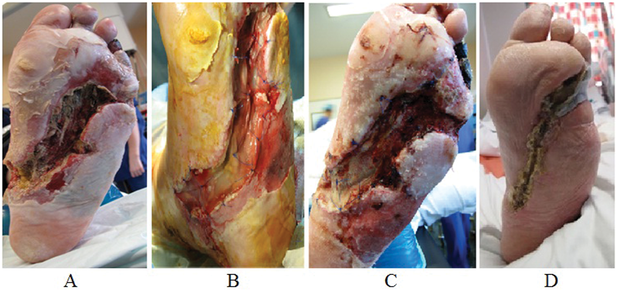
Case 1: A, Ulcer on presentation; B, Application of INTEGRA®; C, Incomplete coverage and patchy granulation tissue 1 week after first application of INTEGRA®; D, Satisfactory healing despite general patient neglect
Unfortunately, his general condition deteriorated, and he presented a month later with further neglect and recurrent falls associated with a left sided cerebrovascular event. The left foot ulcer continued to persist, and a repeat MRI scan revealed underlying osteomyelitis. Following a 6-week course on intravenous antibiotics and a subsequent 4-week course of oral antibiotics, his ulcer healed satisfactorily as demonstrated in Figure 1.
Case 2
A 48-year-old gentleman, not known to be diabetic, presented with a 10-day history of a nonresolving, painful blister affecting the plantar aspect of his right big toe. His previous medical history was unremarkable, with no regular medications. His right big toe was erythematous, sloughy, and edematous with skin loss on the plantar surface (Figure 2). Vascular examination was otherwise unremarkable, with strong posterior tibial and dorsalis pedis pulses palpable, with the rest of his foot appearing well perfused.
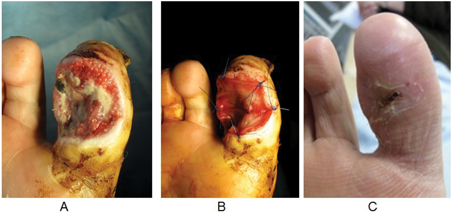
Case 2: A, Ulcer on presentation; B, Application of INTEGRA®; C, Final outcome 3 months following presentation
His blood glucose levels on admission were elevated with significant glucosuria. He was diagnosed with diabetes and started on oral hypoglycemic medications. He was found to have a mild neutrophilia, and MRI imaging identified osteomyelitis limited to the distal phalanyx of his right big toe. Intravenous antibiotic therapy was commenced. A surgical debridement was performed under general anesthetic, with removal of the necrotic/dusky tissues down to the healthy tissues at the level of the plantar flexor tendon. Two days following this procedure, due to the absence of further necrotic tissue and negative wound swabs, a layer of INTEGRA® was applied over the exposed plantar tendon. He was maintained on intravenous antibiotics over 19 days, following which patchy granulation tissue was identified to overlie his plantar tendon. Therefore, a further INTEGRA® layer was applied following a further wound debridement.
Following this second procedure, healthy granulation tissue developed over the plantar tendon and the patient was discharged. After regular follow-up in outpatients’ clinic, his ulcer healed without the need for skin grafting (Figure 2).
Discussion
The management of infected diabetic ulcers requires intravenous antibiotics with surgical debridement of infected and nonviable tissues to aid ulcer healing. The traditional management of osteomyelitis in the diabetic foot advocated surgical debridement and amputation of infected bone. However, recent studies have demonstrated that long-term intravenous antibiotic therapy without surgical amputation can be successful in patients who are systemically well.6,7 Maximizing residual stump length following amputations is an important factor for rehabilitation. 3
INTEGRA® is composed of a collagen–glycosaminoglycan matrix. Similar to other dermal substitutes, it offers wound closure as a mechanical barrier to infection and fluid loss, and it promotes tissue regrowth. 8 Dermal substitutes are increasingly used in the management of burns as an alternative to autograft. A review of the literature in burns by Pham et al 8 identified the use of INTEGRA® to be better than autografts with regard to healing times, but did identify 1 trial that reported unacceptably high rates of infections in these grafts. The review concluded that the evidence with regard to INTEGRA® is limited and therefore no conclusions can be established at present. Recent case reports of the use of dermal substitutes to cover the exposed tendons and bones in diabetic feet have been reported with good healing and preservation of stump length.4,5 A retrospective review of 105 patients who underwent application of INTEGRA® for diabetic foot ulcers by Iorio et al 4 reported its successful use in limb salvage in patients with low risk of amputations. However, they identified that patients with higher risk of amputations may not derive any benefit, with regard to limb salvage, following its application.
Our first patient presented with a severe necrotic ulcer affecting his left foot extending from the plantar surface to the dorsal surface. His ulcer exposed many of his flexor tendons on the plantar aspect of his foot. We had to perform 4 wound debridements and apply 3 sheets of INTEGRA® during these procedures due to its severity. His management was further confounded by his tendency for general neglect, and therefore he was readmitted with further ulceration a month after his initial admission. Following appropriate intravenous antibiotic therapy and treatment of his underlying osteomyelitis, he achieved good recovery from his premorbid state. His management demonstrates the successful use of long-term intravenous antibiotic therapy without extensive amputations, thereby achieving better functional outcomes.
Our second patient presented with a comparatively small ulcer affecting his big toe with exposure of the flexor tendon, which healed completely following our management. The use of a dermal substitute, INTEGRA®, was a useful adjunct in promoting healing over the exposed tendons in our 2 patients.
Footnotes
Authors’ Note
All authors were involved in the management of the patients, review of case notes, and preparation of the article.
Declaration of Conflicting Interests
The author(s) declared no potential conflicts of interest with respect to the research, authorship, and/or publication of this article.
Funding
Dr. Andrew Choong has just been sponsored by part payment of his flight to attend the 81st Annual Scientific Congress of the Royal Australasian College of Surgeons held in Kuala Lumpur, Malaysia, 6th - 10th May 2012 in order to present the work on these Integra® case reports. However, no funding/scholarship was obtained for writing this article or for using Integra® in our patients.