






Abstract
Chronic ulcers are a major public health problem, due to their chronic nature, their poor response to treatment, the high frequency of recurrences, and their affection to the patient’s quality of life. Even with the development of new therapies in the field of chronic wound care, chronic ulcers remain a clinical problem. As a novel branch of research, Catalytic Nanomedicine has offered promising results in disinfection and treatment of chronic wounds through the use of bionanocatalysts, organically functionalized mesoporous nanostructured materials with catalytic properties. Particularly, Cu/TiO2-SiO2 mixed oxide bionanocatalysts have shown favorable results for chronic ulcer healing. In this work, we present the treatment of 15 patients (8 females and 7 males, mean age of 69.59 ± 12.07 years old) affected with chronic ulcers (wound age ranging from 4 months to 10 years old, mean size of 12.94 ± 18.20 cm2) by the administration of Cu/TiO2-SiO2 bionanocatalysts embedded in a nanoemulsion matrix. In all cases, complete epithelialization and healing of the lesions was achieved (healing time from 3 to 35 weeks), without the appearance of side effects. Wound healing time was analyzed in the context of initial wound size, wound’s age, patient’s age, and concomitant conditions, being wound size and patient’s age the main factor affecting the duration of the treatment with the bionanocatalysts.
Introduction
Chronic ulcers are open skin lesions thought to occur due to improper functioning of the venous valves (as in the case of diabetes mellitus), developing mostly along the medial distal leg, 1 and that persist for more than six weeks with no tendency to heal after three or more months. 2 Clinically, chronic ulcers are characterized as solitary or multiple, superficial, excavated, rounded or irregular, slightly painful wounds of variable sizes. Chronic ulcers exhibit centrifugal growth, exudative with or without yellowish-white fibrin, erythematous, bluish or hyperpigmented edges, and reddish background with granulation tissue in the central area; eschar or necrosis are exceptional. 3 Chronic ulcers mainly invade the middle and deep dermis, and rarely deepen the subcutaneous cellular tissue or muscle fascia. 4
Till this day, chronic ulcers constitute a serious health problem in terms of quality of life (pain, discomfort, trophic disorders, etc), and possible complications (local infections, polymedication, bleeding, thrombophlebitis, etc). With an overall prevalence of 1-3% during population’s lifetime, 5 this condition remains a public health problem due its chronic recidivism, with a high requirement of human resources, healing materials, and costly instruments. In addition, the impact of this disease in terms of disability, absenteeism, and labor costs deserves special mention.
For the treatment of chronic ulcers of the lower limbs, numerous therapeutic strategies have been tried, based on the pathophysiology of ulcers, such as reducing edema, promoting re-epithelialization, and correcting comorbidities (e.g. diabetes, smoking, anemia, malnutrition) and/or complications (such as recurrent bacterial infections) that prevent or delay improvement. 6 It is well-known that the type of treatment depends on the etiology of the chronic ulcer, since they can be classified according to it as vascular ulcers (e.g. venous and arterial ulcers), diabetic ulcers, and pressure ulcers. 7 Nonetheless, the quality of the available data on the efficacy of current therapies is insufficient to determine one therapy or dressing as superior over the rest, and decisions must be made individually. 8
In this regard, catalytic nanomedicine has made inroads into new devices that support tissue regeneration in chronic wounds. This new branch of science and technology is based on the use of bionanocatalysts, nanostructured materials composed of pure or mixed oxides that exhibit catalytic properties capable of destabilizing genetic material, and organic functional groups that mimic cellular ligands, thus endowing them with biocompatibility and affinity. 9 Among their applications, bionanocatalysts have been proven to possess microbicidal properties, 10 which can be used to eliminate bacterial infection in chronic wounds. Similarly, through the incorporation of such bionanocatalysts in nanoemulsions (aqueous dispersions of hydrogel particles generated by nanoscale networks that are physically or chemically crosslinked), 11 catalytic nanomedicine has generated devices capable of treating chronic wounds by keeping the moisturized environment required for proper healing. 12
Based on the above, in the present work we report the use of bionanocatalysts made of copper stabilized in a mixed titania-silica network as a treatment for 17 chronic ulcers in 15 patients with different comorbidities. In all cases, application of the bionanocatalysts allowed for complete healing of the wounds, without the appearance of side effects.
Methods
Patient Selection Criteria
During the period of time of the study (January 2017 to December 2018), 51 patients were diagnosed with chronic wounds. Patients were only included to the survey if they gave informed consent. Further inclusion criteria covered the following parameters: patient age over 18 years, patient had at least a chronic wound of more than one-month duration that had been previously treated unsuccessfully, previous treatment was discontinued during the evaluation, patient had a wound that “appeared” to contain biofilm, no other change was to be made to wound care regimen or patient’s care, and patient continued the treatment without withdrawal before wound closure. Patients were included independently of the etiology of the wound: diabetes, vascular, or pressure. The Ankle-Brachial Index (ABI) test was carried out for all patients to determine peripheral artery disease (PAD) through duplex venous ultrasound. Furthermore, the visual analog scale (VAS) was used to evaluate pain. Among the 51 patients, 15 patients were selected for their significant characteristics which represented the total of 51 cases. The 15 patients (8 females and 7 males) were included in the survey, with a mean age of 68.80 years (range, 50 to 87 years).
Cu/SiO2-TiO2 Bionanocatalysts and Nanoemulsion Matrix
The bionanocatalysts were synthesized following the sol-gel method previously described by López et al 13 Briefly, an initial solution was prepared by dissolving copper in a mixture of water and ethanol. The mixed oxide matrix of silica and titania was obtained by the combined dropwise addition of precursors of both oxides into the initial solution. The mixture was kept under stirring for 24 h. The synthesis conditions were set to obtain nanoscale particle sizes and to optimize surface-to-volume ratio. After solvent evaporation, the resulting powder was embedded in a nanoemulsion matrix composed of carboxymethylcellulose and carbomer to improve its cutaneous application. The physicochemical characterization is described elsewhere. 13
Documentation of Wound Status
Photographs were taken before, during, and at the completion of therapy with a digital camera. Wound area was recorded, and depth was measured if feasible. Wounds were considered healed when completely covered with epithelium.
Analysis Parameters
Patient’s records were reviewed for the presences of significant co-morbid conditions, duration of wound presence prior to initiating the application of the bionanocatalysts, location, and size, length of follow-up, and final disposition of the wound.
Statistics
This case analysis is descriptive; statistics comparing healing within the group were not calculated. Continuous data, including age, ulcer evolution, and ulcer area are demonstrated in mean and standard deviation. The result of wound healing time is demonstrated in bar chart.
Case Series
Our experience with the application of the bionanocatalysts has been positive, especially on the chronic ulcer bed, as faster healing has been observed. From the senior author’s experiences, the quality and quantity of the granulation tissue formed are better, which can be seen in the reduction of wound recurrence rates as in the case studies discussed below. Informed consent was obtained from all patients before the usage of their photos in this series. Table 1 summarizes the descriptions of the wounds observed in the 15 patients. For brevity, only the 6 most significant cases are described in detail. Pictures of the healing processes are shown in Figures 1–6.”
Summarized Clinical Histories of the 15 Patients and Wound Description After Physical Examination.

Summarized Clinical Histories of the 15 Patients and Wound Description After Physical Examination.
/ is used to separate data from different ulcers from the same patient.
Etiology was identified according to primary cause (Ep), secondary cause (Es) or no venous cause identified (En), and, in parentheses, the underlying arterial/venous condition or secondary event, as the case may be.
GT: granulation tissue, FT: fibrin tissue, ST: sphaceli tissue, NT: necrotic tissue, SK: skin tissue, TT: tendon tissue. SAH: systemic arterial hypertension, CVI: chronic venous insufficiency, T2DM: type-2 diabetes mellitus.
A 50-year-old female with no significant clinical history presents with a chronic ulcer of 2 years of evolution. Physical examination revealed a pressure ulcer in the internal supra-malleolar region of the right leg with a volume of 1.97 cm3 (3.8 cm × 2.6 cm × 0.2 cm). The wound bed exhibited 90% granulation tissue and 10% fibrin tissue, null exudate, irregular edges, adherence without edge effect, and erythematous and indurated perilesional skin. Pedial and tibial pulses are present, with an ankle-brachial index (ABI) of 1.0. The patient complains of pain, 7/10 according to the visual analog scale (VAS). The patient was started on bionanocatalysts + nanogel with compressive bandages. There was a total reduction of 60.0% of wound size by week 2. Complete epithelization of the wound was noted 7 weeks after the use of the bionanocatalysts. As compared with other cases that underwent standard therapy without bionanocatalysts in our center, we noted that time to wound closure was decreased, with a decreased recurrence rate.
Case 2
A 55-year-old male with a 12-years history of type-2 diabetes mellitus (T2DM), significant metabolic decontrol, and no pharmacological treatment presented two 4-months-old diabetic ulcers in the upper third right leg due to traumatism. On physical examination, the first one is observed to have a surface of 2.24 cm2 (1.6 cm × 1.4 cm), whereas the one located below has a surface of 1.08 cm2 (1.2 cm × 0.9 cm). Both wound beds exhibit 90% granulation tissue and 10% fibrin tissue, scarce exudate, serous regular edges, adherence with edge effect, and healthy perilesional skin. Pedial and tibial pulses were present, with an ABI of 1.1. The patient reports occasional mild pain. The nanogel with the bionanocatalysts was applied in conjunction with compression bandages. Significant improvement was observed after 5 days; similarly, a 50% size reduction was achieved at week 4. Wound closure was attained after 8 weeks.
Case 3
An 87-years-old male patient was recently diagnosed with T2DM and treated with metformin (850 mg/24 h) and linagliptin (5 mg/24 h), and a history of systemic arterial hypertension of 7 years of evolution treated with losartan (50 mg/24 h) and hydrochlorothiazide (12.5 mg/24 h). The patient presents a 3-years-old chronic ulcer in the left leg due to traumatism. He refers to a fear of the healing process since washing with soap and water causes wound bed bleeds and intense pain; he also states he has no hope of recovery. On physical examination, an arterial ulcer is found in the left leg malleolus with a volume of 0.21 cm3 (1.8 cm × 1.2 cm × 0.1 cm). The wound bed exhibits 70% granulation tissue and 30% fibrin tissue, null exudate, irregular edges adhered without edge effect, and indurated perilesional skin with ocher dermatitis. Pedial and tibial pulses are present with an ABI of 0.9. Treatment with bionanocatalysts is applied in conjunction with a compression bandage. Full epithelization of the wound was observed after 3 weeks.
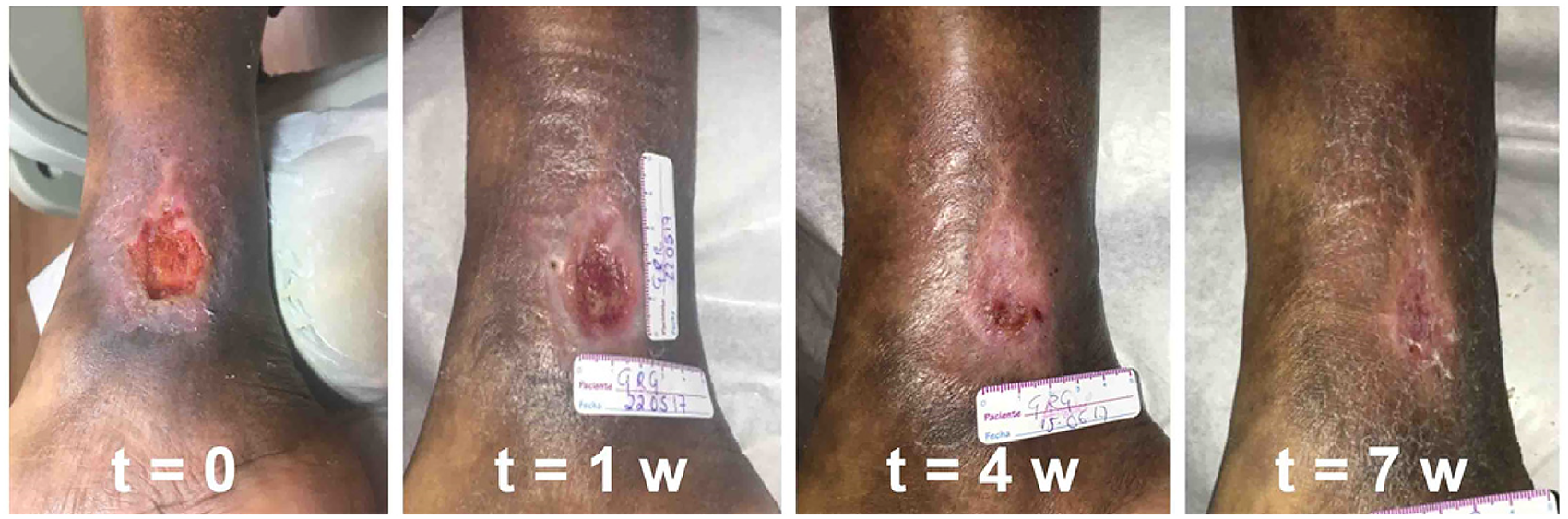
Healing process of patient 1 from 15 May 2017 to 9 July 2017.
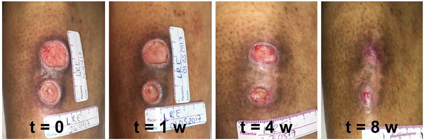
Healing process of patient 2 from 26 April 2017 to 17 June 2017.
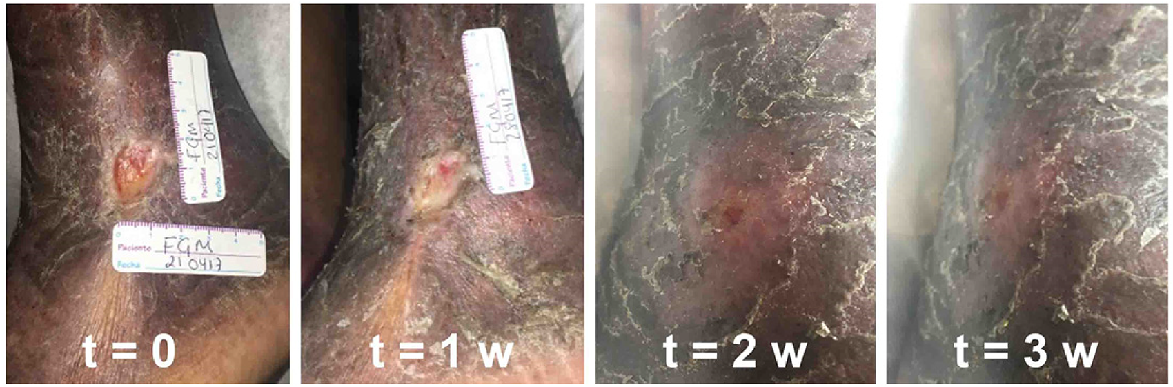
Healing process of patient 3 from 21 April 2017 to 12 May 2017.
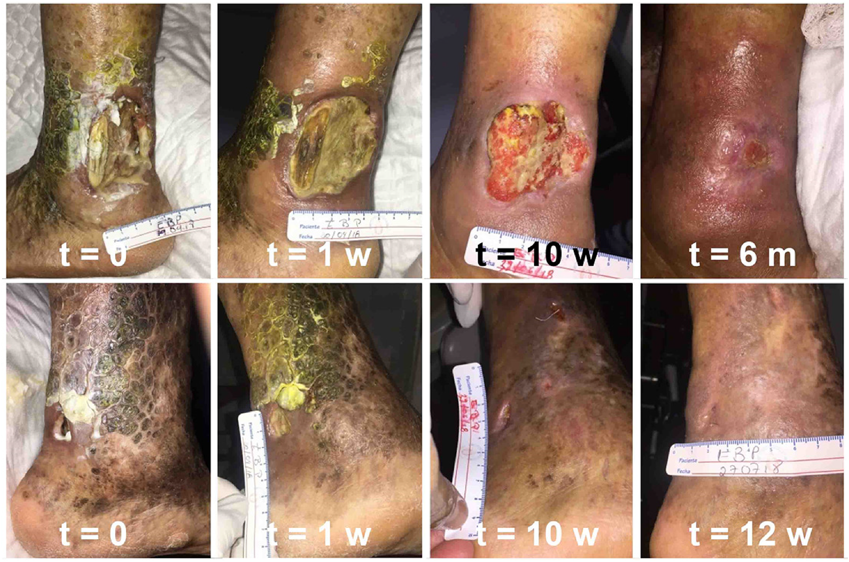
Healing process of patient 7 from 19 April 2018 to 26 October 2018 for both ulcers, first (up) and second (down).
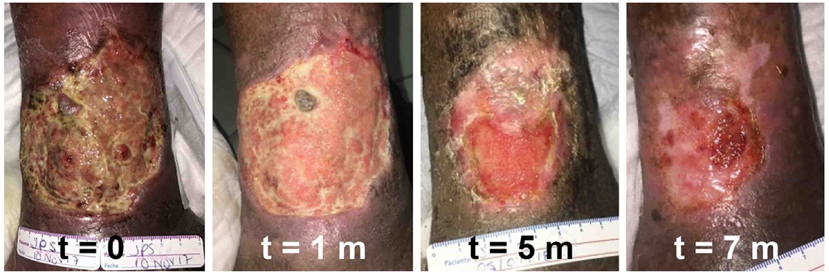
Healing process of patient 9 from 10 November 2017 to 23 July 2018.
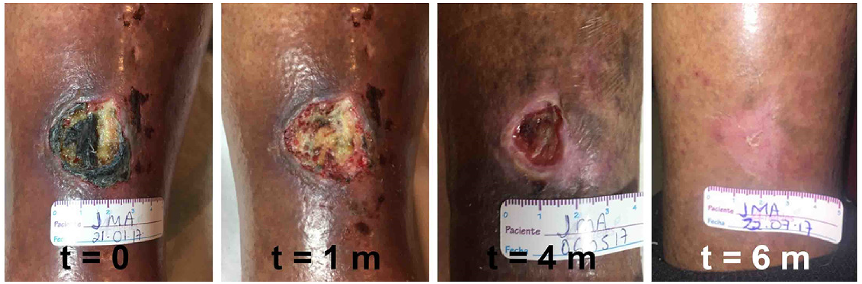
Healing process of patient 10 from 21 January 2017 to 22 July 2017.
A 72-years-old female with a history of chronic venous insufficiency (CVI) and a saphenectomy in the right leg at the age of 58 presented two venous ulcers of 9 months of evolution, one on the inner side and the other on the outer side of the malleolar region of the left leg. At the time of consultation, the patient had never performed any type of healing or cleaning of the affected leg due to disabling pain and intermittent bleeding. On physical examination, the first ulcer had a volume of 43.92 cm3 (6.1 cm × 4.8 cm × 1.5 cm). The wound bed exhibited 60% skin tissue, 30% tendon exposure, and 10% necrotic tissue, with moderate exudate, fetid, uneven undercut edges not adhered without edge effect, and indurated, dirty, erythematous perilesional skin with scales. The second ulcer has a volume of 1.19 cm3 (1.4 cm × 1.7 cm × 0.5 cm), exhibited 90% necrotic tissue and 10% sphaceli tissue, with moderate exudate, fetid, irregular edges not adhered without edge effect, and a perilesional skin with abundant scales. Pedial and tibial pulses are present with an ABI of 0.9. In both wounds, healings were performed daily with bionanocatalysts + nanoemulsion. After 2 weeks, pain and exudate decreased, so the cures started being carried out every 48 h. Full wound closure was observed after 24 weeks for the first ulcer, and after 12 for the second. It is worth mentioning that the patient used compression intermittently as she reported increased pain and edema; nevertheless, once proper compression was carried out, wound closure accelerated.
Case 9
An 80-years-old female patient with a clinical history of CVI and chronic ulcers for 5 years assists consultation with a venous ulcer of 1 year of evolution in the external malleolar region of the right leg. The patient had previously gone to her health center, where the wound was cleaned with Isodine and hydrogen peroxide and carved to bleed; in the absence of improvement, the patient decided to suspend this type of healing. The physical examination shows that the ulcer has a volume of 83.22 cm3 (7.8 cm × 9.7 cm × 1.1 cm), and exhibits 70% fibrin tissue, 20% sphaceli tissue, and 10% granulation tissue, with abundant exudate, irregular edges adhered without edge effect, and dry perilesional skin with ocher coloration. Pedial and tibial pulses were present, with an ABI of 1.0. Healing began every 48 h with bionanocatalysts + nanoemulsion and compression therapy. The wound completely healed after 28 weeks.
Case 10
A 75-years-old male patient with a 10-years-old clinical history of hypertension controlled with irbesartan (300 mg/24 h) and hydrochlorothiazide (12.5 mg/24 h), and a significant risk of malnutrition according to the Nutritional Risk Screening, attends a consultation for the presence of an arterial ulcer of 7 months of evolution due to traumatism. On physical examination, the wound with an area of 8.7 cm2 (2.9 cm × 3.0 cm) is localized in the upper third of the right leg. The bed wound is composed of 80% partially dehydrated necrotic tissue and 20% sphaceli tissue, scarce exudate, irregular edges adhered with edge effect, and erythematous, ocher perilesional skin. Pedial and tibial pulses are present, with an ABI of 0.9. Bionanocatalysts + nanoemulsion treatment with compression bandages is indicated, and after 6 months of treatment, the wound was completely epithelized.
Results
Over 24 months, 17 wounds in 15 patients were treated with Cu/TiO2-SiO2 bionanocatalysts. There were no complications associated with the use of bionanocatalysts. The therapy was used to treat various types of chronic ulcers: vascular (venous and arterial ulcers), diabetic, and due to pressure. All of the wounds were in the extremities. Most of the patients had at least one comorbid condition (Table 2). The most common conditions were hypertension (n = 7), chronic venous insufficiency (n = 6), and type-2 diabetes mellitus (n = 5). Of the 17 wounds, all of them (100%) healed during treatment with bionanocatalysts. No wound required for limb amputation.
Comorbid Conditions Present in Wound Patients.

Comorbid Conditions Present in Wound Patients.
Several patients had multiple comorbidities.
Treatment duration ranged from 3 weeks to 35 weeks. The mean treatment time was 98.82 ± 79 days. For wound size comparison, only wound area was used. Initial wound sizes ranges from 2.16 cm2 to 75.7 cm2 with a mean of 12.94 cm2. Prior to bionanocatalysts therapy, wound duration ranged from 4 months to approximately 10 years, with a median time of 25.2 months. Wound healing time was analyzed in the context of initial wound size, wound’s age, patient’s age, and concomitant conditions (Figure 7).
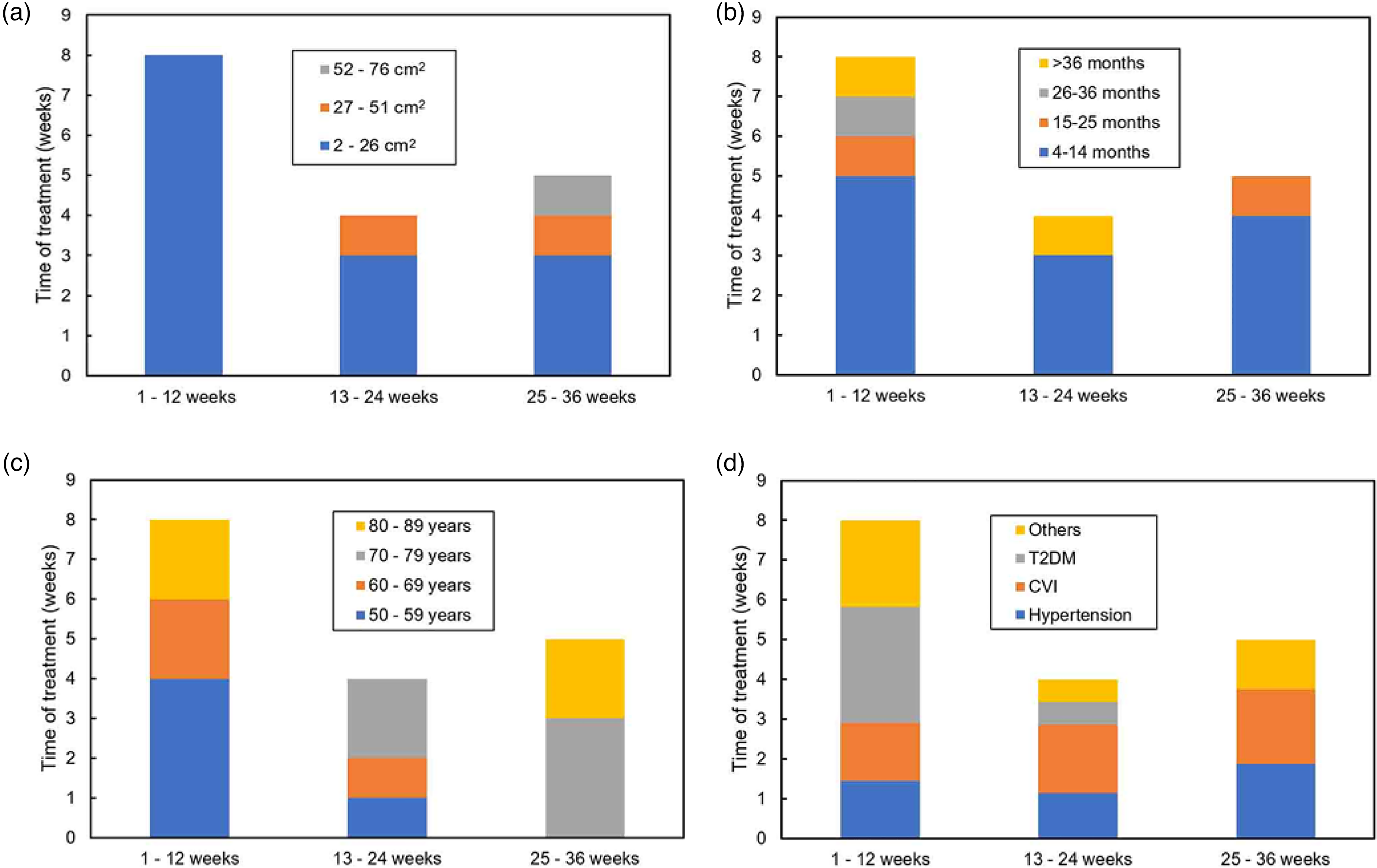
Wound healing time as a function of different parameters: (a) initial wound size, (b) wound age, (c) patient’s age, and (d) concomitant conditions. T2DM: type-2 diabetes mellitus; CVI: chronic venous insufficiency.
For initial wound size, it is evident that the larger the initial area, the longer the recovery time, since wounds that took less than 3 months to heal had areas of less than 26 cm2, while larger wounds (52-76 cm2) required between 25 and 36 weeks of treatment. On the contrary, the time of wound evolution showed no relationship with the required healing time, since short healing times (1-12 weeks) were observed for wounds of all ages. Similarly, young wounds (4-14 months) required various times for healing, with an even distribution among the three classes: 1–12, 13–24, and 25–36 weeks. Regarding the patient’s age, a direct relationship was observed with respect to healing time, with younger patients requiring shorter healing times, while the 70–79 and 80–89 age groups required longer times. In the latter, two exceptions were found for patients 3 and 13; however, this variation may be associated more with wound size, which were at the lowest values (2.24 and 3.91 cm2, respectively). Finally, an evaluation of healing times as a function of the patients’ concomitant conditions showed no apparent relationship associating any one condition affecting vascularization over another as a delayer of healing when wounds were treated with the bionanocatalysts. Patients with the three main conditions, T2DM, CVI, and hypertension showed varied healing times, indicating that it is the aforementioned factors (wound size and patient’s age) that could generate significant effects on healing time. Notably, this does not indicate that vascularization is not important, but rather that, in terms of treatment times, bionanocatalysts are able to treat chronic wounds independently of the pathology that is causing the vascularization problem, with no greater or lesser effect depending on the etiology of the ulcer.
In this case series, we demonstrate the efficacy of bionanocatalysts embedded in a nanoemulsion matrix in catalytic wound healing of 17 chronic ulcers in 15 patients with different microvascularization conditions. In each case, the bionanocatalysts were applied in the wound and covered with compressive bandages without further treatment. In all the cases, the wounds successfully healed, avoiding the need for re-approaches and amputation.
Hypothetic Mechanism of Action
Among the problems for proper wound healing is the formation of biofilms, polymicrobial aggregates composed of different biopolymers that facilitate the adhesion of microorganisms to surfaces, cohesion between them, and resistance to dangerous environmental stimuli, especially to biocidal agents. 14 Biofilms stimulate a chronic inflammatory response, which protects the microorganisms within the biofilm and increases the production of exudate, providing a source of nutrition and aiding in the perpetuation of the biofilm. 15 Nanoemulsions of antimicrobial bionanocatalysts have been shown to be effective against biofilms, 16 and biofilm removal has been associated with improved wound healing. 17 Bionanocatalysts destabilize the microbial cell wall, which allows their introduction and interaction with mitochondria (in the case of eukaryotic microorganisms) and genetic material (prokaryotes and eukaryotes), processes that lead to decrease in ATP production, genotoxicity, and production of reactive oxygen species, which finally culminates in cell death through apoptosis. 18 Reduction of microbial load allows macrophage and neutrophil activity in the area, leading to resolution of the infection and normal tissue regeneration. 19 The observed result is the formation of granulation tissue and reepithelialization of the wound; furthermore, as a result of the decreased chronic inflammation, microcirculation can improve in the region. 20 In addition, the nanoemulsion matrix maintains a moist and warm environment, ideal for healing, which increases the supply of oxygen and nutrients to the wound through angiogenesis, acidifies the area creating a bacteriostatic environment that reduces the risk of infection, facilitates fibroblast cell migration, controls exudate without damaging the perilesional skin, and protects the wound from contamination. 21
Impact of Underlying Conditions in Wound Healing
Through a comparative analysis of the treatment times required for complete wound re-epithelialization, we were able to observe the impact of four important parameters on wound healing: initial wound size, wound age, patient age, and concomitant diseases. Initial wound size was associated with a direct relationship with wound healing time, as previously reported. 22 Similarly, as has been clearly documented, 23 patient age was an important factor, with younger patients showing shorter recovery times than older patients. Interestingly, however, neither the time of wound evolution nor the patient's concomitant conditions appeared to generate a significant effect on healing time when wounds were treated with the bionanocatalysts. This suggests that the same type of treatment (bionanocatalysts + nanogel) can be applied to the wound regardless of the patient’s vascularizing diseases or age of the wound, with only the initial size of the ulcer and the patient’s age being the parameters to be taken into account when generating predictions an defining the timing of application. Nonetheless, a complete treatment plan must include etiology-specific additional therapies to solve the subjacent conditions, since bionanocatalysts only deal with the ulcers (the symptoms), while the primary cause of the ulcer (such as reflux, obstruction, etc.) 24 require for specific treatments (venous ablation, compression therapies, venous stent, etc.) 25 to prevent the recurrence of the ulcers.
Limitations of the Case Series
The results obtained in these case series are not free of limitations. As it is well-known, case series can be prone to bias, limiting its generalizability to larger populations of patients. Nonetheless, the information extracted from the healing processes of the 15 patients evaluated in the present work, as well as the impact of previous conditions on the healing time, allows to hypothesize on the efficacy and mechanism of action of the bionanocatalysts in chronic wound healing. Though, further advanced studies must be carried out to further deep into our knowledge regarding this type of bionanocatalysts.
Future Perspective for Bionanocatalysts in Chronic Wound Healing
Cu/TiO2-SiO2 bionanocatalysts into nanoemulsions offers a novel, low-cost, highly effective treatment for chronic wounds. Its use has shortened the duration of hospitalization and reduced hospital costs associated with ineffective ulcer therapies. The satisfactory results achieved with the treatment in the series of 15 cases meant significant improvements in the patients’ quality of life, eliminating the possibility of future amputations due to complications. Although concerns about the use of nanoparticles, especially titania, 26 have been reported, recent studies have proven their biocompatibility with normal tissues. 27 The patients exhibited no adverse effects related with the application of the bionanocatalysts, proving them to be safe. Similar results have been reported with other patients who underwent the same methodology for the treatment with bionanocatalysts-nanoemulsion. 16
Until now, research has exclusively focused on wounds involving microvascularization. Nonetheless, the microbicidal characteristics of bionanocatalysts, as well as the inherent benefits of their use in nanoemulsions, imply that bionanocatalysts might be used to treat infection-related skin disorders. For example, acne, a chronic inflammatory disease caused, among other factors, to Propionibacterium acnes bacterial colonization of the hair follicles of the face, neck, chest, and back, 28 could be treated with nanoemulsion-embedded antibacterial bionanocatalysts, which would potentially eliminate the infection and provide a moist cure for enhanced tissue regeneration, as described previously. Similarly, ongoing work has shown promising results for catalyzed wound healing in burns by the application of bionanocatalysts. The research is still in early stages; however, it is expected that nanoemulsion-embedded bionanocatalysts will be able to accelerate healing and improve the quality of life of patients with burns.
Conclusions
Antimicrobial bionanocatalysts embedded in a nanoemulsion matrix were successfully used for the treatment of 17 chronic ulcers in 15 patients with different underlying conditions, such as wound size, wound age, patient age, and concomitant diseases. In all cases, complete wound closure was observed in terms of re-epithelization. Wound size and patient age were identified as the main factors to consider when treating chronic ulcers with bionanocatalysts, while wound age and concomitant diseases did not have a significant effect on the course of treatment. Further research should be conducted to improve our knowledge of the use of bionanocatalysts for chronic wounds, especially given the possibility of using them to treat infection-related skin conditions.
Footnotes
Ethical Approval
The authors confirm that any aspect of the work covered in this study that has involved human patients has been conducted with the ethical approval of all relevant bodies and that such approvals are acknowledged within the article.
Declaration of Conflicting Interests
The author(s) declared no potential conflicts of interest with respect to the research, authorship, and/or publication of this article.
Funding
The author(s) disclosed receipt of the following financial support for the research, authorship, and/or publication of this article: This work was supported by the Consejo Nacional de Ciencia y Tecnología, (grant number 1037918); FJPG (CVU 1037018) is supported by a grant from the National Council of Science and Technology.
