
Abstract
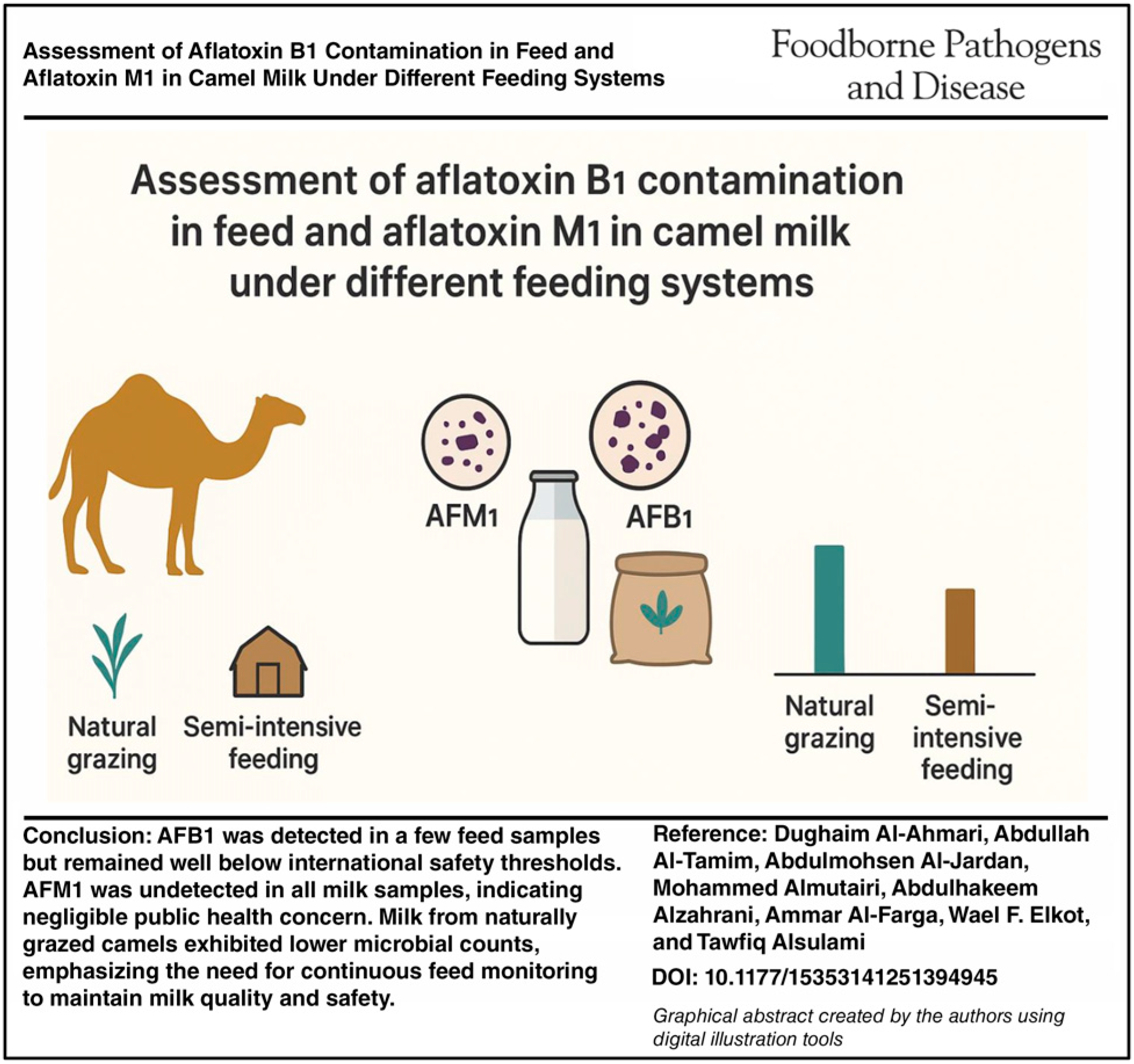
Camel milk is an essential nutritional resource in arid and semiarid regions and has been gaining commercial importance, particularly in the Middle East. Concerns over mycotoxin contamination, particularly aflatoxins (AFs), pose potential health risks and may affect milk quality. Understanding the role of feeding systems on the occurrence of AFs and on milk quality is vital for safeguarding public health and improving dairy industry practices. This study aimed to evaluate the impact of different feeding systems on the chemical and microbiological quality of camel milk and to detect aflatoxin M1 (AFM1) in milk and AF B1 (AFB1) in camel feed. A total of 80 samples were collected from 40 camels across Riyadh and Dammam, Saudi Arabia, between March and May 2024. Camels were divided into two groups based on feeding systems: natural grazing (grasses and legumes) and semi-intensive farm-based feeding (concentrates). Milk samples were analyzed for chemical composition and microbiological quality. Statistical comparisons were made between groups. AFB1 was detected in 1 (5%) of the natural grazing feed samples and in 3 (15%) of the farm-based feed samples; all values were below the Saudi national MRL for AFM1 in milk (0.5 μg/kg), as well as the stricter European Union’s limit of 0.05 μg/kg and the Codex Alimentarius standard of 0.5 μg/kg. AFM1 was below the detection limit in all samples, and consequently, no significant differences between feeding systems could be observed. There was no significant difference in AFM1 levels between the two feeding systems. However, milk from naturally grazed camels exhibited significantly lower microbial counts. Samples from Riyadh showed no AF contamination. The absence of AFM1 in all milk samples suggests minimal public health risk. However, higher AFB1 occurrence in farm-based feed underscores the need for stringent feed monitoring. These findings inform management practices to enhance milk safety in camel dairy systems.
Introduction
The camel (Camelus dromedarius), a key domesticated species, is an important food source worldwide, particularly in the Middle East (Elkot et al., 2024). Camel milk is valued for its nutritional and therapeutic properties, including antimicrobial activity and reported benefits in managing dyslipidemia, diabetes, and certain cancers (Fallah et al., 2020). It is rich in essential minerals and vitamins, containing three to five times more iron and ascorbic acid than cow’s milk (Swelum et al., 2021). While its natural antimicrobial components may reduce susceptibility to bacterial or mold contamination (Elkot et al., 2021), they do not protect against chemical contaminants of environmental origin, such as antibiotic residues and heavy metals, which are linked to management practices, feed, and environmental exposure. Camels are typically raised under two feeding regimes: (1) natural grazing on desert grasses and legumes such as Panicum turgidum, Atriplex leucoclada, and Zilla spinosa, with dry matter contents of 25–35%, or (2) semi-intensive farm-based diets consisting of pelleted concentrates formulated from grains (barley, maize), soybean meal, molasses, and mineral–vitamin premixes (Abrhaley and Leta, 2018). However, camel feedstuffs are vulnerable to contamination by mycotoxigenic fungi and their associated toxins, which pose serious risks to animal and human health (Almoammar et al., 2014; Adriaens et al., 2023). Among these, aflatoxins (AFs) are secondary metabolites of filamentous fungi that frequently contaminate food and feed stored under warm, humid conditions (Abdel-El-Fatah and Daud, 2002; Khalifa and Shata, 2018).
During the detoxification process, AFB1 is biotransformed into AFM1 by the hepatic microsomal cytochrome P450 system, with an estimated conversion ratio of 1:3. Among them, AFM1 is a metabolic derivative of AFB1, which has been classified as a hepatocarcinogen (IARC, 2012). AFM1 is subsequently excreted in milk within 24 h of AFB1 ingestion (Forbisch et al., 1986; Kifer et al., 2020). The presence of AF in milk is inherently connected with AF contamination in the feed. Climate factors, storage practices, and other factors also influence the AF concentration in the feed. These factors may contribute to elevated AFM1 levels in milk, which are typically higher in winter than in summer (Bilandžić et al., 2017; Martin et al., 2016; Sobral et al., 2018; Valitutti et al., 2018).
In Saudi Arabia, camel farming and dairy production are concentrated in Riyadh and the Eastern region (Dammam/Al-Ahsa). Riyadh leads in raw milk production (1.6 billion liters), followed by the Eastern region (1 billion liters; GASTAT). The average daily milk yield of female camels is 6.7 kg, varying by breed and season. Sawani Co., established by the Public Investment Fund, aims to advance the camel dairy sector in line with Saudi Arabia’s Vision 2030. Camel feed is largely imported, increasing the risk of contamination by toxigenic fungi and co-occurring mycotoxins, which can transfer into milk and meat and pose human health risks. Even at permissible levels, multiple mycotoxins may have additive or synergistic effects. Due to the carcinogenicity of AFM1, many countries set strict limits; for example, the European Commission allows a maximum of 50 ng/kg in raw milk. This study therefore investigated AFB1 and AFM1 contamination and the microbiological quality of camel feed and milk in Riyadh and Dammam.
Materials
AFB1 and AFM1 Standard were purchased from Sigma-Aldrich (St. Louis, MO, USA); immunoaffinity columns, AflaM1TM, were from Vicam (USA); and methanol was from Merck (Germany).
Sampling
Forty pluriparous camels (second–fourth parity; 90 ± 15 days in milk) were selected at random (20/region). Each animal contributed one morning milk sample (250 mL) and a composite feed sample (500 g) taken from either (1) grazed pasture within a 200 m radius or (2) the day’s mixed ration batch. Samples were kept at 4°C and analyzed within 6 h (mycotoxins) or 24 h (chemistry, microbiology). Each of the 40 lactating camels provided one milk and one paired feed sample, making 40 milk + 40 feed = 80 total samples. Due to the decline in camel numbers worldwide, in this study, a total of 40 feed samples and 40 milk samples were collected from camel farms located in the Riyadh and Dammam regions between March and May 2024 (20 from natural grazing herds and 20 from semi-intensive farms in each region; see the graphical abstract). Natural grazing camels consumed native grasses and legumes (Panicum turgidum, Stipagrostis plumosa, and Trigonella hamosa) with an estimated 8–10 kg DM/day, whereas semi-intensive herds were supplemented with concentrates (barley, wheat bran, and cottonseed cake) providing 12–14 kg DM/day. Herd size ranged from 25 to 40 camels, with 40–50% lactating females, average body weight 400–500 kg, and daily milk yield of 2 to 3 L under grazing versus 5 to 6 L under semi-intensive feeding. Farmers were mainly family based, with two to three persons caring for the herds, and most had primary to secondary education. The collected milk samples were immediately stored at −4°C until transferred to the laboratory for analysis, using a VI-CAM Series-4EX fluorometer, as described in the manufacturer’s catalog.
Diet description
Supplementary Table S1 lists the ingredient composition (% of DM), proximate analysis of the mixed ration, dominant botanical species, and estimated nutrient composition of the grazed forage.
Methods
Extraction and analysis of AFB1 from the camel feed samples
A 500 g of livestock feed pellet samples was collected from both the control feed pellets, which were free from AFs, and the feed pellets from other farms. A total of 500 g of each feed pellet sample was placed into dried, cleaned, and sterilized polyethylene bags. All collected samples were immediately sent to the toxicology laboratory for analysis and identification of AFB1.
Analytical assays
The qualitative identification of AFB1 in the feeding pellets of the collected samples was performed according to the method of Braicu et al. (2008). Specify Vicam Series-4EX fluorometer (Watertown, MA, USA) and ISO 15917:2018.
Extraction of AFB1
AFB1 was extracted from each homogenized sample (50 g) using 100 mL chloroform and then filtered through filter paper containing 2 g anhydrous sodium sulfate. The extract was reduced to approximately 3 mL using a rotatory evaporator. The extract was then quantitatively transferred to a separatory funnel containing methanol/distilled water (1:1 v/v) and 40 mL of n-pentane. The aqueous methanolic layer was then drained into one beaker, and the pentane fraction collected in another. The aqueous methanolic layer was repeated twice with 50 mL of chloroform. The aqueous layer was then discarded, and the chloroform layer evaporated using a rotatory evaporator.
Adsorption column chromatography
A glass column with dimensions of 22 × 1.5 ID was packed with 10 g of silica gel. Before loading the sample, the column was topped with 2 g of anhydrous sodium sulfate and filled with 20 mL of chloroform. The flow rate was 10 mL/min, and the AFB1 was eluted with 150 mL of chloroform-methanol mixture (97:3 V/V). The extract was then reduced to 1 mL and analyzed using thin layer chromatography.
Principles of AFM1
Milk samples were centrifuged at 4°C and 5000 rpm for 20 min to remove fat, and AFM1 was extracted following the method of Ruangwises et al. (2011). Calibration of AFM1 FL+ was performed using the QSO4 Start-Up Kit. For extraction, 50 mL of milk was mixed with 5 g of sodium chloride and 100 mL of 80% methanol, blended for 60 s, and filtered. A 10 mL of the filtrate was diluted with 40 mL purified water and passed through a 1.5 µm glass microfiber filter. A 10 mL aliquot was then applied to an AFM1 FL+ affinity column at 2 drops, followed by a 10 mL water wash at the same rate. AFs were eluted with 1 mL methanol (1 drop/s) and collected in a clean cuvette. Finally, 1 mL of AFM1 FL+ developer was added, and the mixture was analyzed with a calibrated fluorometer.
Chemical analyses
Total solids (TSs), protein, lactose, ash, pH, and titratable acidity (%) were analyzed following the Official Methods of Analysis by the Association of Official Analytical Chemists (AOAC, 2016) International. Moisture content was determined by drying the samples at 105°C to a constant weight (AOAC Method 925.10). Protein was measured using the Kjeldahl method (AOAC Method 978.04), whereas the ash content was assessed by incinerating preweighed samples in a muffle furnace at 550°C (AOAC Method 930.05). The pH was measured using a digital pH meter (Adwa 1030, Romania), and total titratable acidity was determined by titration with 0.1 N NaOH, using 1% (w/w) phenolphthalein as an indicator.
Microbiological evaluation
Milk samples were analyzed for total aerobic flora, coliforms (total and fecal), psychrotrophic flora, yeast and molds, enterococci, lactococci, leuconostocs, lactobacilli, and Staphylococcus aureus following Wehr and Frank (2004). Total aerobic flora and psychrotrophs were enumerated on Plate Count Agar (Difco, USA) at 30°C/48 h and 7°C for 4 days, respectively. Coliforms were determined on violet-red bile agar at 37°C/48 h and enterococci on Slanetz and Bartley Agar (Bio-Rad, France) at 44°C/48 h. Yeast and molds were counted on antibiotic potato dextrose agar at 25°C for 4 days. Presumptive S. aureus colonies were confirmed by thermonuclease activity on Toluidine Blue O-DNA agar (Sigma, USA) and the coagulase test. Results were expressed as log CFU/mL. Lactic acid bacteria (LAB; enterococci, lactococci, leuconostocs, and lactobacilli) were enumerated according to ISO 27205:2011 on MRS-based media at 30°C/48 h (facultative) or 37°C microaerophilic (lactobacilli).
Statistical analyses
A total of 40 feed and 40 milk samples were analyzed, each measured in triplicate. Results are expressed as means ± standard deviation. One-way analysis of variance with Duncan’s multiple range test (p < 0.05) was applied to replicate values (n = 40 per group). Outliers were tested by Grubbs’ method and excluded only when confirmed. Nondetected (ND) values were treated as half of the detection limit (LOD/2). Statistical analyses were performed using SPSS v20 (SPSS Inc., USA).
Results
Levels of AFB1 among camel feed sample
In this study, 20 camel feed samples and 20 camel milk samples were collected from camel farms in Saudi Arabia to assess the presence of AFs (AFB1 and AFM1). Table 1 illustrates the influence of feed sample source on mycotoxin occurrence. The results showed a significant difference (p < 0.05) in AFB1 concentrations between feed samples from camels raised in traditional environments (natural grazing on grasses and legumes) and those from semi-intensive management systems (on-farm feeding of concentrates). Notably, 90% of feed samples from camels grazing naturally were free from AFB1, whereas 7.5% of the feed samples from on-farm systems contained AFB1, none exceeding the European Union’s (EU’s) regulatory limit of 0.20 μg/kg. Of the 40 samples analyzed, 4 (10%) tested positive for AFB1, whereas 36 (90%) were below detectable limits. The findings suggest that feed contamination was more prevalent in farm-based systems (S3 and S4) than in natural grazing systems (S1 and S2). In addition, no presence of AFs was observed in the samples collected from the Riyadh region.
AFB1 μg/kg Levels in Camel Feed Samples in Saudi Arabia (n = 40)
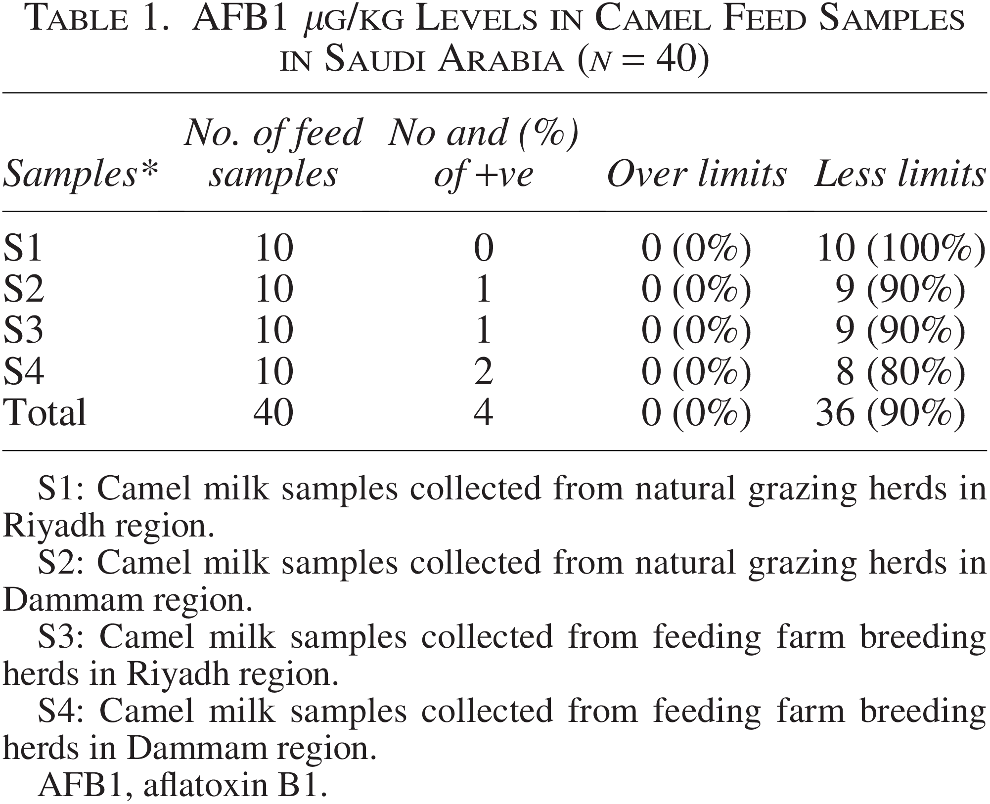
S1: Camel milk samples collected from natural grazing herds in Riyadh region.
S2: Camel milk samples collected from natural grazing herds in Dammam region.
S3: Camel milk samples collected from feeding farm breeding herds in Riyadh region.
S4: Camel milk samples collected from feeding farm breeding herds in Dammam region.
AFB1, aflatoxin B1.
Levels of AFM among camel milk samples
The findings of the present study (Table 2) indicate no statistically significant difference (p > 0.05) in AFM1 concentrations between milk samples obtained from camels raised in traditional environments (natural grazing on grasses and legumes) and those managed under semi-intensive systems (on-farm feeding with concentrates). All of the milk samples obtained from camels that were naturally grazing tested negative for AFM1.
AFM1 μg/ kg Levels in Camel Milk Samples in Saudi Arabia (n = 40)
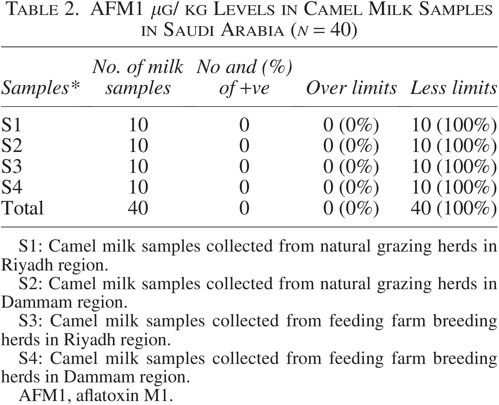
S1: Camel milk samples collected from natural grazing herds in Riyadh region.
S2: Camel milk samples collected from natural grazing herds in Dammam region.
S3: Camel milk samples collected from feeding farm breeding herds in Riyadh region.
S4: Camel milk samples collected from feeding farm breeding herds in Dammam region.
AFM1, aflatoxin M1.
Chemical composition of camel milk samples collected from different regions in Saudi Arabia
Figure 1 shows the chemical composition of camel milk samples collected from different regions in Saudi Arabia. Feeding system was a major determinant of milk composition. Camels grazing on natural pastures produced milk with TS ranging from 11.68% to 12.03%, whereas those fed farm concentrates had higher TS values (12.87–13.10%). In contrast, Figure 2 indicates that pH and acidity (%) did not differ significantly among samples, suggesting that feeding conditions have little effect on the milk’s acid–base stability.
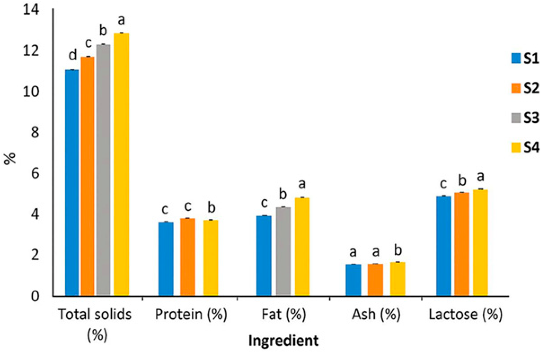
Chemical composition of collected camel milk samples from different regions in Saudi Arabia.
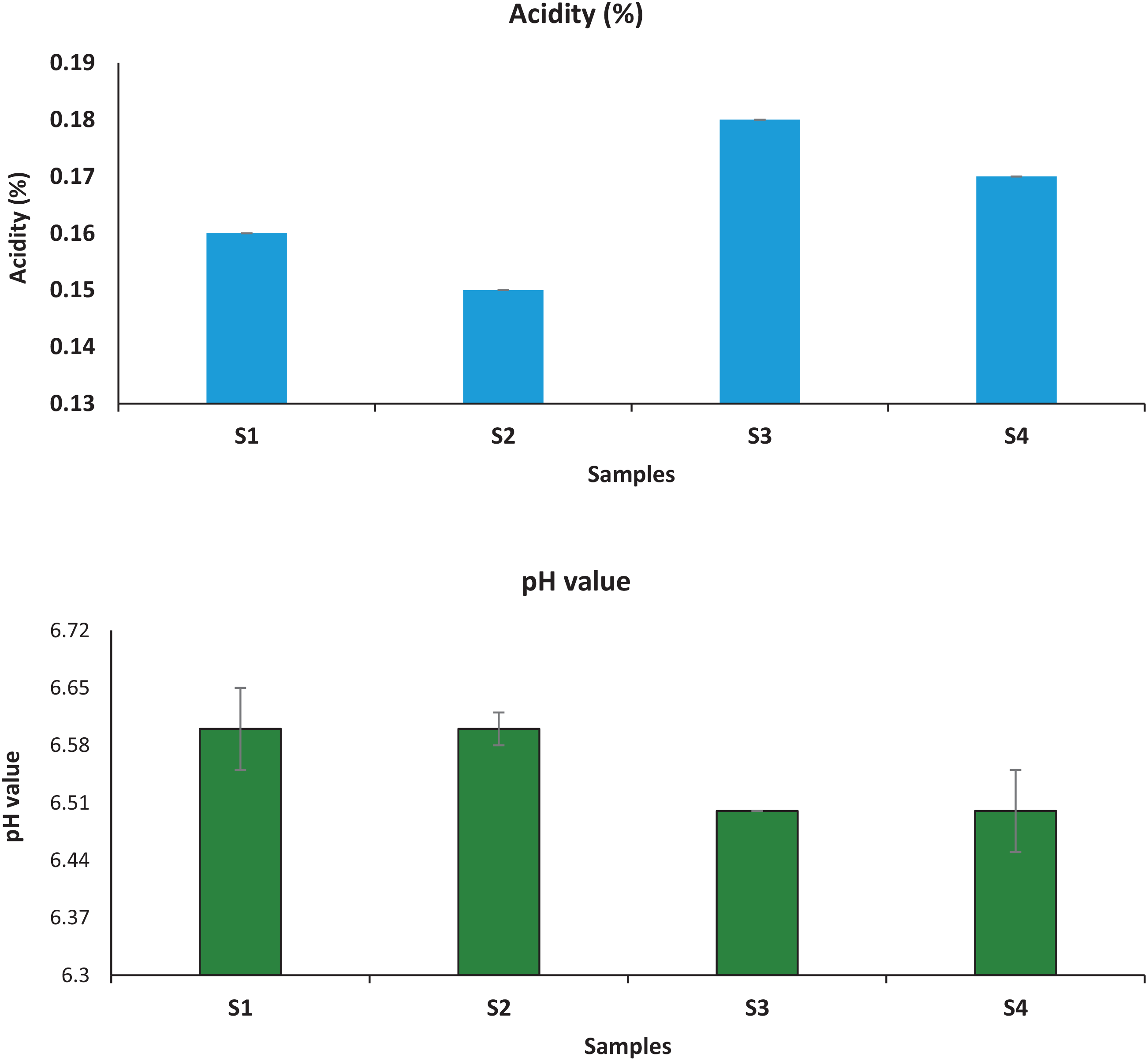
Acidity (%) and pH values of collected camel milk samples from different regions in Saudi Arabia. S1: Camel milk samples collected from natural grazing herds in Riyadh region; S2: camel milk samples collected from natural grazing herds in Dammam region; S3: camel milk samples collected from feeding farm breeding herds in Riyadh region; S4: camel milk samples collected from feeding farm breeding herds in Dammam region.
Microbiological properties of camel milk samples collected from different regions in Saudi Arabia
Microbiological analysis of camel milk samples from different regions in Saudi Arabia (Table 3) showed notable variations. Total aerobic flora ranged from 2.18 log CFU/mL in S1 to 4.95 log CFU/mL in S4, with statistically significant differences across regions. Total coliforms were below the detection limit (<10 CFU/mL) in S1 and S2 but were detected in S3 (2.33 log CFU/mL) and increased further in S4 (3.05 log CFU/mL). Psychrotrophic bacteria, yeast and molds, and fecal coliforms were not detected in any of the samples, confirming their absence within the sensitivity limits of the applied methods. However, S. aureus was not detected in S1 or S2 samples but was present at 2.33 and 3.05 log CFU/mL in S3 and S4, respectively. In contrast, LAB including enterococci, lactococci, leuconostocs, and lactobacilli were found exclusively in S3 and S4 at levels of 2.11–2.65 log CFU/mL. Their occurrence at these sites may reflect environmental influences or compositional factors supporting LAB growth, potentially contributing to natural fermentation and preservation.
Microbiological Quality of Camel Milk Samples Collected from Different Regions in Saudi Arabia (n = 40; mean ± SD)
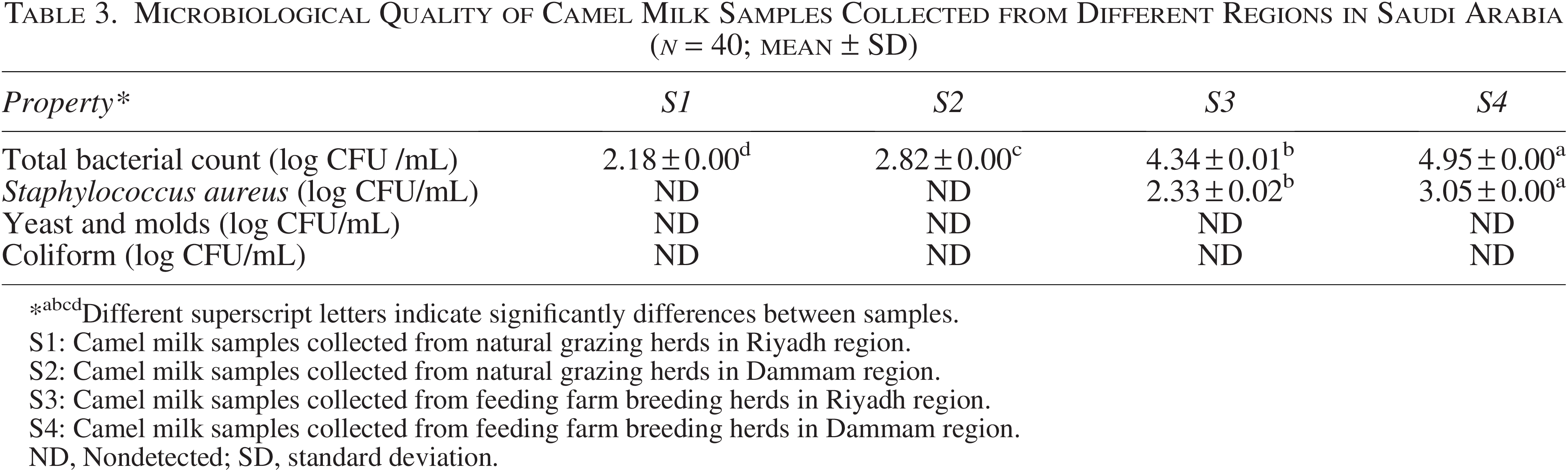
*abcdDifferent superscript letters indicate significantly differences between samples.
S1: Camel milk samples collected from natural grazing herds in Riyadh region.
S2: Camel milk samples collected from natural grazing herds in Dammam region.
S3: Camel milk samples collected from feeding farm breeding herds in Riyadh region.
S4: Camel milk samples collected from feeding farm breeding herds in Dammam region.
ND, Nondetected; SD, standard deviation.
Discussion
The pathological and performance impacts of mycotoxins in camels remain understudied, although cases of mycotoxicosis have been reported in Saudi Arabia (Al-Hiza et al., 2015) and the UAE (Osman et al., 2004). None of the positive samples exceeded regulatory safety thresholds, indicating no immediate health concern. Contamination was more common in farm-based systems (S3 and S4) than in grazing systems (S1 and S2), whereas no AFs were detected in samples from Riyadh. This may reflect a greater risk of fungal growth in stored or processed feeds compared with naturally grazed forage (Kumar et al., 2021). Although AFB1 was detected in some samples, concentrations remained below safety limits, suggesting that current feed management practices are largely effective (Pożarska et al., 2024). Nonetheless, contamination in farm-based herds underscores the need for continued monitoring. Overall, both feeding system and geographic location significantly influenced AF contamination levels.
The detection of AFB1 in some farm-based feeds indicates that it requires strict quality control, proper storage, and regular monitoring (Pożarska et al., 2024). Milk and dairy products, being essential dietary components, especially for children, are increasingly studied for safety, including AFM1 contamination (Bașkaya et al., 2006). Milk composition is influenced by breed, feed, health, housing, and seasonal factors (Bednarski and Kupczyński, 2024). Owing to the carcinogenicity of AFM1, sensitive analytical detection methods are required (Marchese et al., 2018). In this study (Table 2), AFM1 was undetectable in all milk samples, with no differences between camels raised on natural grazing or semi-intensive systems. These findings agree with Khalifa et al. (2023), who reported AFM1-free milk in naturally grazing camels in Egypt but contrast with their detection of AFM1 in semi-intensive systems. Similar low or nondetectable levels were reported in Kosovo, Iran (Fallah et al., 2016), and Saudi Arabia (Bokhari et al., 2017). In contrast, higher prevalence was documented in Egypt (Motawee et al., 2009), Saudi Arabia (Yosef et al., 2014), and Pakistan (Asghar et al., 2018). Such discrepancies may reflect differences in climate, feed quality, management, and animal factors (Asi et al., 2012; Ali et al., 2014). Concentrate feeds are a primary source of AFB1, which is metabolized to AFM1 and excreted in milk (Fallah et al., 2016; Elzupir and Elhussein, 2010). Consequently, the European Commission set a limit of 15 µg/kg AFB1 in camel feed (EC, 2006). The absence of AFM1 in naturally grazing camels may be attributed to the arid climate and low susceptibility of desert plants to fungal growth (Hussain et al., 2010). In farm systems, its absence despite potential feed contamination could be due to the use of antitoxins or the camel’s ability to detoxify mycotoxins (Fallah et al., 2016). Contamination levels were lower than some reports (Alsulami et al., 2023), likely due to seasonal, geographic, and management factors, and confirmed as accurate by our validated analytical methods.
Camel milk samples from Riyadh and Dammam, collected under natural grazing or farm feeding systems, showed notable compositional differences. TSs were higher in farm-fed camels (12.87–13.10%) compared with grazing camels (11.68–12.03%), reflecting the nutrient-rich nature of concentrates. Corresponding increases in protein, fat, and lactose were linked to higher protein, energy, and carbohydrate levels in formulated feeds. In contrast, pH and acidity remained unchanged across feeding systems (Fig. 2), consistent with Elkot et al. (2021), indicating stable acid–base balance. Ash content, however, was significantly higher in grazing camels, likely due to the mineral-rich forage consumed. These differences have practical implications: farm-fed milk, with its higher solids, is better suited for processing, whereas grazing-based milk offers a richer mineral profile for nutrition. Similar patterns have been reported in other dairy species, where pasture feeding enhances mineral content and concentrate feeding improves macronutrients.
Dietary management can tailor camel milk composition for nutritional or industrial use. Total aerobic flora increased progressively (2.18–4.95 log CFU/mL), reflecting handling and environmental factors but remaining within acceptable limits, although stricter hygiene is needed. Coliforms appeared in S3 (2.33 log CFU/mL) and S4 (3.05 log CFU/mL), indicating hygiene-related contamination. Psychrotrophic bacteria, yeast and molds, and fecal coliforms were not detected in any of the samples, confirming their absence within the sensitivity limits of the applied methods. However, S. aureus was not detected in S1 or S2 samples but was present at 2.33 and 3.05 log CFU/mL in S3 and S4, respectively. LAB were detected only in S3 and S4 (2.11–2.65 log CFU/mL), suggesting selective enrichment with possible functional benefits such as spontaneous fermentation and shelf-life extension. No AFM1 was detected, meeting the stricter EU/GCC limit (0.05 µg/kg), far below the Codex reference (0.5 µg/kg). Overall, the findings show negligible toxicological risk and manageable microbial hazards and emphasize the need for better on-farm hygiene.
Generally, the absence of detectable AFM1 in camel milk is reassuring, as AFM1, an AFB1 metabolite classified as a possible human carcinogen, is toxicologically relevant even at low levels. Current limits vary, with the EU and GCC adopting a strict threshold of 0.05 µg/kg compared with the higher Codex reference of 0.5 µg/kg. Our ND values, therefore, indicate compliance with the stricter standard and no immediate consumer risk. Microbiologically, rising aerobic counts and coliforms at some sites point to hygiene lapses, whereas the absence of S. aureus, yeast and molds, psychrotrophs, and fecal coliforms supports overall safety. Collectively, these results confirm acceptable chemical and microbial quality while highlighting the need for improved feed and hygiene practices to further minimize contamination risks.
Conclusion
This study demonstrates that feeding systems strongly influence the nutritional composition and safety profile of camel milk, with concentrate-based diets enhancing solids, protein, fat, and lactose, while grazing increased ash content. Farm-based feeds carried a higher risk of AF contamination, yet all levels were within regulatory limits, and AFM1 was undetected in milk, confirming negligible toxicological risk. These findings emphasize the need for stringent feed quality control, improved storage practices, and routine seasonal surveillance to minimize contamination. Future work should focus on developing practical feed detoxification strategies, assessing seasonal and regional variations, and integrating these insights into policy frameworks to safeguard consumer health and sustain the camel dairy sector.
Study limitations
This study was limited to mid-lactation camels sampled between March and May 2024 and therefore did not account for seasonal or lactation-stage variations in AF excretion. The relatively small sample size (40 animals) also reduced statistical power to detect low-prevalence events such as AFM1 contamination. This limitation reflects both the restricted global camel population and their distribution across small, dispersed farms, unlike other dairy species. Future studies spanning multiple seasons and involving larger cohorts are needed to provide more representative and robust estimates of AF risk.
Authors’ Contributions
All authors are equally credited with contributing to the preparation of this article.
Footnotes
Acknowledgments
The authors extend their appreciation to Ongoing Research Funding Program (ORF-2025–589), King Saud University, Riyadh, Saudi Arabia.
Author Disclosure Statement
The authors declare that they have no conflict of interest.
Ethical Approval
This study did not involve human participants, live animals, or sensitive data requiring ethical approval. Nevertheless, necessary approvals were obtained for the collection of feed and milk samples used in the research.
Data Availability Statement
Data will be made available on reasonable request.
Supplemental Material
References
Supplementary Material
Please find the following supplemental material available below.
For Open Access articles published under a Creative Commons License, all supplemental material carries the same license as the article it is associated with.
For non-Open Access articles published, all supplemental material carries a non-exclusive license, and permission requests for re-use of supplemental material or any part of supplemental material shall be sent directly to the copyright owner as specified in the copyright notice associated with the article.