






Abstract
Raw milk is highly prone to microbial contamination and can carry Salmonella enterica serovars-like zoonotic pathogens. This study follows the One Health-informed framework to investigate the occurrence and potential contamination pathways of Salmonella enterica serovars along the raw milk value chain in Bareilly, Uttar Pradesh, Northern India. A total of 900 samples consisted of raw milk (150), milk bucket swabs (150), milkers’ hand swabs (150), and farm floor swabs (150) collected from one local dairy farm, and one institutional dairy farm, and one retail milk outlet over a 6-months period. Overall, 19 samples (2%) were Salmonella enterica serovars positive, where pathogen occurrence was noted as 4.7% in farm floor swabs, 4.7% in milking bucket swabs, and 2% in raw milk. Salmonella enterica serovars was not detected in udder swabs, towel swabs, and milker hand swabs. Antimicrobial susceptibility analysis demonstrated high Salmonella enterica serovars (n = 19) resistance to ampicillin (73.7%) and tetracycline (68.4%), whereas multiple isolates (48%) exhibited multidrug resistance as well. These findings necessitate upgraded dairy hygiene practices, controlled antimicrobial use in veterinary medicine, and integrated One Health-relevant surveillance to mitigate raw milk consumption-associated public health risks in the studied region.
Introduction
Milk constitutes a major share of human nutrition and contains high-quality proteins, vitamins (A, D, and B complex), essential amino acids, and minerals (phosphorus and calcium) (Gorska-Warsewicz et al., 2019; Yenew et al., 2022). Raw milk is often marketed through informal supply chains in developing countries with limited cold chain and pasteurization facilities. These conditions increase the contamination risk of milk-borne pathogens (Asfaw et al., 2023; Khan et al., 2018,2025).
Salmonella enterica subsp. enterica serovars are key zoonotic foodborne pathogens, that is, known to cause nontyphoidal salmonellosis worldwide. Salmonella enterica serovars are an established source of millions of gastroenteritis cases/year, which significantly increases global foodborne disease burden (WHO, 2018; WHO, 2024; Kumar et al., 2025a). Raw milk is often associated with salmonellosis outbreaks (Weinstein et al., 2025). Infected dairy cattle shed Salmonella enterica serovars in fecal material, which can directly contaminate milking equipment, udder surfaces, bedding material, and the surrounding environment (Yisra and Tigistu, 2025; Loor-Giler et al., 2025). Moreover, the use of contaminated water for cleaning purposes, inappropriate storage conditions, and poor sanitation practices aid in bacterial contamination, survival, and proliferation in dairy farms (Holschbach and Peek, 2018).
The One Health approach emphasizes integrated surveillance and control measures across related sectors (environmental, veterinary, and public health) to attenuate zoonotic disease transmission (Kapoor et al., 2023). The factors like improved animal health management, environmental sanitation, biosecurity measures, optimized antimicrobial usage, and hygienic milking protocols are crucial for pathogen-free farm-to-consumer milk delivery (Marcu et al., 2026).
Antimicrobial resistance (AMR) further increases public health concerns regarding Salmonella enterica serovars contamination in raw milk. Indiscriminate antimicrobial administration in livestock might increase the selection of resistant strains with potential human transmission via the food chain (Ahmad et al., 2021). multidrug resistance (MDR) Salmonella enterica serovars are increasingly reported in humans and animals, which complicate salmonellosis treatment, leading to severe disease outcomes (Kumar et al., 2025b).
The dairy sector has significantly expanded along with increased raw milk consumption in Northern India; however, Salmonella enterica serovars contamination routes along the dairy chain remain underexplored. A detailed understanding of contamination pathways, from farm environments to end consumers, is necessary for designing efficient intervention strategies. Therefore, the current study addressed Salmonella enterica serovars occurrence in interconnected animal–environment–food interfaces within a One Health perspective.
Materials and Methods
Study design and sampling framework
A cross-sectional study was conducted to investigate Salmonella enterica serovars occurrence across the raw milk value chain in Bareilly, Uttar Pradesh, Northern India, which is located 155 miles east of the capital New Delhi (28° N10′N78° 23′E). Raw milk samples were collected from one local middle-scale buffalo dairy farm (10–50 milk-producing animals; May–June), one large-scale institutional buffalo dairy farm (>1800 milk-producing animals; July–August), and one retail milk outlet (December–January; Supplementary Fig. S1). Healthy animals were selected for sample collection based on their physical and udder appearance. Manual milking was performed in small cowsheds during the animal feeding. A total of 900 samples were collected from the abovementioned sources by following U.S. Food and Drug Administration (USFDA) protocols (Andrews and Hammack, 2022). Samples included raw milk (bucket, n = 150), udder swabs (n = 150), milker’s hand swabs (n = 150), farm floor swabs (n = 150), milking bucket swabs (n = 150), and towel swabs (n = 150). Raw milk samples from each farm (one farm/day) were collected on the same day immediately after milking in sterile screw-top bottles (50 mL). Udder and bucket swabs were also collected on the same day (one farm/day). Similarly, floor, milker’s hand, and towel swabs were collected on the same day (one farm/day). Swab samples were collected according to the procedure of Khan et al. (2025). The swabs were immersed in brain heart infusion (BHI) broth (HiMedia, India). Then, the swabs were pressed inside the test tube to remove excess moisture. The moistened sterile swabs were rubbed around the udder and teats (one swab/sample) to collect udder samples. The milker’s palm, finger gaps, and fingers were smoothly rubbed to collect samples of both hands. Bucket samples were collected by rubbing the swabs on the inner surfaces. The floor and towel samples were collected by randomly streaking swabs over a surface area of 15 cm2. Sample swabs were individually placed in sterile BHI-containing (3 mL) screw-cap test tubes (Borosil, India; Supplementary Tables S1 and S2). An icebox was used to transfer aseptically collected samples to the laboratory in the dark. Microbiological examinations were initiated on the sampling day.
Isolation and identification of Salmonella enterica serovars
The protocol of Andrews et al. (2023) was adopted to isolate Salmonella enterica serovars according to the Bacteriological Analytical Manual method of the USFDA, with slight modifications. Briefly, a raw milk sample (25 mL) was homogenized in lactose broth (225 mL, HiMedia) and pre-enriched through incubation (35°C for 24 h). An aliquot of the pre-enrichment culture (1 mL) was transferred to tetrathionate (TT) broth (9 mL, HiMedia) and incubated for 48 h at 35°C.
The aliquots of swab samples (1 mL) were added into sodium pyruvate (1%) and sodium chloride (10%)-containing tryptic soy broth (9 mL, HiMedia) and incubated for 48 h at 35°C. Then, another incubation (48 h at 35°C) was carried out in TT broth. All TT enrichments were streaked onto HE (Hektoen enteric) agar (HiMedia) and again incubated at 35°C for 48 h. Three Salmonella enterica serovars colonies were selected and purified from each HE plate. These presumptive colonies of Salmonella enterica serovars were identified based on their colony morphology and biochemical characteristics as described by Khan et al. (2024).
Molecular confirmation of Salmonella enterica serovars
DNA of Salmonella enterica serovars samples was extracted according to Sambrook and Russell (2001) for PCR identification. The invA gene (389 bp) based identification of Salmonella enterica serovars was performed according to the protocol of Manzano et al. (1998). The following primers [5′-GCTGCGCGCGAACGGCGAAG-3′and 5′- TCCCGGCAGAGTTCCCATT-3′] were used for the DNA amplification. Reference cultures of Salmonella Typhimurium (MTCC 98), Salmonella Enteritidis (E 2094), and Escherichia coli (MTCC 443) served as the positive and negative controls to assess the PCR assay’s specificity. Bacterial strains used in this study are listed in Supplementary Table S3.
Antimicrobial susceptibility testing
Antibiotic susceptibility patterns of Salmonella enterica serovars isolates were assessed by adopting the disc diffusion method (Bauer et al., 1966) and following Clinical and Laboratory Standards Institute (CLSI) (CLSI, 2023) guidelines. Eight commercial antibiotic discs (HiMedia) belonging to seven antimicrobial classes were used in antimicrobial susceptibility testing, including tetracycline (tetracyclines, 30 µg), ampicillin (penicillins, 10 µg), cephalexin (cephalosporins, 30 µg), streptomycin (aminoglycosides, 10 µg), chloramphenicol (phenicols, 30 µg), nalidixic acid (quinolones, 30 µg), ciprofloxacin (fluoroquinolones, 5 µg), and gentamicin (aminoglycosides, 10 µg). Briefly, BHI broth was used to prepare bacterial suspension (0.5 McFarland standard) that was spread onto Muller–Hinton agar plates (HiMedia). The plates were incubated (37°C, 18 h), and then antibiotic susceptibilities were estimated according to the guidelines of CLSI’s clinical breakpoints for Salmonella spp. (CLSI, 2023). An isolate exhibiting resistance to antibiotics belonging to three classes or more is considered MDR.
Calculation of multiple antibiotic resistance index
The multiple antibiotic resistance index (MARI) was calculated by dividing the number of antibiotics facing isolate resistance by the total number of used antibiotics (Khan et al., 2024). A MARI value of >0.2 implied the origin of the isolates from frequent antibiotic usage areas, while a MARI ≤0.2 implied the bacterial origin from less frequent antibiotic usage areas (Khan et al., 2024).
Statistical analysis
The association between the prevalence of Salmonella enterica serovars and sample source as well as the prevalence of Salmonella enterica serovars and sample type was tested using the Fisher–Freeman–Halton exact test at a significance value of p ≤ 0.05. Analysis of resistance-associated variables was calculated using Fisher’s exact test, a p value of ≤0.05 was considered significant.
Results
Prevalence of Salmonella enterica serovars
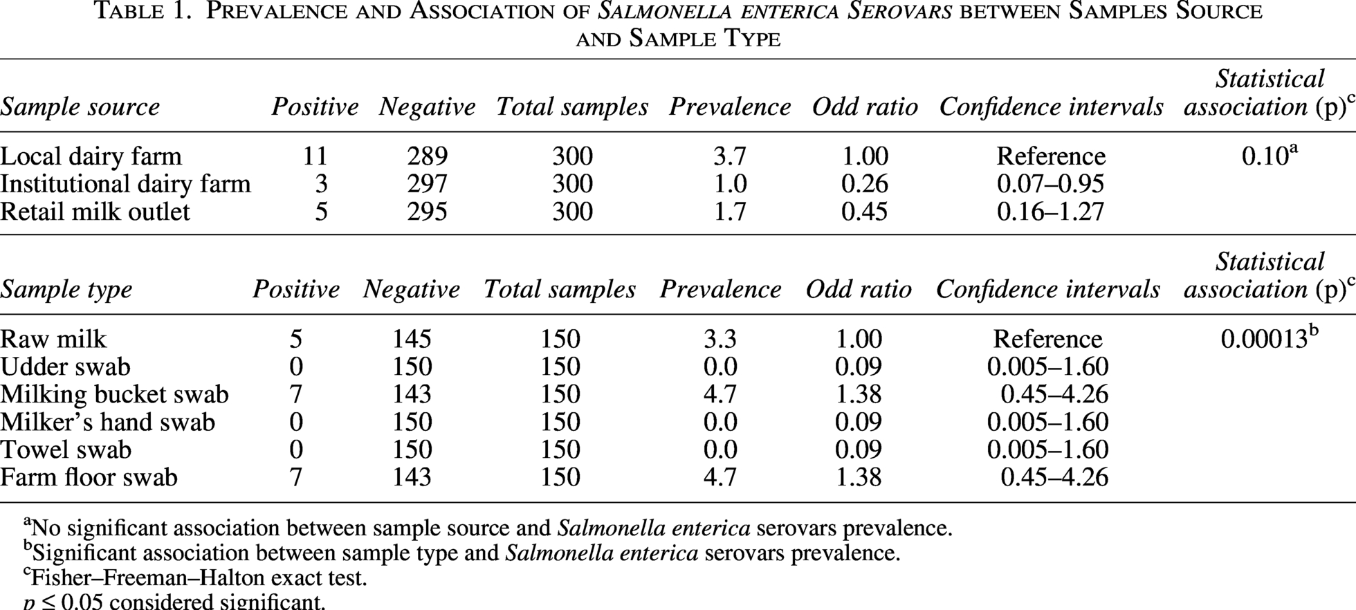
All presumptive Salmonella enterica serovars isolates were confirmed by PCR based on the detection of the invA gene (Fig. 1). A total of 900 milk samples were collected from one local dairy farm, one institutional dairy farm, and one retail milk outlets. Samples were analyzed for Salmonella enterica serovars prevalence along the dairy production and transportation chain (Table 1 and Fig. 2). Salmonella enterica serovars were primarily detected in raw milk samples (3.3%), milking buckets, and farm floor swabs (4.7%), respectively. Swab samples from the animal udder, milker’s hand, and towel remained negative across all sampling sites (Table 1 and Fig. 2). Out of 900 samples, 19 samples were found to be Salmonella enterica serovars positive, constituting an overall prevalence of 2% (Table 1 and Fig. 2).
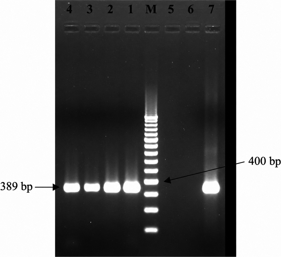
invA gene-based detection of Salmonella enterica serovars isolates using PCR; Lane M = 100 bp DNA ladder, Lane 1 = MS-1, Lane 2 = MS-27, Lane 3 = MS-53, Lane 4 = MS-56, Lane 5 = Escherichia coli MTCC-443 as a negative control, Lane 6 = Salmonella Typhimurium MTCC-98 as a negative control, and Lane 7 = Salmonella Enteritidis E-2094 as a positive control.
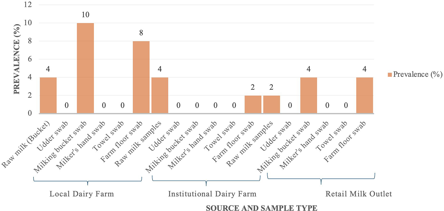
Prevalence of Salmonella enterica serovars in different sources and sample types.
Prevalence and Association of Salmonella enterica Serovars between Samples Source and Sample Type
No significant association between sample source and Salmonella enterica serovars prevalence.
Significant association between sample type and Salmonella enterica serovars prevalence.
Fisher–Freeman–Halton exact test.
p ≤ 0.05 considered significant.
A total of 300 samples were collected from one local dairy farm, where 11 samples (3.7%) appeared positive for Salmonella enterica serovars milking bucket swabs presented the highest contamination (10.0%), followed by farm floor swabs (8.0%) and raw milk samples (4.0%; Table 1 and Fig. 2). Salmonella enterica serovars contamination was low in one institutional dairy farm, where only 3 (1.0%) samples were Salmonella enterica serovars positive out of 300 samples. The pathogen prevalence in raw milk samples and farm floor swabs was noted as 4.0% and 2.0%, respectively (Table 1 and Fig. 2). Salmonella enterica serovars contamination was found to be 2.0% in raw milk samples, 4.0% in milking bucket swabs, and 4.0% in floor swabs among one retail milk outlet sample (300; Table 1 and Fig. 2). The Fisher–Freeman–Halton test established no association between Salmonella enterica serovars’ prevalence, and sampling sites; however, the prevalence of Salmonella enterica serovars was associated with sample type. The results revealed a significant association between the type of sample and Salmonella enterica serovars occurrence (p < 0.00031; Table 1). Raw milk, milking bucket, and farm floor represented important contamination points of Salmonella enterica serovars within the dairy production chain (Table 1).
Antimicrobial susceptibility profiles of Salmonella enterica serovars
All the Salmonella enterica serovars isolates (19) were subjected to antimicrobial susceptibility profile testing. AMR patterns varied among the studied dairy-associated Salmonella enterica serovars isolates (Table 2). The highest resistance of 73.7% was observed against ampicillin (penicillins), followed by 68.4% and 36.9% against tetracycline (tetracyclines) and streptomycin (aminoglycosides), respectively. Salmonella enterica serovars isolates exhibited a moderate resistance of 26.3% and 21% against ciprofloxacin (fluoroquinolones) and cephalexin (cephalosporins), respectively. In contrast, lower resistance percentages of 15.8% and 5.3% were noted against nalidixic acid (quinolones) and chloramphenicol (phenicols), respectively. Notably, none of the isolates (0%) demonstrated resistance to gentamicin (aminoglycosides; Table 2).
Source-Wise Antibiotic Resistance Profiles of Salmonella enterica Serovars
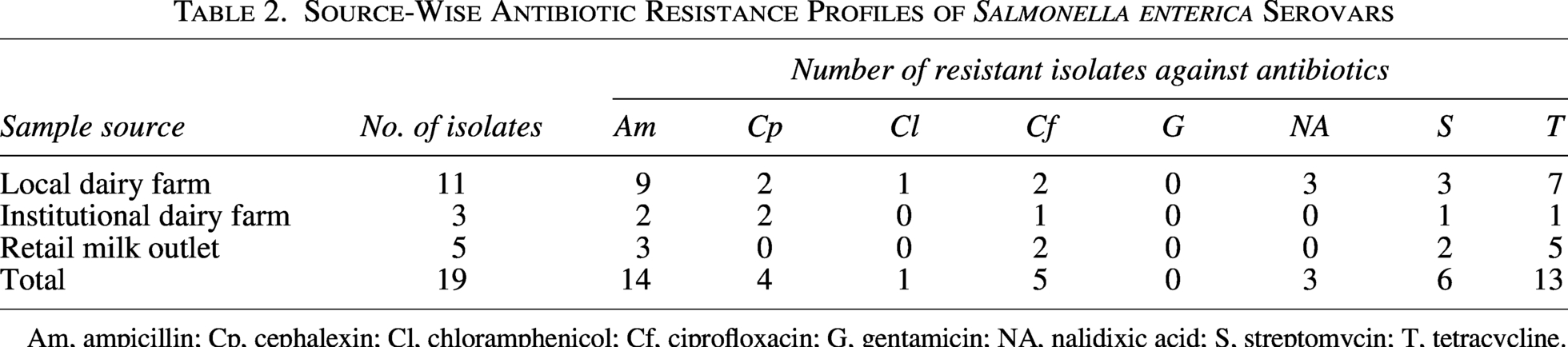
Am, ampicillin; Cp, cephalexin; Cl, chloramphenicol; Cf, ciprofloxacin; G, gentamicin; NA, nalidixic acid; S, streptomycin; T, tetracycline.
Tables 2 and 3 present the source-wise distribution of antibiotic resistance profiles of Salmonella enterica serovars isolates. The highest pathogen diversity and AMR frequency were observed in samples from local dairy farms. Most of the identified isolates from local dairy farms were resistant to ampicillin (81.8%; n = 09/11) and tetracycline (63.6%; n = 07/11; Table 2). Approximately, half of the isolates demonstrated MDR patterns (54.5%; n = 06/11; Table 3). The isolates from institutional dairy farms were comparatively less antibiotic resistant, and only one isolate (33.3%; n = 1/3) exhibited MDR patterns (Table 3). The retail milk outlet isolates also demonstrated considerable resistance profiles and patterns. All five isolates (100%) were found to be resistant to at least two antibiotics, whereas two isolates (40%) showed MDR patterns (Table 3). MARI values revealed that 17 isolates (90%) exceeded the ≥ 0.2 threshold (0.3–0.5), which indicated their origin from extensive antibiotic usage environments (Table 3). MDR patterns were statistically associated with elevated MARI values (≥0.2), while no association was found between MDR and both samples source and sample type (Table 4).
Antibiotic Resistance of Various Salmonella enterica Serovars Isolates
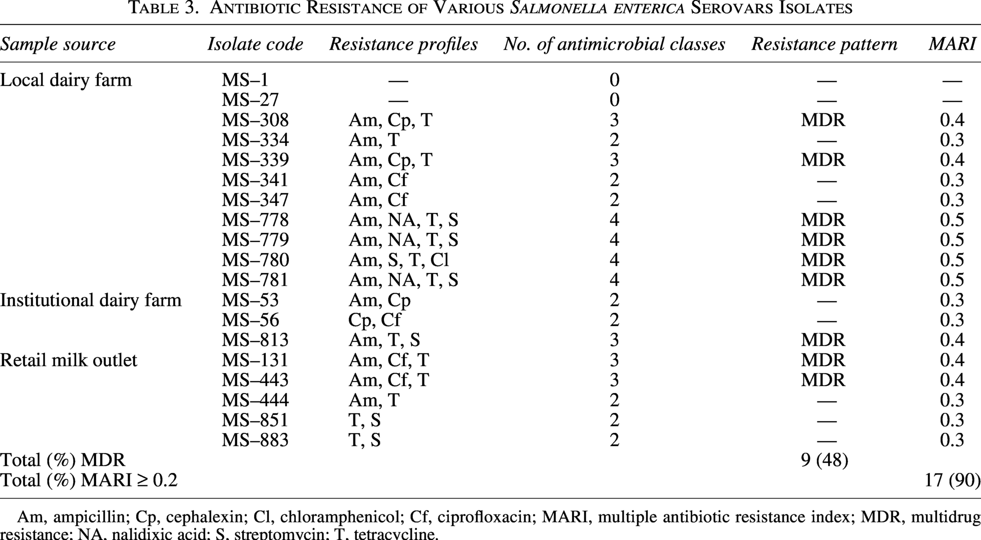
Am, ampicillin; Cp, cephalexin; Cl, chloramphenicol; Cf, ciprofloxacin; MARI, multiple antibiotic resistance index; MDR, multidrug resistance; NA, nalidixic acid; S, streptomycin; T, tetracycline.
Analysis of Resistance-Associated Variables in Salmonella enterica Serovars Isolates
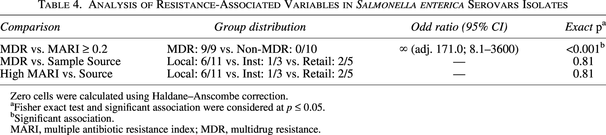
Zero cells were calculated using Haldane–Anscombe correction.
Fisher exact test and significant association were considered at p ≤ 0.05.
Significant association.
MARI, multiple antibiotic resistance index; MDR, multidrug resistance.
Discussion
Salmonella enterica serovars can easily contaminate raw milk, ultimately leading to human salmonellosis (Weinstein et al., 2025; Geinoro et al., 2025). The prevalence of this pathogen in raw milk samples during this study aligns with the globally reported range of 1%–12%, depending on farm management practices and hygienic conditions (van Kessel et al., 2004; Karns et al., 2005; El-Baz et al., 2017; Asefa et al., 2023; Barzegar-Bafrouei et al., 2024; Geinoro et al., 2025). However, higher Salmonella enterica serovars (>30%) prevalence in raw milk has been occasionally reported worldwide as well (Loor-Giler et al., 2025; Adzitey et al., 2025). Several Indian studies have reported varying Salmonella enterica serovars occurrences in dairy products and raw milk. Singh et al. (2018) reported a high (11%) Salmonella enterica serovars contamination in raw milk samples from retail shops and vendors’ home in central India. Similarly, Lingathurai and Vellathurai (2013) described an even higher (13%) Salmonella enterica serovars prevalence in raw milk samples obtained from local farms in Southern India. Contrarily, Khan et al. (2022) reported a low (4.0%) Salmonella enterica serovars presence in raw buffalo milk samples obtained from a local dairy farm in Northern India. The variation in Salmonella enterica serovars prevalence might be due to differential milk handling and farm hygiene practices and seasonal variations (Admasu et al., 2024; Loor-Giler et al., 2025). Generally, Salmonella enterica serovars contaminate raw milk through fecal contamination or animals serving as asymptomatic bacterial carriers. The pathogen contamination could also occur from the environment around the animals. Poor milking and farm hygiene, and unhygienic milk storage and handling, also facilitate Salmonella enterica serovars entry into raw milk (Marcu et al., 2026).
During this study, Salmonella enterica serovars contamination was noted in milking bucket swabs, which confirms their role as a pathogen source. The situation further aggravates with poor environmental and personal hygiene at farms (Asefa et al., 2023; Kahsay et al., 2025). Improper disinfection and cleansing of milk containers aid in milk-borne pathogens’ survival, leading to contamination during milking procedures (Irie et al., 2021). Several investigations have confirmed the role of milking utensils in microbial contamination of raw milk (Aliyo and Teklemariam, 2022; Deddefo et al., 2023). Salmonella enterica serovars detection in farm floor swabs highlights potential environmental pathogen reservoirs in dairy farms, such as manure-containing floors and bedding materials. Environmental contamination is a well-documented risk factor for pathogen occurrence in dairy farms (Marcu et al., 2026).
The present study featured low Salmonella enterica serovars prevalence in large-scale institutional dairy farms, which could be attributed to standardized milking protocols, better management practices, and improved sanitation (Marcu et al., 2026). Indian institutional dairy farms generally follow structured hygiene practices and controlled animal housing and employ trained personnel for milk handling. These factors attenuate microbial contamination risks within these farms. Improved farm management practices are associated with lower microbial contamination in bulk tank milk at large-scale dairy farms (Twomey et al., 2025). Nevertheless, Salmonella enterica serovars detection in farm floor swabs in this study might be caused by environmental contamination, which can occur in well-managed dairy farms as well.
The presence of Salmonella enterica serovars in retail outlet samples could have originated during milk storage, handling, or transportation (Mugabe et al., 2025). Multiple handling steps are involved in retail milk distribution, including transfer between containers and exposure to environmental surfaces, which enhance microbial contamination risk. Several studies have linked unhygienic milk distribution and storage practices with Salmonella enterica serovars contamination in raw milk (Barzegar-Bafrouei et al., 2024; Aiychew et al., 2024; Mugabe et al., 2025). Investigations in India and neighboring countries have reported Salmonella enterica serovars contamination in retail milk outlets. Singh et al. (2018) have documented the contamination of drug-resistant Salmonella enterica serovars in vendor-sold raw milk. Similarly, retail milk samples from Tandojam in Pakistan were found to be contaminated with Salmonella enterica serovars (Baloch et al., 2015). Collectively, poor hygienic conditions, adulteration with contaminated water, and contaminated utensils have been the most common contamination factors. Though overall Salmonella enterica serovars prevalence remained low in the current study, pathogen detection in retail milk highlights associated public health risks.
The high Salmonella enterica serovars resistance to tetracycline and ampicillin and moderate resistance to cephalexin, streptomycin, and ciprofloxacin may suggest notable indiscriminate antibiotic applications in dairy production systems. Nonetheless, these resistance trends must be taken with caution, as AMR in Salmonella enterica serovars can differ among serovars and is influenced by other interacting factors, including genetic lineage, ecological adaptation, and survival fitness, rather than solely by antibiotic exposure (Popy et al., 2025). Studies in developing countries, including India, have reported similar results where antibiotics are commonly administered for prophylactic and therapeutic purposes on dairy farms (Mugabe et al., 2025). High resistance rates of nontyphoidal Salmonella to ampicillin and tetracycline in this study are in line with earlier reports of milk and dairy environments in India, Ethiopia, and Iran (Singh et al., 2018; Barzegar-Bafrouei et al., 2024; Aiychew et al., 2024; Kahsay et al., 2025; Geinoro et al., 2025). Moderate resistance of Salmonella enterica serovars to ciprofloxacin is particularly concerning, as the role of fluoroquinolones is crucial in nontyphoidal Salmonella infection treatment in humans (Kumar et al., 2025b). Lower susceptibility of nontyphoidal Salmonella to fluoroquinolones has been widely reported in the food chain industry in various global reports (Nair et al., 2018). The total sensitivity of the analyzed Salmonella enterica serovars isolates to gentamicin may indicate minimal selective pressure against this antibiotic in the examined settings. Nonetheless, effective antimicrobial stewardship is crucial to maintain the efficacy of essential antimicrobial agents for both veterinary and human medicine (Kumar et al., 2025b).
MDR isolates of Salmonella enterica serovars were predominantly detected in local dairy farm samples, thus establishing their role as environmental reservoirs of resistant pathogens. Small-scale Indian dairy farms often lack effective antimicrobial stewardship programs, which often results in empirical antibiotic administration. MARI (0.3–0.5) values of Salmonella enterica serovars supported these findings and suggested their origin from local dairy farms with extensive antibiotic administrations (Khan et al., 2024). Similar MARI values of Salmonella enterica serovars have been reported in raw milk in other Indian regions as well (Malar et al., 2025). High MDR rates among milk-borne pathogens have been linked to injudicious antimicrobial applications in livestock on Asian rural dairy farms (Veloo et al., 2025). However, institutional dairy farms often adopt better management practices and hygiene protocols along with efficient antibiotic stewardship, which attenuate the prevalence of resistant Salmonella enterica serovars. However, the detection of lower rates of antibiotic-resistant Salmonella enterica serovars in this study might be caused by environmental contamination rather than the colonization of the studied farm. The resistant Salmonella enterica serovars contamination at the retail level could be the outcome of cross-contamination during milk storage, handling, and transportation. Improper sanitation and poor hygienic practices at retail outlets might facilitate resistant bacterial transmission to consumers (Kapoor et al., 2023; Mugabe et al., 2025; Subedi et al., 2025).
According to the One Health perspective, the prevalence of MDR Salmonella enterica serovars in dairy farms poses significant risks to environmental ecosystems, humans, and animal health (Kapoor et al., 2023; Kumar et al., 2025b). Moreover, the environmental dissemination of resistant Salmonella enterica serovars from dairy farms can also occur via farm runoff, manure, and wastewater, which further contributes to the spread of AMR (Talukder et al., 2023; Kapoor et al., 2023; Kumar et al., 2025b; Marcu et al., 2026).
Conclusion
The current study indicates a potential pathogen contamination risk in raw milk and dairy farm environments, along with the occurrence of MDR isolates. According to the One Health perspective, antimicrobial-resistant Salmonella enterica serovars’ presence in dairy environments is a matter of serious concern. Food chains, contaminated milk, and farm waste may contribute to resistant bacterial circulation between the environment, animals, and human compartments, although direct transmission was not investigated in this study. Therefore, improved hygienic milking practices, appropriate antibiotic applications in livestock, and continuous surveillance of foodborne pathogens are mandatory to ensure public health and sustainable clean dairy production.
Authors’ Contributions
Conceptualization and methodology: I.A. and J.A.K. Investigation and writing—original draft preparation: J.A.K. Data curation: J.A.K., F.S.B., F.M.H., L.A.N., and H.H.A. Formal analysis and resources: F.S.B., F.M.H., L.A.N., and H.H.A. Writing—review and editing: H.H.A. and L.A.N. Supervision: I.A. All authors have read and agreed to the published version of the article.
Institutional Review Board Statement
Not required. The study does not include human and/or animal subjects.
Data Availability
All datasets gathered and generated during this research have been included in this article.
Use of Artificial Intelligence Tools Declaration
The authors declare they have not used artificial intelligence in the creation of this article.
Ethics Approval and Informed Consent Statement
This article does not contain any studies with human or animal participants.
Supplemental Material
sj-docx-1-fpd-10.1177_15353141261466779 — Supplemental material for Prevalence and Antimicrobial Susceptibility Patterns of Salmonella enterica Serovars Along the Dairy Value Chain: A One Health-Relevant Surveillance of Dairy Farms
Supplemental material, sj-docx-1-fpd-10.1177_15353141261466779 for Prevalence and Antimicrobial Susceptibility Patterns of Salmonella enterica Serovars Along the Dairy Value Chain: A One Health-Relevant Surveillance of Dairy Farms by Javed Ahamad Khan, Iqbal Ahmad, Fahad S. Bazaid, Fohad Mabood Husain, Leena A. Neyaz, and Hussein H. Abulreesh
Supplemental Material
sj-docx-2-fpd-10.1177_15353141261466779 — Supplemental material for Prevalence and Antimicrobial Susceptibility Patterns of Salmonella enterica Serovars Along the Dairy Value Chain: A One Health-Relevant Surveillance of Dairy Farms
Supplemental material, sj-docx-2-fpd-10.1177_15353141261466779 for Prevalence and Antimicrobial Susceptibility Patterns of Salmonella enterica Serovars Along the Dairy Value Chain: A One Health-Relevant Surveillance of Dairy Farms by Javed Ahamad Khan, Iqbal Ahmad, Fahad S. Bazaid, Fohad Mabood Husain, Leena A. Neyaz, and Hussein H. Abulreesh
Footnotes
Acknowledgment
The authors are grateful to the Director, IVRI, India, and Dr. Ram Swaroop Rathore (ex-principal scientist, retired) for permitting some part of the Ph.D. research work at his laboratory. The authors also express their sincere gratitude to Dr. S.V.S. Malik (Ex-Head VPH) for supporting them throughout the period of work at the VPH division, IVRI, and Dr. R.K. Agarwal (In-charge National Salmonella Center, IVRI, Bareilly) for providing reference strains of Salmonella and untiring continuous support during the investigation.
Author Disclosure Statement
The authors declare no conflicts of interest.
Funding Information
This research received no external funding.
References
Supplementary Material
Please find the following supplemental material available below.
For Open Access articles published under a Creative Commons License, all supplemental material carries the same license as the article it is associated with.
For non-Open Access articles published, all supplemental material carries a non-exclusive license, and permission requests for re-use of supplemental material or any part of supplemental material shall be sent directly to the copyright owner as specified in the copyright notice associated with the article.
