
Abstract
Integrated multi-organ microphysiological systems are an evolving tool for preclinical evaluation of the potential toxicity and efficacy of drug candidates. Such systems, also known as Body-on-a-Chip devices, have a great potential to increase the successful conversion of drug candidates entering clinical trials into approved drugs. Systems, to be attractive for commercial adoption, need to be inexpensive, easy to operate, and give reproducible results. Further, the ability to measure functional responses, such as electrical activity, force generation, and barrier integrity of organ surrogates, enhances the ability to monitor response to drugs. The ability to operate a system for significant periods of time (up to 28 d) will provide potential to estimate chronic as well as acute responses of the human body. Here we review progress towards a self-contained low-cost microphysiological system with functional measurements of physiological responses.
Impact statement
Multi-organ microphysiological systems are promising devices to improve the drug development process. The development of a pumpless system represents the ability to build multi-organ systems that are of low cost, high reliability, and self-contained. These features, coupled with the ability to measure electrical and mechanical response in addition to chemical or metabolic changes, provides an attractive system for incorporation into the drug development process. This will be the most complete review of the pumpless platform with recirculation yet written.
Keywords
Introduction
Under the current paradigm of pharmaceutical innovation, only around 10% on average of drug candidates (5% for oncology) entering clinical trials make their way to regulatory approval. 1 The major issue lies in the disease models used for drug discovery and preclinical development. Most drug screening relies on in vitro cell culture models and uses animal models to predict drug toxicity and efficacy in humans. While traditional cell culture models utilizing multiwell plates enable fast and inexpensive high-throughput screening, the accuracy of the prediction is often compromised due to oversimplified cell microenvironment and tissue structure, and their inability to reproduce the complex interactions among organs and tissues that occur in a living animal. 2 Animal models, on the other hand, are costly, raise ethical issues, and have shown poor predictive power for human response to drugs due to cross-species discrepancies.3,4 Alternative models that can improve the accuracy of preclinical evaluation of drug candidates would reduce drug attrition rates during clinical stages and have a large economic impact.
The development of human cell-based multi-organ microphysiological systems has gained momentum in recent years. These systems, also known as Body-on-a-Chip (BOC) devices, integrate multiple microscale organ models and connect them with microfluidic channels to mimic multi-organ interactions within the body. 5 Advanced in vitro cell culture technology and microfluidics are combined to create a “human surrogate” that could be used to emulate drug absorption, distribution, metabolism, and action in the body. 2 Such human-based systems hold a great potential to better identify drug candidates for clinical trials with humans.
For effective adoption by the pharmaceutical industry, BOC systems need to be relatively inexpensive, easy to operate, and produce reliable results. This review summarizes our recent progress towards self-contained, low-cost BOC systems with functional measurements of physiological responses. A BOC system is considered “self-contained” if the operation of the system does not require any fluid or gas loops outside the device and functional measurements can be performed through non-invasive, non-destructive approaches, such as optical interrogation and in situ electrical recording. The only time that outside intervention is needed is when the system needs medium replenishment for long-term operation, as required for the human body. The review discusses five core aspects of such systems (Figure 1), including:
Controlled culture environment: The development of a blood surrogate with defined chemical formulation as well as the preparation of cell culture substrates with customized surface properties have given BOC systems better control over the cell microenvironment and thus helped generate reproducible results. Pumpless microfluidic platform: Gravity-driven flow and passive fluidic controls have been combined to create fluid recirculation within the BOC model, allowing dynamic organ–organ interactions without the need for external pumps and tubing.6–10 Such pumpless microfluidic platforms form the basis of a self-contained BOC system, making low-cost operation of a large number of BOC units possible. Functional measurements: On-chip integration of biosensors through adoption of microfabrication technologies has enabled non-invasive in situ functional measurements of electrical activity, contractile force, and barrier integrity.7,9–12 Along with metabolic analysis, these functional measurements have demonstrated high sensitivity in evaluating responses to drugs compared to morphology and viability analysis commonly used in drug toxicity screening.
7
Advanced single organ models: Advanced single organ models, utilizing novel tissue engineering methods, human stem cell technology and microfabrication,12–17 provide sophisticated organ mimics as functional units for BOC systems to predict human responses to drugs. System integration: Integration of multiple single organ units and functional measurement tools into one pumpless system has been demonstrated in several BOC systems. The integration, guided by residence time-based scaling and physiologically based pharmacokinetic (PBPK) models, enables physiologically relevant organ–organ interactions on chip, enhancing the predictive power of BOC systems for human responses. Self-contained low-cost Body-on-a-Chip (BOC) systems hold great potential for high-content, high-throughput drug screening. There are five core aspects of such systems: (1) controlled culture environment with defined culture medium formulations and surface chemistry helps with generating reproducible conditions and extending the depth of functional analysis. (2) Pumpless microfluidic platforms are the basis of a self-contained BOC system; (3) non-invasive functional measurements are enabled with integrated biosensors; (4) advanced single organ models provide improved organ mimics for BOC systems. Image shows a three-dimensional model of gastrointestinal (GI) macrovilli,
74
reproduced from Esch et al.
74
with permission from Springer. (5) Integration of multiple organ units based on proper scaling rules and integration strategies recreates physiologically relevant organ–organ interactions.
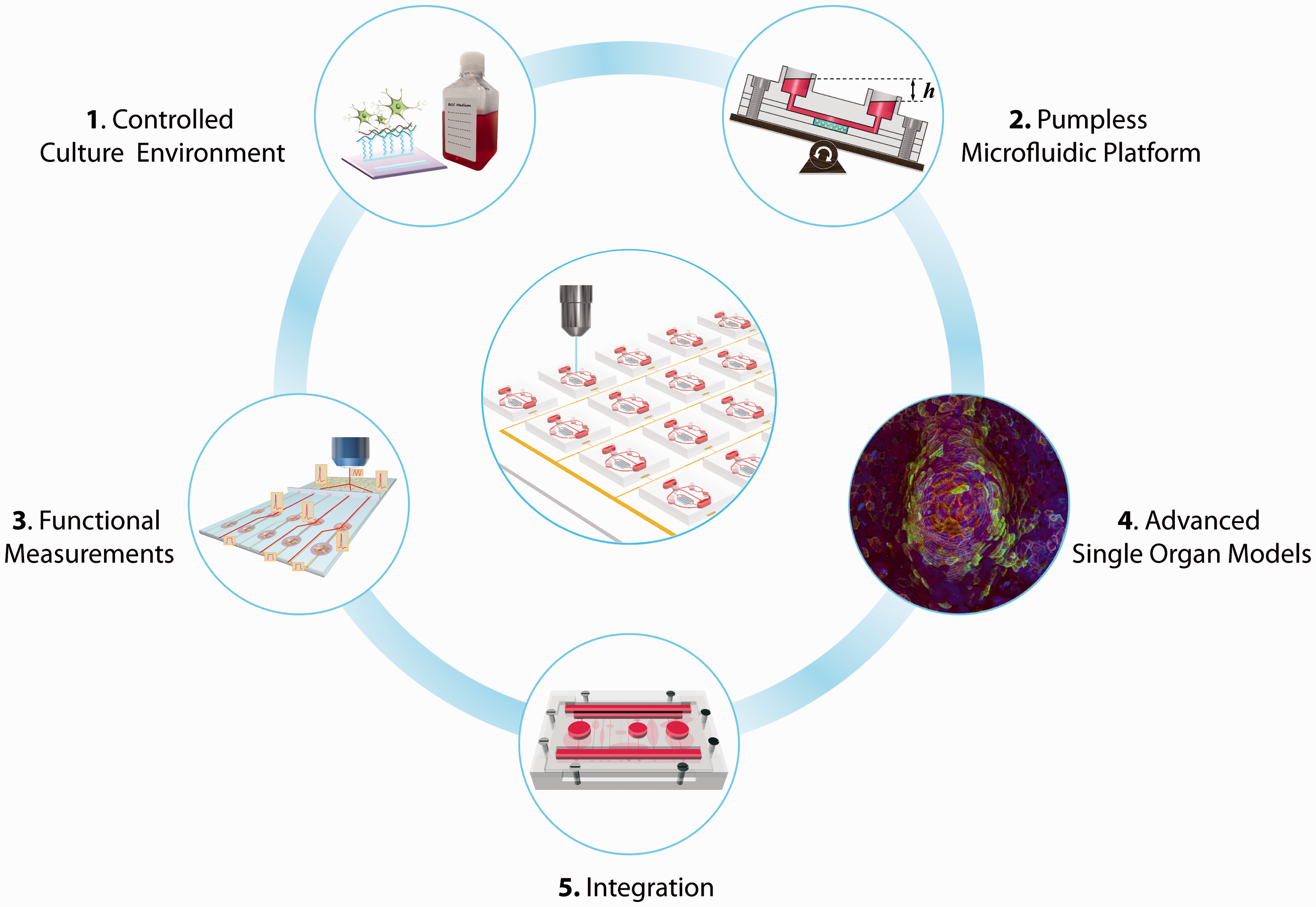
Development in all five areas provides essential elements for building self-contained, low-cost BOC systems for drug development.
Controlled culture environment
A drastic change in the cell microenvironment occurs when cells are isolated from the body and placed into a petri dish.2,18 Mimicking the chemical composition of the cellular interspace and the cellular anchorage in vitro helps to offset some of the effects (i.e., phenotypic changes) triggered by the environmental shift. 19 Standardized serum-free culture medium formulations enable precise control over the chemical environment surrounding the cells.2,18 Customized surface composition of cell culture substrates that favors the anchorage of specific cell adhesion molecules prevents phenotype loss and improves specific cellular functions.7,20–23 These strategies allow one to experiment with BOC systems under more reproducible conditions.
Defined cell culture medium formulation
To fully control the chemical microenvironment of cell cultures in vitro, well-defined culture medium is essential. 2 Human or animal sera, typically supplemented in culture medium as a source for a variety of biologically important components, can vary considerably in composition from batch to batch. The elimination of serum from cell culture medium is critical to achieving full chemical definition. These undefined supplements can be replaced with a chemically well-defined formula that provides similar effects in supporting cell growth and maintenance. In 1995, the Hickman group implemented such an approach in experiments with hippocampal neurons. 21 Further research has led to the successful adoption of defined and serum-free medium formulations for (i) single cultures of skeletal myotubes, 22 cardiomyocytes, 23 and motoneurons20,24; (ii) dual cultures of motoneurons with skeletal muscle (to form neuromuscular junctions), 17 sensory neurons (to recreate the stretch reflex arc),16,25 or oligodendrocyte progenitor cells (to model myelination) 26 ; and (iii) multi-organ cultures with four different organ types. 7
Surface modifications
The cell microenvironment is also influenced by the functional groups present on the surface of culture substrates, especially for planar cultures, such as those grown on microelectrode arrays and microcantilevers for functional measurements. The surface chemistry controls the adsorption and activity of cell adhesive proteins, 27 influencing cell–surface interactions such as adhesion and migration.28–30 Silane chemistry, an effective and widely used surface modification technique, has been used to produce self-assembled monolayers (SAMs) of cytophilic and cytophobic surfaces on glass coverslips and BioMicroelectromechanical Systems (bioMEMS) devices.7,22,23,25,30–43 With deep ultraviolet excimer laser lithography, SAMs can be patterned on cell culture substrates to control cell adhesion and alignment. 44 These patterned silanes can produce defined tissue structures that enable high-content functional analysis.23,33,43,45,46 For example, chemical patterning of SAMs was used to engineer neuronal networks. 46 The directionality of the network architecture was controlled with carefully designed discontinuities in chemical patterns on which hippocampal axonal growth was directed and “feed-forward” structures were created. In this way, the engineered chemical pattern pathways led to a layered architecture of neuronal networks where communication could travel within each layer and with directed communication pathways between layers. These defined neuronal networks allowed comprehensive network analysis when combined with extracellular electrical activity recording techniques. 46
Pumpless microfluidic culture platforms
BOC systems utilize microfluidic technology to interconnect individual organ units and facilitate nutrient and metabolite exchange between chambers. Several multi-organ systems have been developed on microfluidic platforms.47–51 To achieve reliable and precise fluid transport, BOC microsystems often require extensive support from external pumps and connecting accessories, which consume significant laboratory space and complicate system assembly and operation. Developing a pumpless microfluidic platform is a key step towards building self-contained BOC systems.
In 2010, Sung et al. proposed a novel design of microfluidic devices that utilized gravity-induced flow and a rocking motion to recirculate cell culture medium. 6 Microfluidic components, including three organ chambers and connecting channels were built on a single 4 cm x 4 cm chip. The integrated chip was placed on a tilting rocker plate to allow gravity to drive medium through the cell culture chambers of the device. Medium recirculation between two reservoirs was achieved by reversing the tilt direction periodically (Figure 1 (2)). This pumpless design using gravity as the driving force offers several key advantages: (i) it is space saving. Elimination of external pumps and tubing allows one to arrange self-contained BOC devices side by side closely to accommodate more units on a rocker plate. With additional stacking trays, tens to hundreds of units in parallel operation can be easily achieved on a single rocker. This pumpless solution holds great potential for relatively high-throughput application of microphysiological systems in pharmaceutical industries. (ii) It reduces unwanted drug adsorption on surfaces. Removal of connecting tubing also minimizes unwanted drug adsorption on the device by significantly reducing the exposed surface area. By decreasing the amount of drug adsorbed on device surfaces, more of the drug is available to interact with the organs on the chip. (iii) It is bubble-free. Air bubble formation is a notorious issue that afflicts most microfluidic cell culture systems. Air bubbles are detrimental to cells and generally lead to failure of a culture system. Yet they can be prevented in a gravity-driven system due to their buoyancy. This clears a major obstacle towards long-term, reliable operation of BOC systems. (iv) It is user-friendly. With all fluidic components integrated in a single platform, the assembly and operation of the devices are greatly simplified, and sterility is more easily sustained than when external tubing is utilized. (v) It provides easy access for liquid handling. The pumpless platform uses open access reservoirs that are similar to culture plate wells. This feature can facilitate automated liquid handling to remove samples and replenish medium. Overall, the gravity-driven pumpless platform provides a simple solution to achieve long-term reliable recirculating perfusion in BOC systems. By designing the channel dimensions and the tilt angle, the pumpless culture platform can achieve a wide range of flow rates that are suitable for BOC systems, from submicroliters to milliliters per minute. It should be noted that with highly integrated fluidic components on chip, the ratios of flow rates among organs are fixed once the chips are fabricated, although the overall flow rates are still adjustable through modifying the tilt angle. Such integration may reduce some flexibility for prototyping, but will benefit the long-term reliability and throughput of BOC operation.
The pumpless platform has evolved into several versions, and has been applied to various single-organ12,13,15 and multi-organ systems.7–10 Details will be discussed later in this review. It should be noted that the original pumpless platform using gravity-driven flow and a rocking motion creates a reciprocating mode of recirculation rather than a continuous unidirectional perfusion. Such reciprocating recirculation causes little deviation in the pharmacokinetic profiles of drugs compared to closed-loop recirculation. 6 Similarly, the resulting bidirectional flow has not had adverse effects on the metabolism of liver tissue constructed from primary cells. 15
The reciprocating flow induces bi-directional shear stress (SS), which could potentially affect SS-sensitive cells, such as endothelial cells and epithelial cells. Several modified versions have thus been developed to better accommodate SS-sensitive cells and tissues. Wang et al. introduced a “step chamber” in a microfluidic blood–brain barrier (BBB) model, 12 in which the barrier cell culture plane was offset by a distance from the channel plane to minimize the magnitudes of bi-directional SS on the cell surface. Such modification allows brain microvascular endothelial cells (BMECs) to survive and maintain their unique BBB phenotype under a reciprocating circulation. Esch et al. designed a set of passive valves and a backflow channel to create a self-contained unidirectional pumpless system. 9 The system supported gastrointestinal (GI) tract tissue in coculture with liver tissue for 14 d. In contrast to culture in a bidirectional system, the GI tract epithelium retained its barrier function in the unidirectional flow system, as evidenced through transepithelial electrical resistance (TEER) measurements. 9
Overall, the development of the “pumpless” platform is a contribution toward effective adoption of BOC systems in drug discovery research. The “pumpless” platform is a low-cost, relatively high-throughput solution to achieve reliable long-term operation of BOC systems. It can be applied to both barrier and non-barrier tissues, easily adaptable for single organ and multi-organ microsystems, and versatile in integrating on-line analysis tools.
Functional measurements
Compared to morphology, viability, and biochemical analysis that are traditionally used in drug screening studies, measurements of organ-specific functions, such as cardiac electrical activity, muscle contraction, and intestinal barrier permeability, could provide valuable functional information and produce more sensitive evaluation of drug responses. 7 Several in situ biosensors, such as microfabricated microelectrode arrays (MEAs) and microcantilevers, have been developed and incorporated into cell culture platforms to provide non-invasive functional readouts.7,9,11,12 These technologies can be readily integrated into microfluidic BOC systems and combined with optical interrogation and metabolic analysis to provide comprehensive functional assessment of drug responses in a self-contained BOC system without interrupting the cultures.
Extracellular electrical activity recording
Electrical propagation in the human body is exclusive to electrically conductive tissues such as nerves, muscles, and cardiac tissues. Functional analysis of those tissues has been extended beyond mere viability analysis and optical measurements (such as for cardiomyocyte beat frequency, BF) with extracellular electrical recording techniques, which have greatly advanced our knowledge of electrophysiology of populations of neurons. Among them, MEA-based multichannel recording systems are a non-invasive and high-throughput approach that provides comprehensive electrical signal recordings from conductive tissues (Figure 2(a)). The microfabricated nature of the MEA’s also makes it easier to be integrated into microfluidic BOC systems for electrical activity analysis.
Functional measurements for Body-on-a-Chip systems. (a) Extracellular electrical activity recording with microelectrode arrays (MEAs) for conductive tissues, such as myotubes,
77
cardiac microtissues,7,23 and nerves.7,20 (b) Measurement of force generation by muscles7,22 using a microfabricated cantilever-based system. Illustration reproduced from Smith et al.
84
with permission from World Scientific Publishing. (c) Barrier function analysis through transepithelial electrical resistance (TEER) monitoring and permeability studies for barrier tissue, such as skin,
13
gastrointestinal tract,71,72 and blood–brain barrier.
12
(d) Analysis of hepatic enzymatic activity and immune response.8,9,15 *Organ-specific functional measurements that have been incorporated in a pumpless microfluidic system.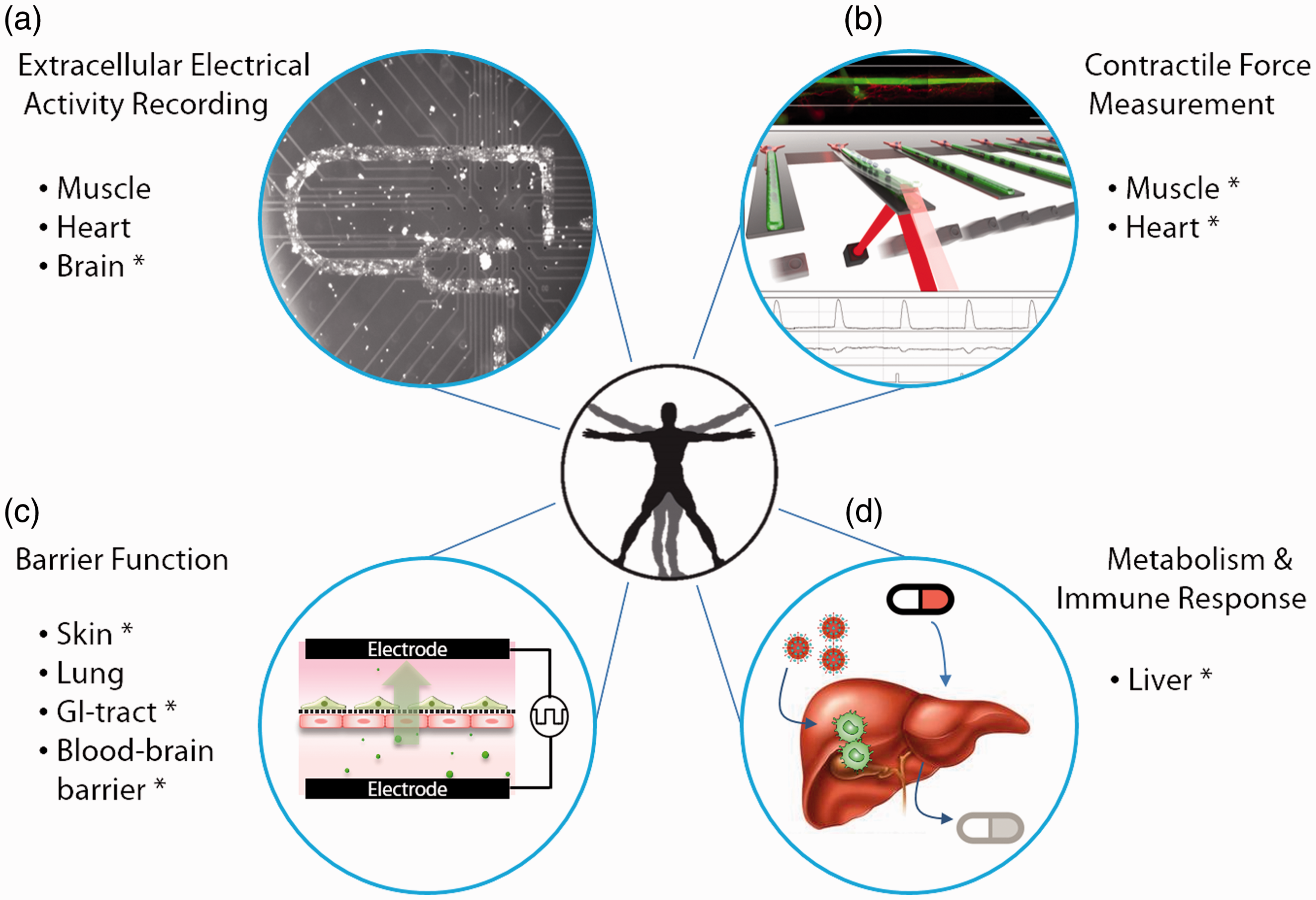
The utility and ease of use of cell-based MEA recordings have enabled researchers to replace the time-consuming and cumbersome patch clamp technique for extracellular electrical activity recording. The sensitivity of toxin detection based on the electrical response of cardiac cells recorded using MEA’s systems has been found comparable to those using standard patch clamp techniques. 11 Combining the MEA recording ability with surface patterning techniques that facilitate tissue alignment further expands the complexity and depth of electrical properties that can be measured (Figure 2(b)). This strategy has been applied to human stem cell derived cardiomyocytes to form aligned cardiac microtissues and enabled the measurement of a variety of critical parameters in assessing cardiac electrical response to drugs, including conduction velocity, rhythm generation (frequency and amplitude), action potential (AP) length (QT interval), and the length of the refractory period after an AP. 23 The differential side effects of sotalol, norepinephrine, and verapamil on cardiac electrical functions presented in that combined system were in line with published clinical data, 23 indicating great potential for the systems to evaluate cardiac electrical response to drugs.
Extracellular electrical activity measurements via MEAs have also been used to study the network architecture and electrophysiological behavior of hippocampal networks engineered with surface chemistry patterning. The extracellular recordings via MEAs have been used to measure the communication pathways both within layers and among layers. For example, long-term potentiation-like activity was induced in hippocampal networks to facilitate network analysis. 46
Contractile force measurement
A primary functional output of muscle tissues is their contractile force, which can be measured in a number of ways in vitro. 52 An approach developed by the Hickman group that combines microfabricated cantilevers and optical interrogation to measure cardiac or skeletal muscle force output (Figure 2(b)) is of particular interest due to its compatibility with microfluidic cell culture platforms, high precision, and ability to directly calculate force.
The force measurement is based on a microfabricated cantilever beam made of single-crystal silicon, whose dimensions and mechanical properties are well defined.34,38 Myocytes are cultured on top of the cantilevers, such that mechanical forces generated within the tissue is transferred to the cantilever beam causing a bending of the silicon cantilever. To measure the force, a laser is directed at the tip of the cantilever at an angle, and the reflected beam is directed onto a photodetector. As the cantilever bends, the reflected beam shifts on the photodetector, and this shift is measured. 32 The geometry of the system, the sensitivity of the detector, and the mechanical properties of the cantilever all go into the calculation of the force from these measurements.31,39
This cantilever-based force measurement system was first developed to assess the physiological characteristics of skeletal muscle, including contractile force, time to peak force and time to half relaxation (the time needed for the muscle to generate peak force and to relax to half of the peak stress, respectively). 31 An array of 32 cantilevers was fabricated on a single 1.5 cm x 1.5 cm chip to increase the throughput of measurements.31,32,34,38 The geometry of the cantilever was designed such that myoblasts on a cantilever fuse into a single functional skeletal muscle unit, a myotube. Each myotube can be investigated individually, allowing for measurement of myotube-level changes as well as changes to the population of myotubes. For example, data from this system can distinguish whether an effect occurs on the entire population of myotubes or only a subset. The effects of chronic drug treatments and long-term exercise protocols have been measured with respect to force and fatigue using this system. 40 While initial experiments were carried out with primary embryonic rat myotubes,31,34,38,39 C2C12 mouse-based cells, 32 and primary adult rat myotubes, 40 this system has successfully performed force measurements on primary human myotubes. 22
A similar system optimized for cardiomyocytes has also been developed. 23 Cantilevers were customized for cardiomyocytes such that the cardiomyocytes aligned along the length of the cantilever to match in vivo syncytium structure. In this model, several drugs with cardiac effects on various aspects of the mechanical and electrical function were tested in multiple dosages, and these drugs demonstrated effects as expected from in vivo data. This system has been incorporated into a pumpless microfluidic device and monitored cardiomyocyte force over a 14-d period in a four-organ coculture system. 7
Transepithelial and transendothelial electrical resistance (TEER) monitoring
Barrier tissues such as the gut, BBB, and pulmonary tissues are lined with monolayers of epithelial cells or endothelial cells, separating either the body from the external environment or the blood from tissues. 53 Neighboring cells are bound together mainly through intercellular junctions, such as tight junctions, forming barrier layers that regulate mass transport across the interface. A variety of approaches can be used to evaluate barrier functions, including immunostaining of tight junction proteins as well as permeability studies. Among them, the transepithelial/ TEER measurement is the simplest. It can be performed non-invasively on live cells with quantitative and real-time readouts. 54
The TEER value reflects the paracellular ionic conductance of cell monolayers and is a sensitive and reliable indicator of the barrier integrity. 54 The values are determined by measuring the electrical resistance or impedance over a spectrum of frequencies, normalized to the effective monolayer area. A four-point probe method 54 is commonly used in custom-designed and commercial TEER measuring systems, such as Endohm chambers and the “chopstick” electrode systems developed for transwells. Those systems measure ohmic resistance by utilizing a pair of electrodes on the different sides of the cell monolayer to apply square-wave current of microamps, and then use another pair of electrodes to record the voltage drop across the monolayer.
A uniform current density throughout the cellular monolayer is critical to ensure a valid TEER reading. In the “chopstick” system, the eccentric and suspended positioning of the electrodes tends to give uneven distribution of current flow across the cell layer that can result in 20% to 40% higher readings for large transwells (e.g., six-well transwells). 55 The Endohm chambers adopt a fixed electrode geometry to minimize variation and a pair of concentrically centered circular disk current electrodes to achieve uniform current field.
A similar design to the Endohm chambers has been employed for Organ-on-a-Chip (OOC) systems with customized microelectrodes or thin-film microelectrodes 56 (Figure 2(c)). Voltage-sensing Ag/AgCl pellets and annular current electrodes have been integrated into the pumpless platform to monitor the growth and barrier tightness of BBB 12 and GI tissue 9 constructs. Resistance measurements using these on-chip electrode systems have been compared to measurements from two commercial systems. The electrodes were also wired to give a real-time readout, without disrupting the culture. On-chip TEER monitoring can provide a non-invasive quality control measure of barrier integrity prior to the use of barrier tissues for drug permeability testing.
Metabolic and immunogenic measurements
Synthesis of plasma proteins, ammonia detoxification, drug metabolism, and immune response represent important hepatic functions. Previous in vitro models have demonstrated the possibility to quantify these functions (individually) from human primary hepatocytes in coculture with non-parenchymal cells (NPCs).57–61 However, the maintenance of stable function through time in a microfluidic pumpless system has been a challenge. A multicellular three dimensional liver model on the pumpless platform was developed and maintained for up to 14 d under flow and functional measurements were carried out through the experimental time non-invasively. 15 The daily synthesis of albumin and urea were quantified from the medium exchange samples through the biochemical reactions of immunodetection (enzyme-linked immunosorbent assay, ELISA) and nucleophilic addition, both with final colorimetric determinations. The metabolism of drug compounds or xenobiotics by the hepatocytes is a tool that aims to improve the molecule physicochemical properties to facilitate elimination from the body. The liver is the major metabolizer in the body. Stable phase I metabolic enzyme activities were confirmed in the microfluidic platform throughout the 14 d period by evaluating the enzyme activities of 1A1 and 3A3 isoforms with a luminescence assay. A fluorescent-based reaction can also be used in these microfluidic devices avoiding the sampling step from the reservoir. 62 Immune response studies of the hepatic platform were also possible with the coculture of human primary hepatocytes and hepatic NPCs upon the induction of Kupffer cells with lipopolysaccharide. An induced secretion of the cytokine IL-8 was quantified through a biochemical reaction using immunodetection (ELISA) and maintained for 14 d under flow demonstrating stable immune capacity in the system. 9 All the parameters measured above reflect important hepatic functions among all others.
The platform consisted of human primary hepatocytes cocultured with human hepatic NPCs. The applied flow improved stabilization of the production of albumin and urea as well as cytochrome p450 1A1 and 3A4 enzymatic activities. 15 The combination of a functional hepatic module together with other organs is of great importance for drug screening, not only for prodrug effects, but for the effect of the produced metabolites after the drug has been metabolized. Neither the addition of a GI tract module to the liver 9 nor the addition of the other three organs into a liver module in a 4-Organ system 7 affected the hepatic metabolic function.
Drug studies in such microfluidic models can be used to create PBPK models to predict the molecular fate of other compounds in the human body. 63 It should be noted that to obtain accurate metabolic parameters from these studies, it is important to minimize or carefully characterize the absorption and adsorption of drugs to the microfluidic devices. Drug compounds with hydrophobic characteristics are known to have high affinity for absorption into polydimethylsiloxane (PDMS), a commonly used material for microfluidic devices, or adsorbed on the surface.64,65 The pumpless BOC system inherently benefits from the elimination of external tubing which reduces non-specific adsorption. Other strategies to reduce drug absorption and adsorption include PDMS surface modification, 66 alternative materials for device fabrication,7,8 and pinhole-free parylene coating.12,15 Quantification of the predrug and metabolites can be carried out through mass spectrometry to further characterize the absorption and adsorption in the systems.
Advanced single OOC models
Human cell-based miniaturized organ models are the functional units of a BOC microsystem used in simulating human responses to drugs. Advances in human stem cell technology and tissue engineering have driven rapid development of OOC models for both barrier and non-barrier tissues.53,67 Those technologies have enabled efficient generation of high-fidelity human cells and in vivo-like cellular microenvironments to produce authentic metabolic and functional activities and simulate drug responses in the organs.12,13,15 Rapid adoption of microfabrication technology in biological contexts has also allowed integration of various biosensors for in situ functional analysis for OOC models.
Barrier tissues
BBB-on-a-Chip
The BBB, mainly composed of BMECs and overlying astrocytic foot processes, constitutes a dynamic physical and metabolic barrier between the blood and the brain. Most central nervous system (CNS) drug candidates have failed due to poor brain penetration across the BBB. 68 High-fidelity in vitro models of the BBB would boost the development of neurotherapeutics by increasing the efficiency of brain drug permeability screening.
Wang et al. have developed a microfluidic BBB model with in vivo-like barrier properties based on the pumpless platform. 12 The BBB-on-a-Chip (BBBoC) microsystem consists of a cell insert carrying BBB constructs and a pumpless culture platform providing luminal perfusion. The BBB constructs were prepared by coculturing human-induced pluripotent stem cell (hiPSC)-derived BMECs with primary astrocytes on two sides of a porous membrane of the cell insert. The constructs were then transferred onto the pumpless device for long-term maintenance. The adoption of the stand-alone cell insert simplifies dual-side cell seeding, ensures uniform membrane cell coverage critical to establishing barrier integrity, and separates the preparation of premature BBB constructs from long-term maintenance of mature BBB on chip.
The barrier properties of the BBBoC model were validated by TEER measurement, immunofluorescence staining and permeability studies. The BBBoC integrated electrodes for in situ monitoring of TEER values and is the first microfluidic BBB model that has demonstrated sustained TEER levels within the range of reported in vivo values (1500 to 8000 Ωcm2). 69 It formed continuous networks of tight junction proteins (zonula occludens-1 and claudin-5). The permeability toward large molecules (fluorescein isothiocyanate [FITC]-dextran tracers) and small molecule model drugs (caffeine, cimetidine and doxorubicin) was demonstrated to be comparable to in vivo values. Such a microfluidic BBB model providing in vivo-like barrier properties could be a useful single-organ model for CNS drug permeability screening as well as a functional BBB unit to be integrated in the BOC systems.
In addition to modeling the BBB itself, Brown et al. has also included pericytes and neurons in a three-dimensional culture setting to recreate the neurovascular unit for studies where the mutual interaction between the BBB and CNS is of interest. 70 This model and other 3D BBB–CNS are also reviewed in detail in the current issue.
Human skin equivalent (HSE)-on-a-Chip
Biomimetic in vitro skin models can be useful tools to predict skin responses to new drugs and evaluate the efficacy of transdermal drug delivery. Abaci et al., in collaboration with the Christiano group at Columbia University, have developed a microfluidic skin model based on the pumpless platform. 13 Full thickness HSEs with both epidermal and dermal components were grown on porous support membranes with air on one side and with microfluidic perfusion on the other side. Establishment of a stable air–liquid interface that allows maturation and terminal differentiation of HSEs is usually challenging in a gravity-driven flow system. HSE-on-a-Chip has overcome this challenge by designing and carefully leveling the microfluidic housing to keep the medium levels from both reservoirs lower than the bottom layer of the HSE at all times. The system was able to maintain skin constructs on chip for three weeks with sustained barrier function validated by transdermal transport analysis. Immunohistochemistry stains revealed differentiation and localization of keratinocytes and establishment of all epidermis sub-layers after a week of on-chip culture. The keratinocytes at the epidermal–dermal interface remain proliferative up to three weeks. HSE-on-a-Chip also reproduced the toxic effects of doxorubicin on skin cells and structure, suggesting it could be a useful model for skin drug testing. Notably, the model used 36 fold less amount of culture medium and cells compared to conventional transwell models.
GI tract model
Replicating the barrier function of the GI tract epithelium accurately is important when using the tissue for evaluating drug candidates. In order to achieve physiologically relevant drug bioavailability profiles, the cellular barrier must be intact with similar permeability as in vivo.
The GI tract epithelium is a multi-cell type barrier tissue that regulates the uptake of orally administered drugs. Mahler et al. have developed a multi-cell type GI tract model that consists of epithelial cells, mucous-secreting goblet cells and M-cells. 71 This model was used to simulate the oral uptake of nanoparticles of different sizes. 72 The authors showed that differently sized nanoparticles preferred different routes of transport across the GI tract epithelium. Those routes were provided by the different cell types, demonstrating the importance of building a GI tract model that contains the major cell types found in vivo.
GI tract epithelial cells are also SS sensitive cells, and align under the fluidic flow. It is critical to create in vivo-like fluid flow conditions to establish and maintain GI tract epithelial barrier integrity as measured by TEER values. GI tract epithelial cells did not develop high TEER on a pumpless platform with reciprocating flow, but achieved and retained a high TEER level when the system was outfitted with a set of valves that created unidirectional flow. 9
Another aspect of the GI tract model that has been recreated on-chip is its three dimensional architecture, consists of macrovilli that greatly increase the absorptive surface area of the gut. The gut macrovilli was replicated in a hydrogel 73 and on microfabricated porous polymer membranes 74 using microfabrication techniques.
Chen et al. have recently developed an ex vivo colon model by recellularizing the acellular human colon matrix with primary colon cells, including epithelial cells, endothelial cells, and myofibroblasts. 75 In that study, they established a primary culture of colonic epithelial cells (hCECs) that were originally harvested from patient samples and immortalized with human telomerase reverse transcriptase (hTERT). These cells were able to form organoids with microcypt-like structure when cultured in 3D hydrogel. They can be a long-term stable source for hCECs for GI tract models for BOC systems.
Non-barrier tissues
Heart-on-a-Chip
Cardiac adverse effects are one of the main reasons for drug failure. The current preclinical and clinical models fail to predict the toxicity in advance, reflected in the number of withdrawn drugs due to cardiotoxicity observations after market release. 76 Trying to overcome this problem, a human cardiac system was engineered to study basic physiology and toxicology of the two key cardiac functions: electrical conduction and contractile force. In Stancescu et al. 23 a stable function of the human embryonic stem cell derived cardiomyocytes was achieved by combining surface modifications and a serum-free medium formulation. Cardiomyocytes were cultured on BioMEMS components to enable the electrical activity measurements from microelectrodes and the contractile force from cantilever chips. This system was challenged with three cardiotoxic drugs affecting different mechanism of action (chronotropic and inotropic effects): sotalol, norepinephrine, and verapamil. Functional responses were dose-dependent and were as expected from human clinical observations.
Muscle-on-a-Chip
The mechanical properties of the muscle have been of great interest for a broad field in science (i.e., bioengineering, neuromuscular studies and pharmacology). A human in vitro muscle platform was developed for studying the contractile forces of single myotubes. Human myotube formation has been successfully reproduced in vitro from human skeletal muscle stem cell/progenitors. 77 Myotubes showed stable electrical properties similar to differentiated myotubes after two weeks under controlled culture conditions (serum-free and chemically modified surfaces). 77 Myotube contractile force has been studied in this culture on cantilever chips upon electrical stimulation. The myotube culture was characterized for increasing its contractile force with maturity and showed a physiologic response of fatigue under a continuous electrical stimulation. 22
CNS and Peripheral Nervous System (PNS)-on-a-Chip
The underlying mechanisms of the nervous system (NS) are incompletely understood. Solutions to NS-related diseases are affected by the incomplete puzzle. 78 In vitro cultures for studies of the nervous system are a challenge due to the delicate culturing conditions required. Hickman and others have shown over a number of years the controlled culturing of several neuronal types under serum-free conditions and for long-term periods (weeks to months). Hippocampal neurons, 79 motoneurons, 24 sensory neurons, 80 and HiPSc-derived cortical-like neurons 20 have been fully characterized with stable electrical function. The integration of this neuronal culture with other cells types and with bioMEMS chips are in progress towards building a CNS and PNS-on-a-chip. While these systems have not been incorporated into microfluidic devices yet, the progress outlined below shows great promise.
The communication between neurons in the CNS occurs within complex network architectures. The behavior of neurons within this natural environment, particularly the neuronal plasticity, can be studied by controlling the cellular network architecture and the conduction path (communication) using chemical surface patterning and MEA chips.45,46 Participation of other cell types in the neuronal communication is also of interest. Supporting cells such as myelinating cells are involved in both the development of cell–cell connections as well as play a critical role in protecting and maintaining neuronal communication. Models for both CNS and PNS myelination have been established to promote neuron–neuron and neuron–muscle communication and to study diseases involving demyelination and injury repair.26,81,82 A higher degree of neuronal communication is the reflex arc circuit (CNS–PNS link). The sensory and the effector portions have been successfully reproduced in vitro under controlled conditions. The use of bioMEMS chips in different sections of this circuit helped validate the functional communication under electrical or chemical challenges.16,17,25,37,83–85
Liver-on-a-Chip
The liver is of great importance to the drug development industry due to its metabolic capacity and being a recurrent target for drug-induced toxicity.86,87 Esch et al. constructed multi-cellular 3D liver tissues that consisted of primary human NPCs (Kupffer cells, stellate cells, and fibroblasts) and primary human hepatocytes. When placed into a microfluidic cell culture device and subjected to gravity-driven flow that changes direction periodically, this tissue responded with an increase in metabolic activity. 15 The observed result is similar to what others have seen with pumped systems, 88 confirming that pumpless systems can replicate tissue function well.
BOC system integration
Multiple OOC models and on-chip functional analysis tools can be ultimately integrated onto a microfluidic platform to build a “human surrogate” that can be used to improve predictions of human response to drug candidates. The biggest advantage of such BOC systems over traditional multiwell plate cell culture models is the ability to mimic the complexity of organ–organ interactions in vivo. In addition to diffusion-mediated interactions that can be easily achieved by submerging all organ units in a common medium, such as in the “wells-within-a-well” coculture system, 89 the microfluidics-based BOC systems allow for precise control over fluid transport between organs and thus are able to provide time-dependent dynamics of organ interaction. To recapitulate physiologically relevant drug dynamics and actions, and effectively interpret on-chip data into human clinical parameters, proper scaling rules and integration strategies are need. 63 Shuler has suggested scaling rules based on blood residence time in each organ, which controls overall chemical conversion in a scale-independent manner. Others have proposed alternative approaches, such as allometric scaling and functional scaling.90–92 Residence time-based scaling is advantageous in replicating organ–organ interactions by taking into account the rate of chemical conversion in the tissue. PBPK models have also been proposed and applied to guide the design and assist the data interpretation of BOC systems.2,6,9,10,18 One might purposely make a BOC system non-physiological by removing or changing the size of certain organs to investigate mechanisms or disease conditions.
A full realization of a perfect “human surrogate” is not yet achievable, and may not be necessary for the purpose of drug screening. The time and money it could cost to build such a model may not yield sufficient advantages over simpler systems. Multi-organ systems with two to 13 organs have been demonstrated on pumpless microfluidic platforms (Figure 3).6–10 In 2010, Sung et al. published a pumpless system that contained three tissues: liver, bone marrow, and colon cancer tissue (Figure 3(a)).
6
The actions of the cancer drugs 5-fluorouracil (5-FU) and uracil were recreated within that system, showing that metabolites generated in the liver tissue can recirculate and achieve the expected outcomes in the tumor tissue compartment. Lee et al. developed a liver-tumor model using a pumpless format.
10
With that two-organ model, they reproduced the simultaneous metabolism and tumor cytotoxicity action of a flavonoid luteolin, and demonstrated the importance of using a proper scaling approach for multi-organ system design, as well as the power of combining a pharmacokinetic/pharmacodynamics (PK/PD) model with on-chip experiments in interpreting on-chip data and gaining mechanistic insights.
Currently developed self-contained multi-organ systems. Integrated Body-on-a-Chip (BOC) systems that support coculture of two to 13 organs have been developed on pumpless microfluidic platforms. (a) A liver–tumor–marrow three-organ system. Reproduced from Sung et al.
6
with permission from the Royal Society of Chemistry; (b) a modular system for the coculture of GI tract epithelium and 3D primary liver tissue with in situ transepithelial electrical resistance (TEER) measurement capacity.
9
(c) A palm-size system for 4–5-organs with integrated electrical activity and contractile force sensors for non-invasive functional read-outs.
7
(d, e) BOC systems for 10 or more organs.
8
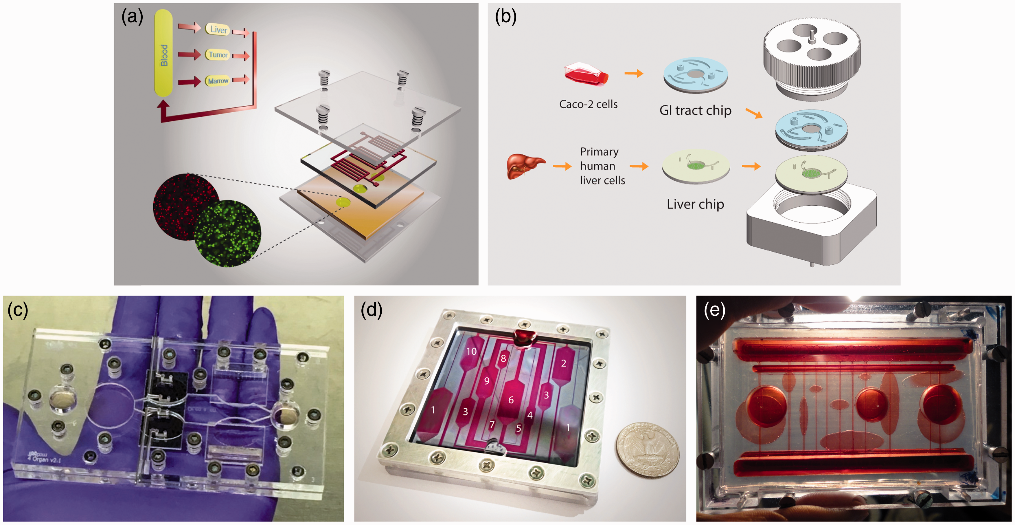
Two-organ systems of GI tract and liver have been developed previously on a platform that was operated with pumps to estimate the bioavailability of acetaminophen, 48 and to evaluate the effects of 50 nm polystyrene nanoparticles on the GI tract and on liver tissue. 93 The latest version of the GI tract–liver system developed on a pumpless platform features a modular design. 9 The GI tract and liver tissues were cultured on separate microfluidic chips, and after maturation those chips were combined with each other to yield the two organ system (Figure 3(b)). Modular designs offer advantages because they allow the preparation of different tissues with special maturation requirements, such as tissues constructed from primary or stem cell sources, at their own pace before assembly into a single BOC system.
In 2016, Oleaga et al. reported a multi-organ model with four organs: liver, heart, muscle, and brain. 7 The different cell types were located in communicating compartments inside a pumpless microfluidic system sharing the same culture medium with defined composition. Cellular morphology, viability, and function were measured for each cell type. The relevance of the multi-organ system as an in vitro platform for the study of systemic toxicity was achieved after the validation with five known drugs that exert side effects at high concentrations. The system responded in concordance with previous toxicity reported in in vivo and in vitro studies. 7 Miller and Shuler presented a whole-body model on a pumpless microfluidic platform, which consider explicitly thirteen organs (14 chambers) (Figure 3(c)). 8 Their work demonstrated the feasibility of constructing, operating, and maintaining a simple, self-contained, multi-organ microphysiological system with a large number of cell compartments for a sustained period with the capability of measuring cellular functions. They used a layered design to separate barrier and non-barrier types of cell cultures. The barrier chamber layer allowed for direct access that could be used to force chemical or biological reagents to pass through barrier cells before exposing the non-barrier tissues. These platforms represent the next generation of in vitro systems towards the BOC.
Concluding remarks
There have been many OOC and BOC models developed over the last decade, and we believe more of such microfluidics-based organ and whole-body models will be developed. We anticipate such systems will grow more sophisticated and will be a paradigm-shifting technology that will be adopted by pharmaceutical and chemical companies in reducing and replacing animal models. We also believe that self-contained, low-cost BOC systems, because of their simplicity and multiplex features, will become the workhorse of drug development. Under the new paradigm of pharmaceutical innovation, the time and cost to bring a new medicine to patients will be significantly reduced as the clinical approval success rate improves by adopting these simple but powerful BOC models.
Footnotes
Author Contributions
YIW, CO, JJH, and MLS devised the manuscript. YIW wrote the Introduction and the sections on pumpless microfluidic platforms, TEER monitoring, single organ models for the BBB and the skin, and the introduction for the Functional measurements and the Advanced single OOC models sections. CO, CL, and YIW wrote the section on controlled culture environment. CL, CM, and YIW wrote the section on extracellular electrical activity recording. CL and YIW wrote the section on contractile force measurement. CO wrote the sections on metabolism and immune response analysis and single cell models for the heart and muscles. MBE wrote the sections on single organ models of the liver and GI tracts and contributed to review on pumpless platforms. CO and CL wrote the CNS and PNS-on-a-Chip section. YIW, CO, MBE, and PM wrote the Body-on-a-Chip system integration section. MLS and YIW wrote the concluding remarks. YIW, MBE, CO, CL, PGM, JJH, and MLS contributed to editing the manuscript.
Acknowledgment
The preparation of this mini-review has been supported by the National Institutes of Health (R44 TR001326-0A1), and is based on work accomplished under a grant from the National Center for Advancing Translational Sciences at the National Institutes of Health (UH2TR000516).
Declaration of Conflicting Interests
C Long, C McAleer, JJ Hickman (chief scientist), and ML Shuler (CEO/president) are also associated with Hesperos, Inc. (Orlando, FL) which is commercializing this technology. JJ Hickman has larger than a 5% equity in Hesperos, Inc., and C Long, C McAleer, and ML Shuler have less than a 5% equity in Hesperos, Inc. YI Wang, C Oleaga, MB Esch, and PG Miller have no conflicts of interest or financial ties to disclose.