






Abstract
Background:
Traumatic arterio-enteric fistula is predominantly seen after penetrating trauma with only 21 reported cases documented in the past 25 years. They may present in an acute or delayed manner with upper or lower gastrointestinal bleed. A detailed clinical examination with requisite imaging can help in detecting such injuries.
Case Description:
Case 1: A 20-year-old gentleman, presented with penetrating stab injury to the gluteal region with bleeding per rectum. Imaging revealed evidence of injury to the inferior rectal artery which was found to be communicating with the extraperitoneal portion of the rectum. He was managed with a combination of endovascular and open surgery with a successful outcome. Case 2: A 29-year-old gentleman, presented in a delayed manner 2 weeks after a gunshot wound to the gluteal region, which was managed operatively in another hospital. He developed a massive lower gastrointestinal bleed 2 weeks after presentation. Imaging revealed evidence of a pseudoaneurysm of the inferior gluteal artery which had a fistulous communication with the gastrointestinal tract leading to bleeding. It was managed by endovascular techniques successfully.
Conclusion:
Arterio-enteric fistulas following trauma are rare phenomena and they need a high index of suspicion for diagnosis. Once diagnosed, they can be managed based on their location and patient physiology by interventional techniques, surgery, or a combination of the two.
Keywords
Introduction
Arterio-Enteric Fistula (ArEF) is a rare entity. The reported incidence of primary ArEFs (caused by underlying disease) is 0.004-0.007% while it is 0.69-2.36% in the case of secondary ArEFs (caused by prosthetic vascular grafts). 1 They are commonly seen in the setting of major vascular surgeries in the abdomen and in the setting of malignancies, either due to direct erosion of the bowel wall or following irradiation. Traumatic ArEFs are a much rarer entity with very few cases reported in the literature. They are usually caused by a penetrating injury when the injury is inflicted on a retroperitoneal structure or the stomach adjacent to a blood vessel. Management of Traumatic ArEFs poses a significant challenge to the trauma surgeon as they are difficult to diagnose. Further, they can lead to catastrophic hemorrhage as there is no tamponade effect. Moreover, their treatment is also challenging as the patient may be in extremis due to shock. They are usually managed surgically by exploration and operative repair. 2 We report on 2 patients, both of whom suffered penetrating injuries with subsequent enteric bleeding; one in an acute manner and the other with delayed presentation. Both were managed successfully with a combination of Damage Control Interventional Radiology (DCIR) and surgery. We also present a brief review of the literature on Traumatic ArEFs along with the management options available.
Case Report
Case 1
A 20-year-old gentleman presented 1 hour after sustaining penetrating assault with a sharp object. On arrival his airway was patent. In breathing, he had tenderness and subcutaneous emphysema on the left side of the chest. However, air entry was equal on both sides with no signs of respiratory distress. He had blood pressure (BP) of 106/68 mm hg and a pulse rate (PR) of 108/min. His Glasgow Coma Scale (GCS) was 15/15. His chest x-ray was normal. His extended Focused Assessment with Sonography in Trauma (eFAST) examination did not reveal any intra-abdominal or pleural fluid. On secondary survey 2 penetrating wounds were detected, one in the left lateral chest 3 x 2 cm in dimension in the left 6th intercostal space and another in the right para-sacral area 2 × 1 cm in dimension (Figure 1A). There was no evidence of any active bleeding from either of the wound site. The patient’s vitals soon began to deteriorate with tachycardia increasing to 130/min and blood pressure falling to 80/60 mm hg. A left intercostal drain was placed due to suspicion of tension pneumothorax. However, there was no gush of air or blood and his hemodynamics did not improve. The patient was re-evaluated clinically and a log roll and digital rectal examination were performed. Active bleeding per rectum was noted. He was resuscitated with blood and blood products and, once stabilized, underwent contrast-enhanced computed tomography (CECT).

A, Showing site of penetrating injury. B, Axial and sagittal contrast enhanced CT images showing a presacral hematoma with active extravasation of contrast which is seen extending into the rectal lumen (arrows). C, Left internal iliac artery angiogram shows faint contrast blush from the a branch of the inferior rectal artery (arrow) and post embolization angiogram showing non-opacification of the inferior rectal artery and its branches (dashed arrows).
CECT revealed free air in the extraperitoneal perirectal space suggestive of extraperitoneal rectal perforation. Intravenous contrast extravasation was noted from one of the rectal arteries into the rectal lumen (Figure 1B). However, again the patient’s hemodynamics deteriorated after CT (BP: 90/50, PR: 132/min). Blood transfusion was started and a decision was made for angiography and embolization. Arterial access was established through the right femoral artery and a 6F arterial sheath was placed using the Seldinger technique under fluoroscopic guidance. Angiography of the left internal iliac artery was performed with a 5F RDC (Renal Double Curve) catheter and selective angiography of the inferior rectal artery was performed with a Progreat microcatheter. Contrast blush was seen from a branch of the inferior rectal artery. Since the branch vessel could not be cannulated, the inferior rectal artery was embolized with a slurry of gel foam. There was no evidence of contrast blush or pseudoaneurysm in the right internal iliac artery and inferior mesenteric artery angiograms. Post-procedure, his hemodynamics improved (Figure 1C). Thereafter, he was taken to the operating room (OR) for management of extraperitoneal rectal perforation for which a trephine diversion loop sigmoid colostomy was done. His postoperative period was unremarkable. The intercostal drain was removed on postoperative day2. Stab sites were observed for any signs of infection and he was subsequently discharged on post-injury day 5. A Barium enema study was done 6 weeks later which showed no contrast extravasation, following which restoration of bowel continuity was done.
Case 2
A 29-year-old gentleman was referred to us 2 weeks after a history of a gunshot wound to the gluteal region having already undergone laparotomy and repair of extraperitoneal rectal perforation with diverting proximal colostomy at a nearby hospital. His primary survey was normal and detailed evaluation revealed a gluteal wound measuring 0.5 x 0.5 cm in dimension with imaging revealing a left ischial tuberosity and sacral fracture. The patient was managed with routine wound care measures and had an uneventful hospital stay for 2 weeks. 2 weeks after admission, he had an episode of massive rectal bleed. After resuscitation with blood and blood products, a CT Angiography was done. This revealed a pseudoaneurysm of the left gluteal artery with a pre-sacral hematoma reaching up to the rectum (Figure 2A). The patient was immediately shifted to the angiography suite where DSA confirmed CT findings. Arterial access was established through the right femoral artery and a 6F arterial sheath was placed using Seldinger’s technique under fluoroscopic guidance. Angiography of the left internal iliac artery was performed with a 5F RDC (Renal Double Curve) catheter which showed a pseudoaneurysm arising from the inferior gluteal artery which bled into the alimentary tract. The branch supplying the pseudoaneurysm was selectively cannulated and coils embolization was done. There was a non-opacification of the pseudoaneurysm in the post embolization angiogram. There were no further episodes of bleeding thereafter and the patient was discharged successfully (Figure 2B). After a month of recovery, he was readmitted and underwent a restoration of bowel continuity.

A, Axial contrast enhanced CT and coronal maximum intensity projection images showing a contrast filled outpouching (pseudoaneurysm) arising from the inferior gluteal artery (white arrows). A presacral collection (asterisk *) is seen closely abutting the rectum (hash #) as well as causing lytic destruction of the underlying bone (black arrow). B, Left internal iliac angiogram showing pseudoaneurysm (arrow) from the inferior rectal artery. Post coil embolization, there is non-opacification of the pseudoaneurysm. The coils (dashed arrow) are seen in the inferior gluteal artery.
Discussion
There are 5 potential sites of bleeding in patients with hemorrhagic shock in trauma: chest, abdomen, pelvis, retroperitoneum, and external bleeding. In the abdomen, blood loss can occur either into the peritoneal cavity or intraluminal in the gastrointestinal tract (GIT). Bleeding inside the peritoneal cavity can be identified with the help of FAST and clinical abdominal signs. Traumatic ArEFs present a distinctive problem as the bleeding is intraluminal to the GIT. It is difficult to diagnose because there may be no clinical abdominal signs at presentation and FAST is also usually negative. Further, in an acute trauma setting and with the patient in hemorrhagic shock, it is very difficult to differentiate between a retroperitoneal bleed and bleeding into GIT. In acute cases, they may present with hemorrhagic shock, hematemesis, hematochezia, or melena. While in delayed cases, ArEFs may present with occult bleed from the GIT and/or sepsis. ArEFs usually have a warning sign in the form of a “herald bleed.” 2 This is followed by torrential bleeding which could be life-threatening. In an acute case, early imaging with CT may help, but it is always a dilemma for the treating physician to send a hemodynamically unstable patient to CT. In such cases, initially, the patient is taken to OR for exploratory laparotomy. During exploratory laparotomy (EL) if no intraperitoneal or retroperitoneal source of bleed is identified and GIT is filled with blood clots, then upper GI endoscopy or Lower GI colonoscopy may be performed based on the site of bleeding. Furthermore, the patient can then be taken to CT for the identification of the bleeding source. In both our cases, we were fortunate that the distal part of the GIT was involved and early signs in the form of hematochezia were evident. Further, the mechanism of injury and the presence of a penetrating wound in the gluteal region helped us in the early suspicion of the traumatic ArEF. The first case also highlights the importance of log roll examination and Digital rectal examination (DRE) in a trauma patient. DRE has been of utility in diagnosing anorectal injuries. However, in cases of hemorrhagic shock where the source of bleed is not obvious, it is very important to perform a DRE as was seen in our case.
The management of Traumatic ArEFs remains challenging as very few cases are reported in the literature. The first case of ArEF was reported in 1818. A French soldier had swallowed a bone and developed chest pain several hours later. Five days later, he suffered fatal hematemesis. 3 There was another case report published in 1988 where following road traffic injury the patient developed delayed ArEF and expired due to bacterial aortitis. Here, however, the ArEF was discovered post mortem. 4 We extensively searched Google Scholar and Pubmed using keywords such as “traumatic arterioenteric fistula and aorto enteric fistula” and found only 21 reported cases of ArEFs with the details of their management over the last 25 years (Table 1). The rarity of such cases being reported may be attributed to the fact that they pose a diagnostic challenge as many of them remain asymptomatic. The most common mode of injury was penetrating (14/25), while the blunt mode of injury was seen in only 7/25 cases. Fifteen patients survived to discharge, while 5 patients succumbed to the injury and in one case the outcome was not known. A majority of these cases involved major vessels such as aorto-esophageal, superior mesenteric artery-duodenal, aorto-gastric and renal artery-gastric. Despite extensive literature search we failed to find any published cases of rectal artery-rectal fistula. Most of the cases have been treated with a combination of endovascular and surgical techniques.
Details of Reported Cases of Traumatic Arterio-Enteric Fistula Over Last 25 Years.
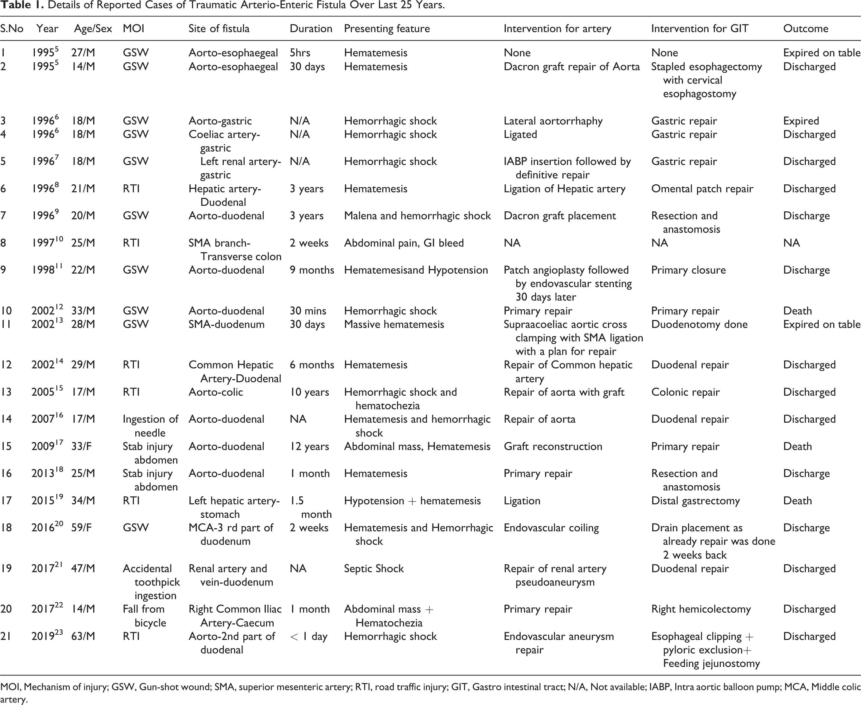
MOI, Mechanism of injury; GSW, Gun-shot wound; SMA, superior mesenteric artery; RTI, road traffic injury; GIT, Gastro intestinal tract; N/A, Not available; IABP, Intra aortic balloon pump; MCA, Middle colic artery.
Endovascular procedures in the form of coiling, balloon occlusion, and/or stenting have helped in the management of the vascular component of ArEF, while the enteric component is dealt with surgically. Endovascular methods were initially limited to hemodynamically stable patients. However, now their use has been extended in hemodynamically unstable patients as well for early control of bleeding, especially in those areas which are difficult to access surgically. This extension of endovascular interventions in bleeding patients has been termed as Damage Control Interventional Radiology (DCIR) and is an integral part of the Hybrid Operating Room system. However, this approach is associated with the risk of further compromising the physiology of the patient if sufficient expertise is not available and the procedure becomes too time-consuming. Hence, a judicious decision based on the physiology of the patient should be taken along with simultaneous resuscitation with blood products. The PRESTO (Prompt and Rapid Endovascular Strategies in Traumatic Occasions) dictum of “start soon and finish sooner” has been the motto in such scenarios. 24 Ours is a level 1 trauma center with availability of expertise and a blood bank with an established Massive Transfusion Protocol. In our first case, the site of bleeding was difficult to access surgically as it was in extraperitoneal perirectal space. The surgical approach to this artery would be similar to the approach of abdominoperineal resection. However, the artery is positioned deep in the pelvis and may not be approachable through the abdomen. In such a case, one may have to approach it through the perineum with a risk of damage to the sphincters and a permanent colostomy. Hence, we took an attempt to control the bleeding endovascularly even though the patient had borderline hemodynamic parameters. In the second case, the patient had an instance of delayed bleed with an evident pathology diagnosed on CT angiography. Rapid access to interventional facilities allowed us to avoid a complicated open surgical procedure.
The enteric part of ArEFs is managed by surgical exploration. In both our cases, the injury was in the extraperitoneal part of the rectum. There has been a concept of 4 “D” s (debridement, diversion, distal rectal wash, and drainage) in the management of rectal injuries. 25 However, the utility of drainage, debridement, and distal wash has not been proven and is no longer recommended. Diversion in the form of ileostomy or colostomy is the recommended treatment.26,27 However, this method too has been challenged, especially in small penetrating extraperitoneal injuries. 28 In such cases, it has been treated non-operatively without any diversion and if any abscess develops, it is drained trans-anally. In our first case, we opted to follow our institutional protocol for extraperitoneal rectal injuries which are in line with the 2016 EAST (Eastern Association for the Surgery of Trauma) guidelines for the management of extraperitoneal rectal injuries. 26
Conclusion
To conclude, while ArEF is a rare complication following trauma, a high index of suspicion should be maintained. Endovascular techniques can be used successfully in such cases and can significantly decrease morbidity associated with extensive surgical procedures.
Footnotes
Authors’ Note
J.D.J., H.A., and N.H were involved in manuscript preparation. J.D.J. and V.K. were involved in data collection. A.K. and A.G. reviewed the manuscript critically. The consent for publication has been duly informed to the patients and written consent has been obtained for the same. Ethical approval was not required for this study.
Declaration of Conflicting Interests
The author(s) declared no potential conflicts of interest with respect to the research, authorship, and/or publication of this article.
Funding
The author(s) received no financial support for the research, authorship, and/or publication of this article.
