






Abstract
Basilar artery occlusion (BAO) is one of the most devastating types of ischaemic stroke and is identified by using computed tomography (CT) angiography. Marfan syndrome is an autosomal dominant disorder involving multisystem connective tissue, and the neurological complications are relatively rare. In this article, we report a case of a young Marfan syndrome patient complicated with BAO ischaemic stroke. The patient was an 18-year-old man with right hemiparesis, aphasia and impaired consciousness. CT angiography of the brain showed an occlusion distal to the basilar artery. Endovascular therapy including intravenous thrombolysis and mechanical thrombectomy (MT) was administered to this patient inside the therapeutic window. The patient had a favourable clinical outcome after endovascular therapy. Marfan syndrome may be a rare cause of ischaemic stroke with BAO. In addition, our report provides some evidence that can be used as a reference when planning therapeutic strategies for BAO patients with Marfan syndrome.
Background
Basilar artery occlusion (BAO) is a rare disease found in approximately 1% of patients with ischaemic stroke, but it has a high mortality rate, especially in the absence of early reperfusion. 1 Mechanical thrombectomy (MT) for BAO is associated with high rates of successful reperfusion, which is a strong predictor of a favourable outcome. 2
Marfan syndrome is an autosomal dominant disorder involving multisystem connective tissue, such as that in the musculoskeletal, ocular and cardiovascular systems. Neurological complications are relatively rare and generally are ischaemic or haemorrhagic events. 3 In this article, we described a young adult with Marfan syndrome who suffered from acute ischaemic stroke caused by embolic occlusion of the basilar artery and was successfully treated with intravenous thrombolysis and MT. To our knowledge, this is the first case of BAO resulting in a Marfan syndrome–related ischaemic stroke treated with endovascular recanalization.
Case Presentation
An 18-year-old man who presented with right hemiparesis, aphasia and impaired consciousness was admitted to the hospital within 7 minutes after onset. He had not been definitely diagnosed as Marfan syndrome, and no aortic dissection was found at the time of presentation, but his mother had Marfan syndrome and died of aortic dissecting aneurysm, and his grandmother also had aortic aneurysm, according to his father. On admission, his blood pressure was 136/87 mmHg. He was tall and slender with long limbs (Figure 1). His Glasgow Coma Scale (GCS) score was 6 points, and his National Institute of Health Stroke Scale (NIHSS) score was 20. An ECG showed a sinus rhythm of 70 bpm. An emergency non-contrast CT scan of the brain revealed a hyperdensity sign in the basilar artery territory, which indicated an acute thrombus. Subsequent CT angiography revealed an occlusion distal to the basilar artery (Figure 2A and 2B). The patient’s height was 190 cm, and his arm length to height ratio was 1.07. He had spider-like fingers, and he had a positive thumb sign (thumb sign: the distal phalanx of the thumb can protrude beyond the ulnar border.). CT scan and digital subtraction angiography in the acute phase. (A) Non-enhanced CT scan showed hyperdensity sign in the basilar artery, suggestive of intravascular thrombus. (B) CT angiography demonstrated occlusion in the distal segment of the basilar artery, which was confirmed later by digital subtraction angiography. (C) Digital subtraction angiography revealed distal occlusion of the basilar artery and reduced flow signal within the bilateral posterior cerebral artery (arrows). (D) After mechanical thrombectomy, the basilar artery was recanalized.

The patient was immediately given intravenous thrombolysis with alteplase at a dose of 0.9 mg/kg after the CT scan within the therapeutic window. After discussing the risks and benefits, the decision regarding treatment was made by the stroke team and his father. It was obvious that he was a candidate for endovascular recanalization, so he was transferred to the interventional neuroradiology unit. Digital subtraction angiography was performed approximately 3.5 hours after the onset of symptoms and showed distal occlusion of the basilar artery with no artery dissection (Figure 2C and 2D). The procedure was performed under general anaesthesia. First, an 8-F sheath was placed in the right femoral artery. Then, an 8-F Navien catheter was placed in the left vertebral artery, where there was an occlusion just distal to the basilar artery, and MT was performed with a Solitaire stent retriever. Thrombolysis in cerebral infarction (TICI) 3 of basilar artery recanalization was eventually achieved.
The patient was admitted to the intensive care unit, and after 4 weeks, he was transferred to the rehabilitation clinic. CT angiography of the brain showed complete recanalization of the basilar artery. Magnetic resonance imaging (MRI) showed cerebral infarction in the bilateral cerebellum and pons (Figure 3). Furthermore, the patient was evaluated by transthoracic echocardiography and confirmed to have an aortic root aneurysm (47 mm diameter of the aortic root) and coronary sinus bulging. The ocular examination showed that the patient had myopia and slight ectopia lentis. In addition, he was definitively diagnosed with Marfan syndrome through genetic testing after discharge. His short-term outcomes were favourable, with the right hemiparesis almost completely resolved (Figure 1), an NIHSS score of 3 and a modified Rankin scale (mRS) score of 2 after 4 months. A CT scan was performed 24 hours after mechanical thrombectomy, and magnetic resonance imaging (MRI) was performed 3 months after the initial presentation. (A) Non-enhanced CT scan showed hypodensity in the cerebellum and pons, which indicated cerebral infarction. CT angiography of the brain performed 4 weeks after onset revealed complete recanalization of the basilar artery. (C-D) Hyperintense signals on MR T2WI images were detected in the pons and bilateral cerebellar hemispheres, and hypointense signals were observed on T1WI in the same areas (arrow).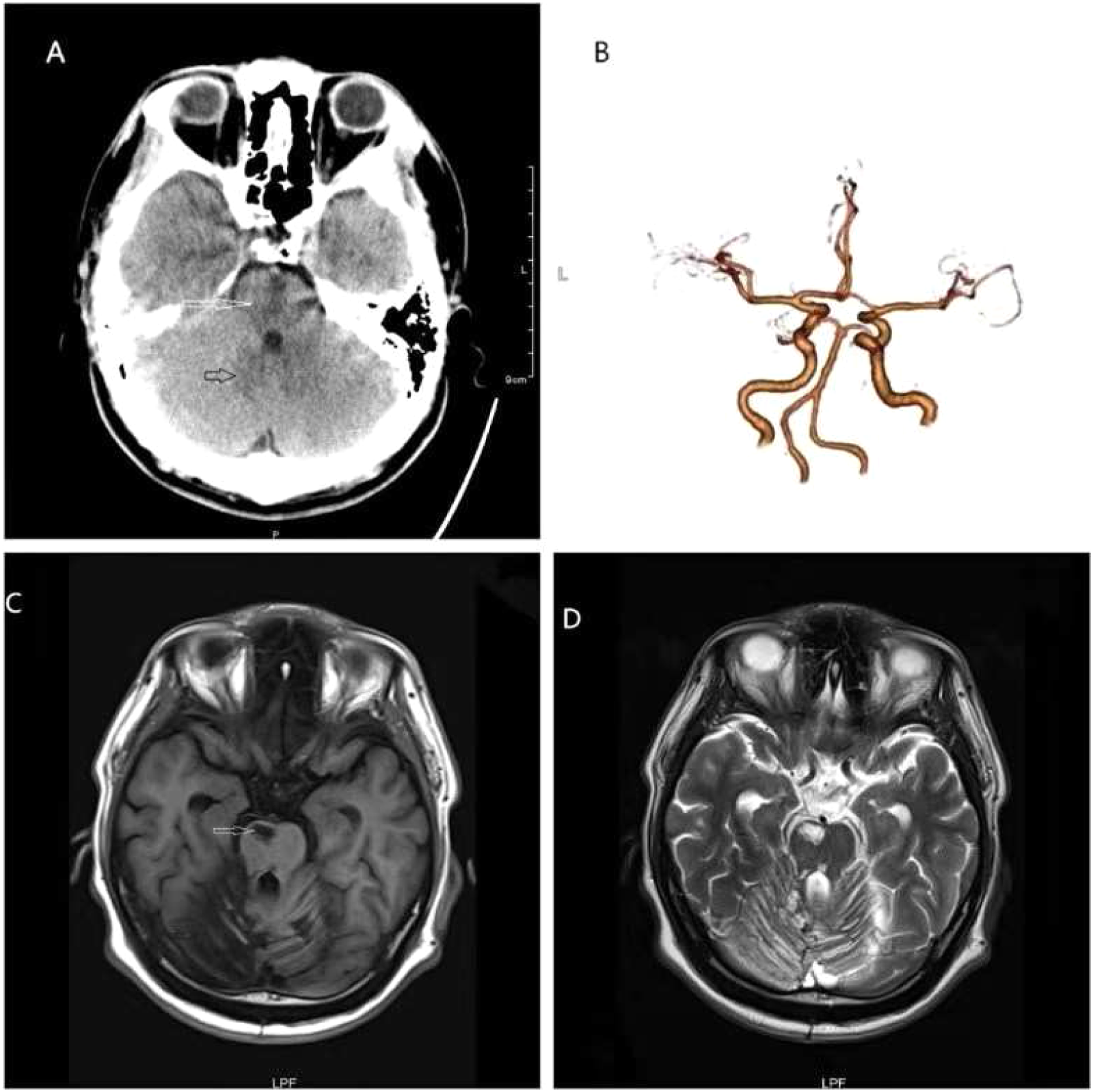
Discussion and Conclusion
BAO is a type of ischaemic stroke that usually presents with symptoms of acute impaired consciousness, hemiparesis and cranial nerve dysfunction. 4 Our patient with BAO was also diagnosed with Marfan syndrome after further examination in the hospital. The pathophysiological mechanism underlying the ischaemic stroke in this patient with Marfan syndrome is unclear. Wytik showed that cardiogenic embolism was found in most Marfan patients with ischaemic stroke, although the association with arterial dissection was not significant. 5 Maski reported a rare spontaneous intracranial artery dissection with cerebral infarction in a young Marfan syndrome patient and suggested that the infarction was due to the cerebral vasculature itself. 6 In this case, the patient had no evidence of atherosclerosis, cerebral aneurysm, cerebral arterial dissection or other known risk factors. Transthoracic ultrasound showed an aortic root aneurysm and found no emboli of cardiac origin. Aortic root aneurysm is characterized by slower blood flow and endothelial dysfunction, which cause thrombi to form easily, and aortic root size is an independent determinant of larger stroke volume. 7 Therefore, we speculate that the emboli of the basilar artery might originate from aortic root aneurysm.
Basilar artery occlusion stroke is known to have bad outcome with a high rate of mortality. The endovascular therapy has been demonstrated as effective as medical therapy in patients with stroke due to BAO. 8 Alemseged also showed that endovascular revascularization is associated with a favourable outcome in patients with BAO with good collaterals and less extensive occlusion, even >6 hours after onset. 9 In our case, endovascular therapy was administered to the patient and resulted in favourable clinical outcomes. To our knowledge, few Marfan syndrome patients treated with recanalization therapy have been previously reported in the literature. Elia reported one Marfan syndrome patient with chronic aortic dissection undergoing thrombolysis for acute ischaemic stroke with a near-complete recovery of neurological deficit and favourable long-term outcome. 10 Anna Gójska-Grymajło also reported that a young Marfan syndrome patient complicated with brain stem infarction had no complications after intravenous thrombolysis and had a good outcome. 11 To our knowledge, this is the first case report showing that basilar artery recanalization can be achieved safely and with a good clinical outcome in a Marfan syndrome patient using MT with intravenous thrombolysis. This case report may provide some evidence that can serve as a reference when planning therapeutic strategies for BAO patients with Marfan syndrome.
In summary, our case report shows that stroke and embolic phenomenon can occur in Marfan patient, and emboli may be from an aortic root thromboembolic source. Endovascular therapy may be safe and with a good clinical outcome in a Marfan syndrome patient with BAO. However, further studies are needed to evaluate the safety and efficacy in a patient with acute ischaemic stroke of Marfan syndrome.
Footnotes
Declaration of Conflicting Interests
The author(s) declared no potential conflicts of interest with respect to the research, authorship, and/or publication of this article.
Funding
The author(s) received no financial support for the research, authorship, and/or publication of this article.
Authors’ Note
Informed consent has been obtained from the patient for publication of the case report and accompanying images.
