






Abstract
Anxiety disorders are highly prevalent in patients with alcohol use disorder. The purpose of the present study was to examine the neural correlates of behavioral inhibition in alcohol-dependent patients (ICD-10: F 10.2), and in healthy controls and to determine the influence of anxiety on these processes. Therefore, behavioral responses (reaction times; error rates) and event-related potentials of 16 patients with alcohol dependence syndrome and 16 age-and gender-matched healthy controls were recorded while the participants performed an auditory go/no-go task. The patient group was stratified according to their self-rated trait anxiety (STAI) with scores above and below median. We hypothesized that patients suffering from alcohol dependence would show reduced no-go P3 amplitudes involved in response inhibition compared to healthy subjects. In patients with alcoholism and high trait anxiety the decline of no-go P3 amplitudes was expected to be less distinct.
The estimation of effect size based on the reaction times of patients with high and low anxiety ratings revealed a cohen's d of 0.61 indicating a small effect. High trait anxiety ratings were also associated with slightly enhanced no-go P3 amplitudes in central brain regions (Mean no-go P3 amplitude at Cz: 10.43 μV) compared to patients with low anxiety scores (Mean 8.98 μV). The effect size (cohen's d) revealed a small effect. Using the Mann-Whitney-U-test for independent samples of the comparison of high- and low-anxious patients, however, did not reveal any significant differences concerning no-go P3 amplitudes. Patients with alcohol use disorder and healthy controls did not differ significantly with regard to reaction time, error rate and no-go P3 amplitudes.
This study suggests that no-go P3 amplitudes in patients with alcohol use disorder might be affected to some degree by habitual anxiety. The results emphasize the importance of monitoring trait anxiety in studies regarding cognitive functions in subjects with alcohol use disorder.
INTRODUCTION
Alcoholism may be associated with a range of cognitive dysfunctions 1 including impaired executive functions, reduced verbal, mnestic, perceptual-motor, and visual-spatial abilities. 2,3 Cognitive dysfunctions in psychiatric diseases are frequently associated with electrophysiological anomalies. 4 There are numerous reports examining electrophysiological deficits in patients with alcohol use disorder, mostly focusing on attention processes and context updating capacities. Studies have found that the electrophysiological responses to target stimuli are of significant lower voltage in abstinent alcoholics than in nonalcoholics. 5,6 However, not all studies were able to replicate these findings. 7
Epidemiological studies revealed high prevalence rates of mental disorders in persons with alcohol use disorder. 3 A comorbid anxiety disorder seems to be present in about 9–30%, and a comorbid affective disorder in about 6–24% of all subjects with alcohol use disorder. 9 Anxiety appears to be linked to larger neurophysiological responses: auditory P300 amplitudes were enhanced in normal volunteers exposed to anxiety provoking situations 10 as well as in anxious individuals. 11 A study with college students indicated that subjects with higher general anxiety and worry have enhanced evoked potentials compared with phobic and nonanxious control subjects during a cognitive task. 12 In addition, threat-related faces elicited faster latencies and greater amplitude of early ERP components in highly anxious individuals than in low-anxious individuals. 13 Another study reported on higher expectancy or vigilance towards negative stimuli in subjects with high state anxiety scores or high combined state and trait anxiety scores. 14 The authors concluded that this may indicate the presence of valence-related vigilance biases in anxiety.
At least some cognitive disturbances which often are observed in patients with alcohol use disorder are also common in patients with affective disorders and anxiety, e.g., executive deficits. 15,16 One major component of executive control is the ability to inhibit behavior or responses that are inappropriate in the current context. 17 Response inhibition, as assessed through go/no-go tasks, can be defined as the act of withholding or terminating a behavioral response and is considered to be governed by a cognitive inhibitory process. 17 The go/no-go paradigm has been widely used to assess impulsivity and control processes. 6,18 The tasks require the subjects to respond to one type of stimuli (go condition) but to withhold the response to the other (no-go condition). The go task seems to represent response execution processes. The no-go condition includes the active inhibition of the prepared response. 6
Electrophysiologically, the response inhibition process seems to be associated with a negative deflection, which reaches a frontocentral maximum about 200 ms after the presentation of the stimuli. 18 More recently, it has been shown that N2 is enhanced by the necessity to respond to low-frequency stimuli, regardless of which kind of response was required. 19 Based on these results the authors concluded that N2 reflects response conflict rather than inhibitory control. Another ERP component related to no-go tasks, P3, is a positive peak which is observed around 300–600 ms after stimulus onset, the so-called no-go P3. 6 There is evidence that no-go P3 is related to response inhibition. 20
Several studies have demonstrated that a mild dose of alcohol selectively reduces the ability of drinkers to inhibit their behavior while leaving unaffected their ability to activate behavior. 21 These findings support the assumption that behavioral inhibition is particularly sensitive to the impairing effects of alcohol. In addition, patients with alcohol use disorder often show difficulties with tasks in which inhibitory capacities are critical. 22 These cognitive deficits are often combined with electrophysiological abnormalities, e.g., a reduced no-go P3 potential. 5,6 Furthermore, there is evidence that patients activate inappropriate brain circuitry during cognitive processing. 6
Neurobiological anomalies associated with alcoholism may also be influenced by comorbid disorders and may in fact be a marker for a subtype of alcoholism respectively. 23 Hill and colleagues 7 found that female alcoholics showed reduced P300 amplitudes when a comorbid lifetime diagnosis of depression was present. Male alcoholics and female patients without affective disorder did not show any P300 variations. Bauer et al. 24 could not replicate these results: analysis of alcoholics with a high depression level compared to a low depression level did not reveal significant P300 differences between these groups. This study indicated that anxiety did influence ERPs: women reporting high anxiety showed significantly smaller P3 amplitudes than nonanxious alcoholics. A study of Enoch and colleagues 25 showed reduced P300 amplitudes in patients with alcohol use disorder and enhanced amplitudes in patients with lifetime anxiety disorder. In patients with comorbid alcohol use and anxiety disorder P300 amplitudes were also reduced. At present the results concerning the influence of anxiety on cognitive functioning in alcohol use disorder are still rather inconsistent.
The purpose of the present study was to examine the neural correlates of behavioral inhibition in patients suffering from alcohol dependence and in healthy controls and to determine the influence of anxiety on these processes. We recorded behavioral responses and event-related potentials (ERPs) while the participants performed a go/no-go task. We hypothesized that patients suffering from alcohol dependence would show reduced no-go P3 amplitudes involved in response inhibition compared to healthy subjects. In patients with alcoholism and high trait anxiety the decline of no-go P3 amplitudes was expected to be less distinct. Referring to the response conflict hypothesis 19 we did not expect to provoke a no-go-related N2 because go and no-go trials were presented equally often.
MATERIAL AND METHODS
Subjects
Sixteen detoxified male alcoholics (ICD-10: 10.2) participated in the study, ranging in age from 34 to 56 years. All alcoholic patients were inpatients at the Department of Psychiatry and Psychotherapy, Ludwig-Maximilians-University of Munich. Exclusion criteria were stroke, head trauma, anoxia, encephalitis, learning disability, polysubstance abuse, and comorbid neurological or psychiatric disorder (except depression). The average duration of abstinence was 12.6 days. Three patients were on medication: two on antidepressive medication (selective serotonine reuptake inhibitor), the other patient on anticonvulsive drugs. The results were compared to those of healthy controls (N = 16; mean age = 39.3 years; range: 30 to 55 years) with no known history of neurologic and psychiatric disorder or hearing problems. Three patients and two healthy subjects, who were supposed to participate in the study, did not understand the instructions of the go/no-go paradigm and consequently were not included in any analysis. The two groups were matched for age and verbal intelligence. 26 The years of education differed significantly between the two groups with significantly higher education in the control group compared to the patient group. Fifteen of the participating patients were right-handed, 1 left-handed. In the control group, 14 healthy controls were right-handed, 2 left-handed. The handedness was classified according to a modified version of the Edinburgh Inventory of Handedness. 27 Present state and trait anxiety was assessed by means of the state-trait anxiety inventory (STAI-X 28 ). Anger was assessed using the state trait anger expression inventory (STAXI 29 ). Furthermore, the subjects were asked to fulfill the Beck Depression Inventory (BDI 30 ). A written informed consent was obtained from each participant before the participation. The study was approved by the local ethical committee. Each healthy volunteer was paid € 25 for participating in the study.
In patients compared to controls clinical scales revealed enhanced depression scores as well as state and trait anxiety ratings. The overall anger ratings and anger control scores did not differ significantly from group to group. The demographic and clinical characteristics of the sample are presented in Table 1.
Demographic and clinical characteristics of the sample
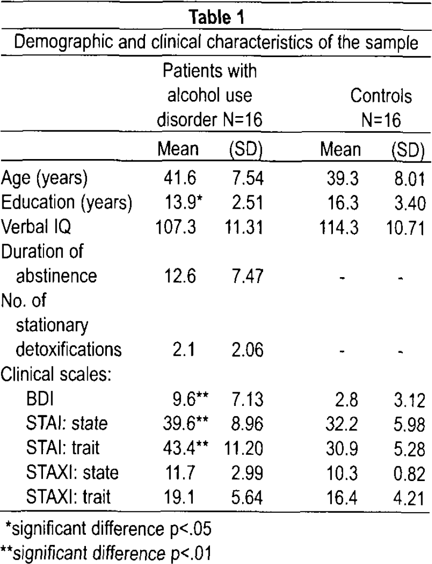
significant difference p<.05
significant difference p<.01
The trait anxiety ratings of patients significantly exceeded the means for the corresponding age group of the normative sample (Mean 34.61 / STD 8.91). To determine the influence of habitual anxiety on inhibitory performance in alcohol use disorder, the patients were assigned to different groups according to their ratings on the STAI trait based on median split in one group comprising patients with high self-rated habitual anxiety (STAI > 43; Mean 52.8 / STD 5.01; alc/anx+) versus another group comprising patients with low scores on this scale (STAI ≤41; Mean 34.0 / STD 6.50; alc/anx-).
The alcoholic patients with high (alc/anx+) and low trait anxiety (alc/anx-) scores were comparable regarding their age (alc/anx+: Mean 39.4 / STD 7.31; alc/anx-: Mean 43.8 / STD 7.57; Z = −1.59; p  .113), years of education (alc/anx+: Mean 13.9 / STD 2.71; alc/anx-: Mean 13.9 / STD 2.49; Z
  -.27; p = .791), verbal IQ (alc/anx+: Mean 106.6 / STD 9.40; alc/anx-: Mean 108.2 / STD 15.09; Z = -.29; p
  .774), duration of abstinence (alc/anx+: Mean 13.9 / STD 8.03; alc/anx-: Mean 11.4 / STD 7.19), and number of stationary detoxifications (alc/anx+: Mean 2.9 / STD 2.36; alc/anx-: Mean 1.4 / STD 1.51).
Experimental Procedures: Go/No-Go Paradigm
The subjects participated in a cued go/no-go paradigm (see Figure 1). The auditory stimuli consisted of sinus tonus (duration of 40 ms, pressure level: 100 dB) of three differential pitches delivered binaurally via headphones.
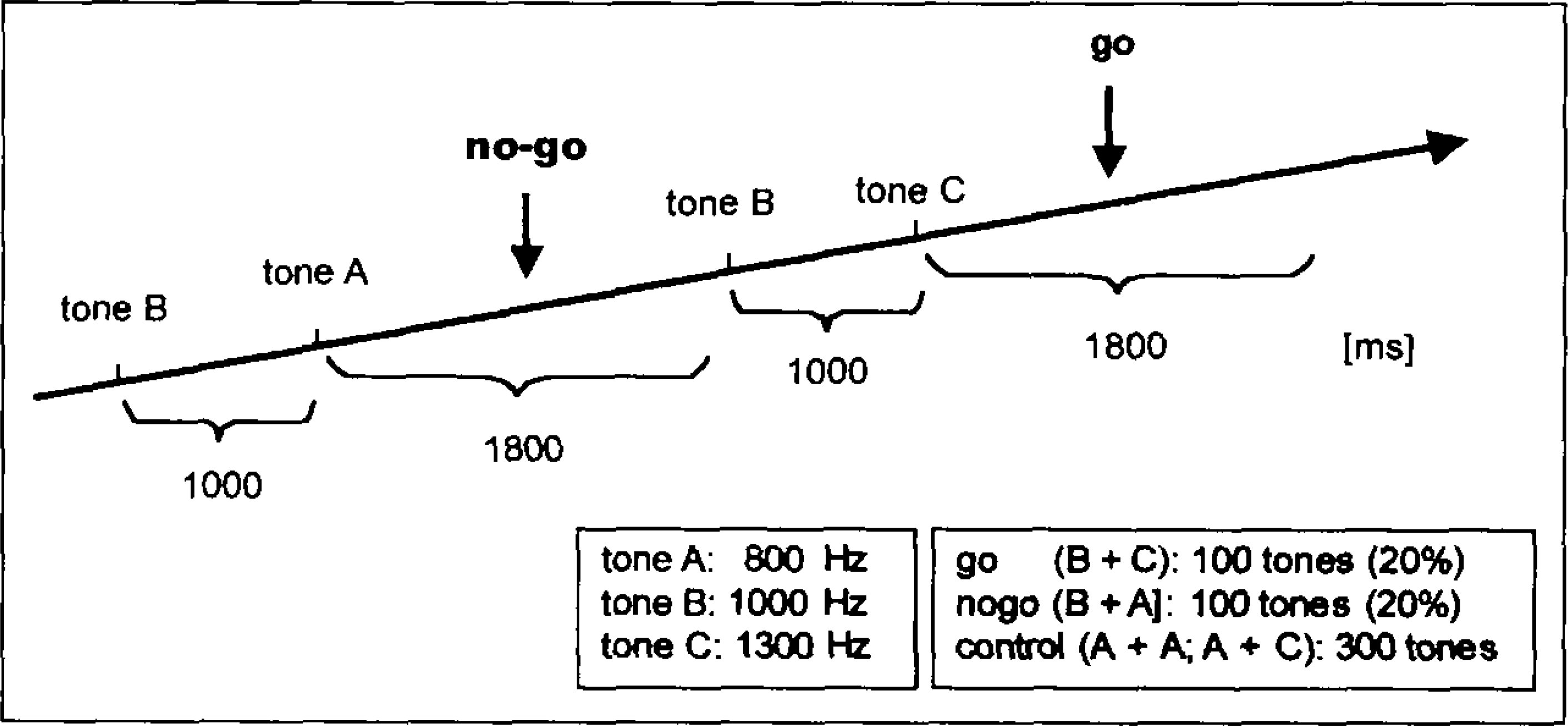
Auditory go/no-go paradigm.
The tones were presented in pairs at intervals of 1000 ms. The tone with the middle frequency served as cue indicating that a button press was required when it was directly followed by the tone with the high frequency (go condition). The prepared behavioral response was to be inhibited if the cue was followed by the tone with a low frequency (no-go condition). Furthermore, there were two control conditions starting with the low frequency tone indicating that no response was required. The conditions were presented in pseudo-randomized order. All conditions were presented 100 times with an interstimulus interval of 3 to 6 seconds. One experimental run took about 25 min.
Analysis of Behavioral Data and EEG
Reaction times, errors of omission and commission were computed. Any response delayed by more than 650 ms after the stimulus was counted as error.
The evoked potentials were recorded in the context of a project concerning simultaneous acquisition of EEG and fMRI. Sixty-one electrodes were placed on the scalp according to the International 10–10 System, all referring to Cz (sampling rate: 5000 Hz). Eye movements were recorded from a channel placed beneath the right eye. Impedances were maintained below 10 kΩ. The EEGs were acquired with an amplifier unable to saturate by MR activity (Brain Products, Munich). Eye movements and eye blinks as well as cardioballistic artifacts were excluded using the automatic artifact correction of the BESA 5 soft ware package (MEGIS Software GmbH, Gräfelfing, Germany). Further analyses were done with Analyzer Software (Brain Products, Munich). The data were re-referenced to an average reference, filtered with a 30 Hz low-pass filter (slope 24 dB/oct), segmented separately for the different conditions (go; no-go; control; sampling epoch: 750 ms; baseline: 100 ms), and baseline corrected. Epochs containing artifacts (amplitude higher than ± 90 μV) were rejected. The artifact detection was done on Fz, FCz, Cz, and Pz. Three participants' data (1 alcohol patient and 2 control subjects) with excessive artifacts (less than 30 trials remaining after artifact rejection) were dropped from the analyses. One EEG of a healthy control was lacking due to technical problems during acquisition. The no-go P3 was defined as the largest relative maximum of the ERP at electrode Fz, FCz, and Cz in the search window of 230 to 450 ms after the presentation of the respective stimuli.
Statistics were obtained using the routines in the SPSS 14.0.1 program. A t-test for independent groups was used in order to compare the no-go P3 amplitudes across patients and the control group and the behavioral data, respectively. We employed the Mann-Whitney-U-test for the comparison of no-go P3 amplitudes, reaction times and error rates of patients with high and low trait anxiety. The significance level was 0.05, p-values between 0.05 and 0.1 were marked as a trend. The magnitude of effect independent of sample size was measured by calculating cohen's d.
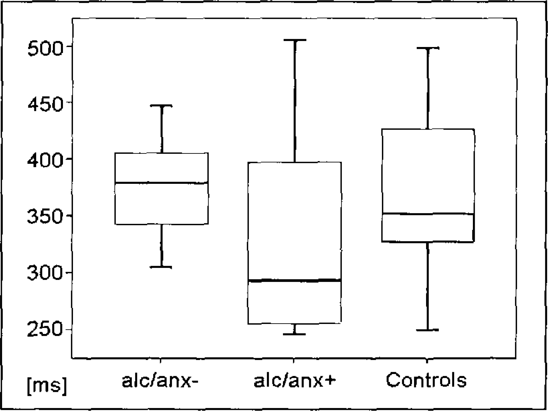
Reaction times for control group and patients with alcohol use disorder and low (alc/anx-) and high (alc/anx+) STAI trait ratings.
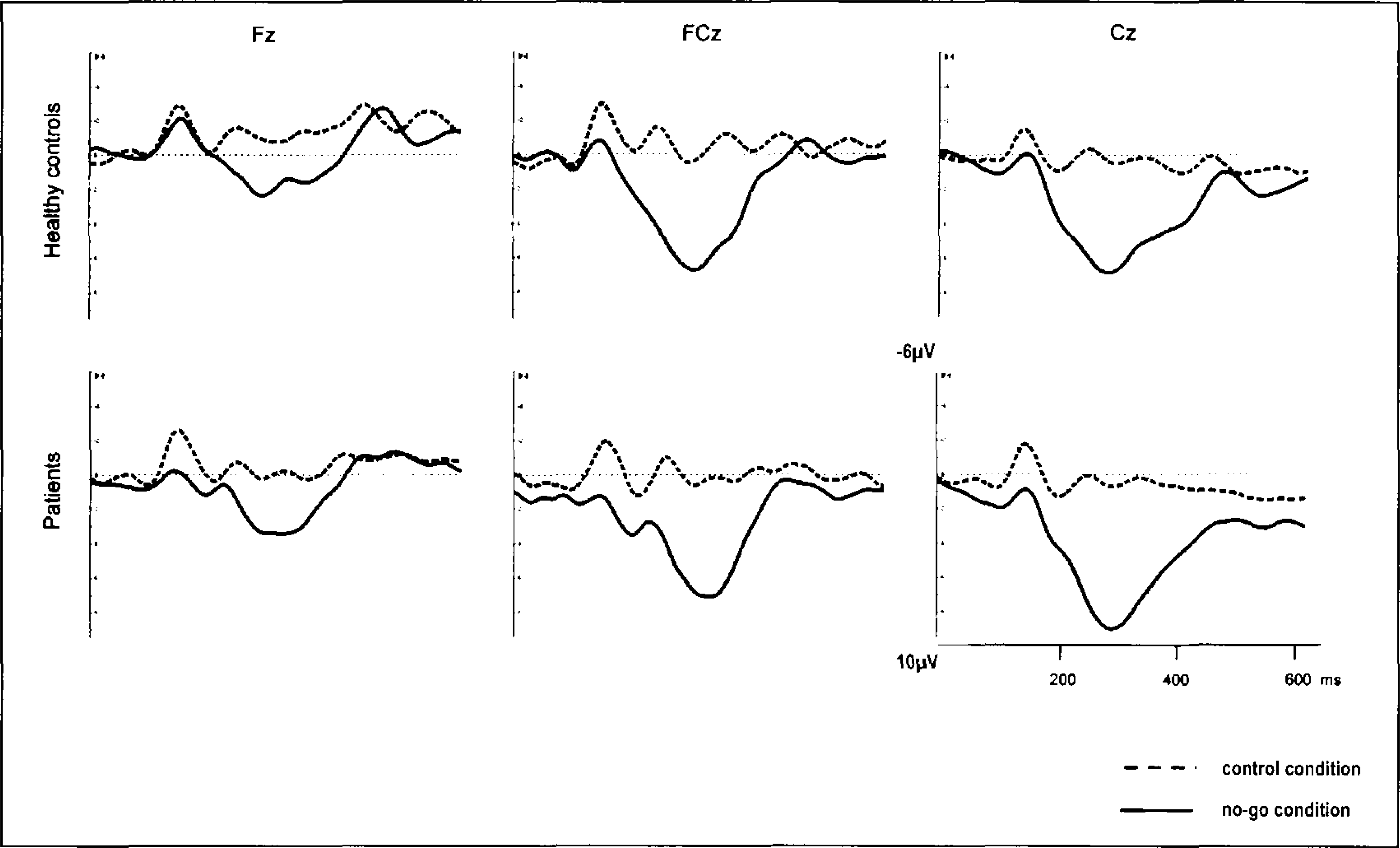
ERP waveform for control group and patients with alcohol use disorder at frontal and central electrode positions. Grand mean waveforms for control condition (dotted line) and no-go trials (black line).
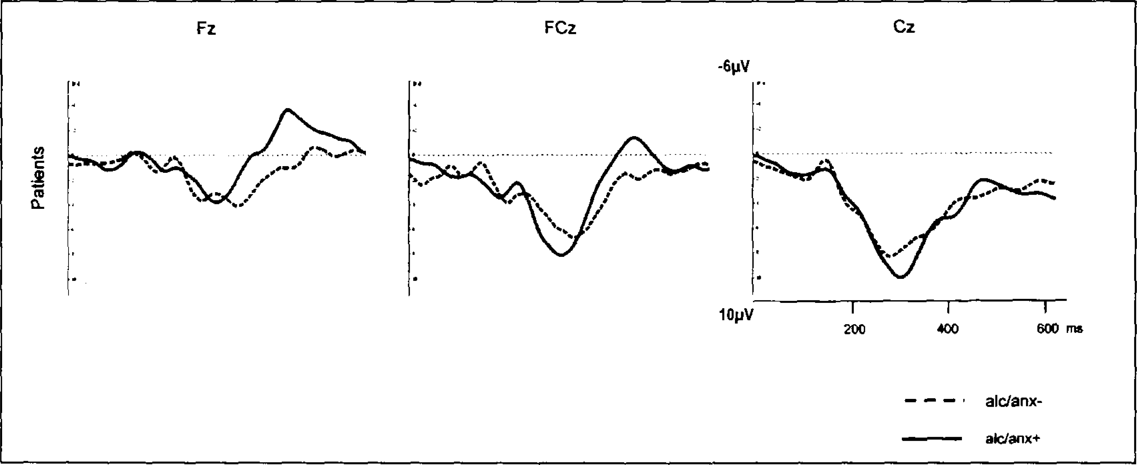
ERP waveform for no-go condition for patients with alcohol use disorder and low (alc/anx-) and high (alc/anx+) STAI trait ratings at frontal and central electrode positions.
RESULTS
Behavioral performance
The average reaction times for the go trials were 370.2 ms (STD 79.10) and 352.9 ms (STD 75.26) for the control group and the alcohol addicts, respectively. The groups showed no significant difference with respect to reaction time (T  -.625, p
  .537). We detected no significant differences between the two groups with regard to omission error rate (T
  -.394, p
  .697) and commission error rate (T
  .394, p
  .697).
Patients with low trait anxiety ratings showed slightly decelerated reaction times (Mean 375.5 ms / STD 45.37) in comparison to patients with high STAI trait scores (Mean 330.3 ms / STD 94.38), although the difference was not significant (Z = −1.26, p = .208; see Figure 2). The cohen's d (d = 0.61) effect size was 0.29. An effect size of 0.29 indicates a small effect. 31
ERP data of patients and the control group
The statistical analysis of no-go P3 amplitudes did not reveal significant group differences between alcohol patients and healthy controls at Fz (patients: Mean 5.52 μV / STD 2.4 μV; healthy controls: Mean 4.26 μV / 2.5 μV; T = 1.395, p  .174), FCz (patients: Mean 8.70 μV / STD 5.3 μV; healthy controls: Mean 8.83 μV / STD 4.3 μV; T = -.070, p
  .944), and Cz (patients: Mean 9.66 μV/STD 4.4 μV; healthy controls: Mean 8.19 μV / STD 4.9 μV; T
  .855, p = .400), respectively. However, the patient group exhibited slightly enhanced no-go P3 amplitudes at Fz and Cz (see Figure 3).
The comparison between alcohol patients with low trait anxiety rates (mean no-go P3 amplitude at Fz: Mean 5.90 μV / STD 2.0 μV, at FCz: Mean 8.47 μV / STD 4.63 μV, and at Cz: Mean 8.98 μV / STD 3.80 μV) and patients with high STAI trait-ratings (mean no-go P3 amplitude at Fz: Mean 5.10 μV / STD 3.0 μV, Z  -.46, p = .643, at FCz: Mean 8.97 μV / STD 6.33 μV, Z
  -.23, p
  .817, and at Cz: Mean 10.43 μV / STD 4.95 μV, Z
  -.81, p = .418) revealed a slightly enhanced contribution of central areas in patients with alcohol use disorder as well as high self-rated trait anxiety (see Figure 4). Although the no-go P300 in alc/anx+ in Cz was about 1.5 μV higher than in alc/anx-the differences failed to be significant. The cohen's d (d = 0.33) effect size was 0.16. An effect size of 0.16 can be interpreted as a small effect.
31
-.81, p = .418) revealed a slightly enhanced contribution of central areas in patients with alcohol use disorder as well as high self-rated trait anxiety (see Figure 4). Although the no-go P300 in alc/anx+ in Cz was about 1.5 μV higher than in alc/anx-the differences failed to be significant. The cohen's d (d = 0.33) effect size was 0.16. An effect size of 0.16 can be interpreted as a small effect.
31
DISCUSSION
In this study we attempted to examine the possible influence of anxiety on the ability to inhibit behavioral responses in patients suffering from alcohol disorder. Therefore, the entire patient group was split into two subgroups with high (N = 8) and low trait anxiety (N = 8), respectively. The results showed that the no-go P3 amplitudes were enhanced in patients with alcohol use disorder and high self-rated habitual anxiety compared to alcoholics with low trait anxiety. The average amplitudes were about 1.5 μV smaller in patients with low anxiety ratings than those with high STAI trait scores indicating a small effect (cohen's d). However, using a Mann-Whitney-U-test the difference failed to be significant due to the small sample size. In accordance with these results, we also found differences regarding behavioral performance: alcohol addicts with high trait anxiety tended to respond faster than patients with low trait anxiety ratings.
The results are in line with reports about enhanced electrophysiological responses in high-anxious subjects compared to nonanxious controls. 11,12,14 In addition, larger electrophysiological responses have been found during the presentation of disorder-associated stimuli or negative stimuli in phobic subjects 32 and panic disorder. 33 The results have been interpreted as indicator that the processing of personal fear-associated stimuli significantly induced negative affect and increased arousal in phobic patients. However, other studies reported a reduced electrophysiological reactivity compared to controls in patients with anxiety disorders. 34
Enoch and colleagues 25 produced reduced P300 amplitudes in patients with alcohol use disorder and enhanced amplitudes in patients with lifetime anxiety disorder. However, contradictory to our results, they found reduced P300 amplitudes in patients with comorbid alcohol use and anxiety disorder. The reason for the discrepancy might be the difference in definition of anxiety: in the study of Enoch and colleagues 25 psychiatric lifetime diagnoses for anxiety disorders were made according to the DSM-III-R, including phobia, panic, and generalized anxiety disorder. The STAI trait questionnaire we used denotes relatively stable individual differences in anxiety proneness and refers to a general tendency to respond with anxiety to perceived threats in the environment. A precise localization of brain structures underlying altered electrophysiological responses would be possible with a combined acquisition of event-related potentials and BOLD-responses. 35
The comparison of electrophysiological data of patients with alcohol use disorder and controls did not reveal any significant differences between groups concerning no-go P3 amplitudes. Frontocentral reactions during response inhibition even seemed to be slightly more pronounced in patients than healthy subjects. Comparing performance data of alcohol use patients and the control group revealed that groups did not differ regarding reaction times and error rates. The reason for the discrepancy between the present findings and that of former studies appears to be the high anxiety in the alcohol addicts of our study.
There are several caveats to our results. The sample size of participants with alcohol use disorders with high and low trait anxiety, respectively, were small (N  8). For this reason, the results should be considered preliminary. Other factors like comorbid psychiatric disorders might be responsible for discrepant findings and should be carefully considered. Altered P300 amplitudes, for example, have been reported in a number of psychiatric disorders
36,37
and personality traits.
38,39
Hill and colleagues
7
suggested that the comorbid depression might be responsible for the reduced P300 amplitudes in women suffering from alcohol use disorder in their study.
8). For this reason, the results should be considered preliminary. Other factors like comorbid psychiatric disorders might be responsible for discrepant findings and should be carefully considered. Altered P300 amplitudes, for example, have been reported in a number of psychiatric disorders
36,37
and personality traits.
38,39
Hill and colleagues
7
suggested that the comorbid depression might be responsible for the reduced P300 amplitudes in women suffering from alcohol use disorder in their study.
The results emphasize the importance of monitoring trait anxiety in studies regarding cognitive functions in subjects with alcohol use disorder.
Footnotes
ACKNOWLEDGMENT
Parts of this work were prepared in the context of the MD thesis of Evangelos Karamatskos at the Faculty of Medicine, Ludwig-Maximilians-University, Munich.