






Abstract
Reoperations for deteriorated stentless bioprostheses are quite challenging procedures. Calcification of the aortic annulus and of the subcoronary root makes often impossible the removal of the failed valve, living a complex Bentall operation or a high-risk transcatheter aortic valve implantation valve-in-valve procedure as the only options, particularly in cases of small-size prostheses. The Perceval sutureless prosthesis (LivaNova PLC, London, UK) can be a valid alternative for failed stentless valve replacement. We report our experience with 3 complex cases of degenerated Sorin Pericarbon Freedom prosthesis treated successfully by means of Perceval sutureless implantation and demonstrating the reproducibility and the safety of this surgical approach.
Introduction
Stentless aortic bioprostheses are undoubtedly related to excellent hemodynamic performances and to faster and complete left ventricular mass regression, when compared to stented valves; therefore, they can be very helpful in cases of expected patient/prosthesis severe mismatch. 1 Despite these recognized benefits, stentless valves have limited popularity among surgeons, mainly due to their more demanding implantation technique and to the increased risk of reoperation secondary to valve deterioration. Indeed, the finding of a heavily calcified aortic annulus and/or root at the reoperative surgery turns the implant of a new prosthesis into a very high-risk procedure. This problem may occur with small-size stentless prostheses (<25 mm), implanted in subcoronary position, like the Sorin Pericarbon Freedom (SPF) valve. In the case of prosthesis degeneration, a complex Bentall procedure remains the only surgical option, as alternative to a very high-risk and complex transcatheter aortic valve implantation valve-in-valve (TAVI-ViV) approach. A sutureless prosthesis implantation may represent a third and feasible strategy, ensuring excellent results by a fast, straightforward, and low-risk operative procedure. We report a case series of 3 patients who underwent aortic valve redo surgery at our unit between February and October 2016, for the failure of an SPF prosthesis, and were successfully treated with implantation of a LivaNova Perceval sutureless (LPS) prosthesis.
Case Series
Case 1
A 63-year-old male with a body surface area (BSA) 1.84 m2 presented at our outpatient clinic with dyspnea and fatigue, after a recent hospitalization for acute heart failure. The patient had an history of ischemic heart disease, treated by percutaneous coronary intervention, with associated aortic valve disease. In 2008, he had undergone multiple coronary artery bypass grafts (CABG) with arterial conduits associated to aortic valve replacement (AVR) using a size 23 stentless SPF prosthesis. The medical history revealed hepatic cirrhosis HBV related, previous Hodgkin lymphoma, mild-degree chronic renal insufficiency, thrombocytopenia, hyperuricemia, and chronic gastritis. The surgical history consisted of multiple surgical oncological procedures including partial thyroidectomy and total prostatectomy for a carcinoma, a colon polypectomy for adenoma, several cutaneous basal cell carcinomas, and an odontogenic cyst removal. The echocardiographic examination at the time of the reoperation revealed a hypertrophic and moderately enlarged left ventricle with preserved ejection fraction (EF). The stentless aortic prosthesis was partially calcified and severely insufficient; the left cusp was torn and prolapsed into the left ventricular outflow tract (Fig. 1). During resternotomy, an accidental, wide right ventricular tearing required prompt right femoral artery–femoral vein cannulation and emergency extracorporeal circulation (ECC), with effective repair of the lesion. After complete mediastinal dissection and preparation, the aorta was cross-clamped and cold cardioplegic solution administrated. The aortic annulus and the subcoronary root appeared completely calcified, and a wide left prosthetic cusp tearing with partial detachment was present. Next, the 3 prosthetic leaflets were completely removed. After accurate sizing of the calcified valve annulus, a size S LPS prosthesis was implanted. The procedure was uneventful, with aortic cross-clamp time of 56 minutes. The patient was moved from the intensive care unit (ICU) to the ward after 3 days and discharged home on the 14th postoperative day (POD) in good clinical condition. The echocardiographic evaluation obtained at hospital discharge revealed a normally functioning prosthesis with no perivalvular leaks and max/mean transvalvular aortic gradient of 19/10 mmHg. At the X-ray postoperative control, the implanted Perceval prosthesis was well visible (Fig. 2). The last available echocardiographic follow-up showed a normal left ventricle and a mean gradient of 8 mmHg.
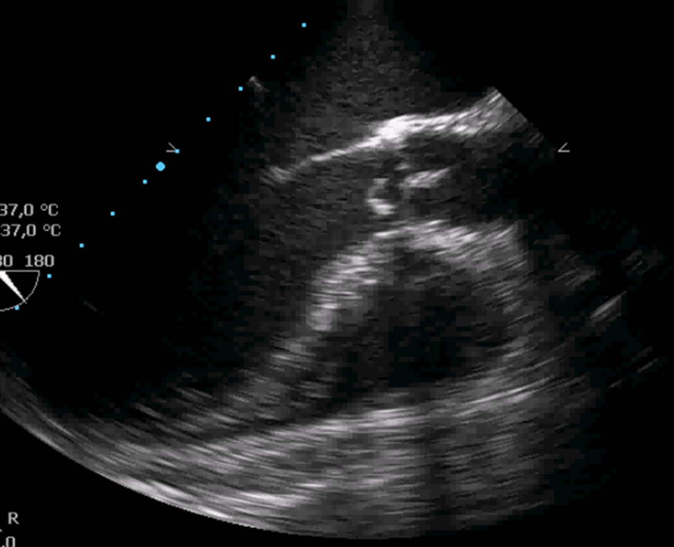
Preoperative echocardiography showing the torn left cusp prolapsing into the left ventricle.
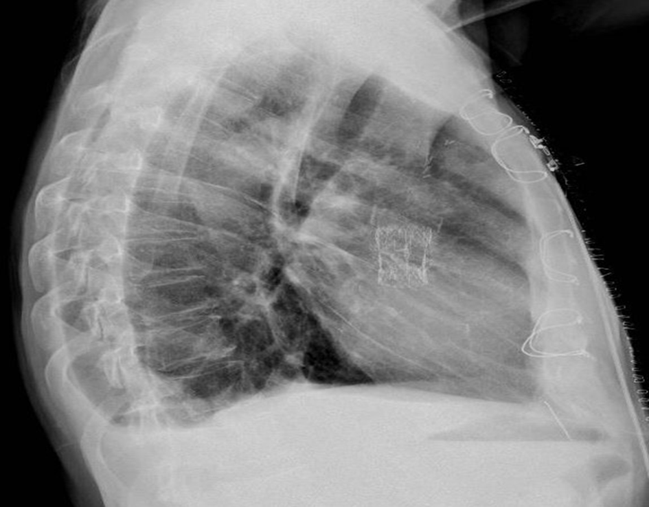
Postoperative X-ray after Perceval implantation.
Case 2
A 68-year-old male, with a BSA 2.03 m2, heavy smoker, with history of arterial hypertension and dyslipidemia, was symptomatic for dyspnea on exertion. In 2005, he had undergone AVR for a severely calcified aortic valve stenosis with a size 25 stentless SPF prosthesis. The surgical history consisted of a cholecystectomy and a partial gastrectomy with neoplasm removal. The echocardiographic examination showed moderate left ventricular hypertrophy with preserved EF. The stentless SPF prosthesis was severely stenotic and moderately insufficient, with extensive annular and cusp calcifications. The redo procedure was uneventful, and after accurate prosthetic cusps removal, a size S LPS prosthesis was successfully implanted with a cross-clamp time of 41 minutes. At the transesophageal echocardiographic control, the LPS valve was well expanded and perfectly positioned, with absence of perivalvular leaks (Fig. 3). After 2 days in the ICU, the patient was moved to the ward and discharged home on the 8th POD in satisfactory clinical condition, with a max/mean transvalvular aortic gradient of 30/22 mmHg at predisharge echographic control. The echocardiographic evaluation at 6-month follow-up revealed a significantly lower max/mean gradient (19/12 mmHg). At the last available follow-up control, 3 years after surgery, the patient was in good clinical status and the mean transvalvular gradient was 10 mmHg.
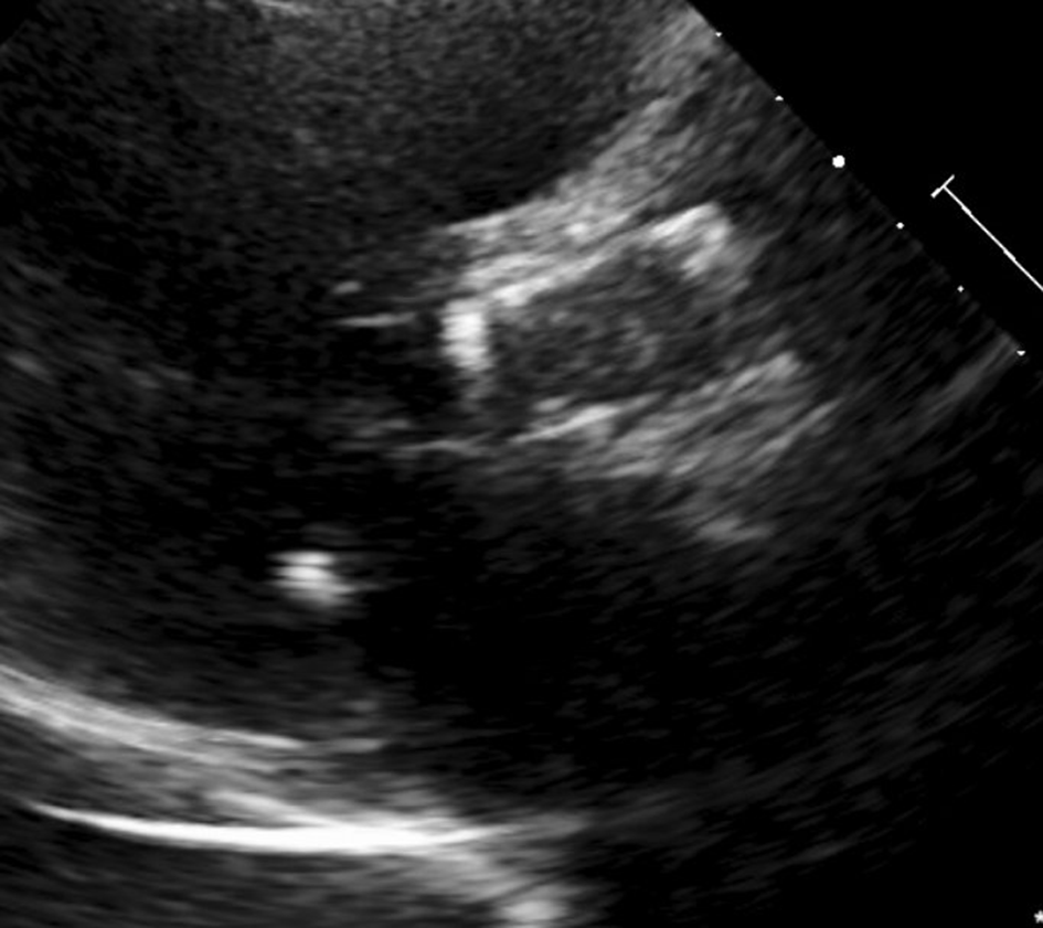
Postoperative transesophageal echocardiography showing the Perceval valve in place.
Case 3
A 75-year-old female, severely obese (BSA 2.27 m2), smoker, hypertensive and dyslipidemic, was severely symptomatic for dyspnea and fatigue (New York Heart Association Class III) . In 2004, she had undergone the implantation of a size 25 stentless SPF prosthesis for severe mixed aortic valve pathology with aortic leaflets fibrosis and retraction. The medical and surgical history consisted of hypothyroidism in medical treatment, monoclonal gammopathy, diabetes, chronic obstructive pulmonary disease, permanent atrial fibrillation, and a previous surgical appendectomy. At the echocardiographic examination, the left ventricle was severely hypertrophic with normal EF. The stentless prosthesis appeared degenerated and calcified with severe stenosis. The coronary angiography revealed a critical stenosis of the left main coronary artery. The patient underwent a CABG on the left anterior descending artery and a prosthetic replacement with a size S LPS prosthesis. A vein graft was used for CABG due to the very poor flow of the small-size mammary artery. As for the 2 cases previously described, the aortic annulus and the root were heavily calcified and of quite small size. The cross-clamp time, prolonged by the complex CABG procedure, was 88 minutes (pure AVR time was 38 minutes). The ICU stay was quite long, mostly due to a prolonged respiratory insufficiency requiring tracheostomy and mechanical ventilation, and a supervening septic state treated with antibiotic therapy. The patient recovered completely after 40 days and was moved to the ward. The following postoperative course was uneventful and she was discharged home in good clinical condition. The echocardiographic findings before discharge revealed a normal functioning LPS prosthesis without leaks and a max/mean gradient of 24/14 mmHg. At the last available follow-up, the patient was asymptomatic and the mean transvalvular gradient was 12 mmHg.
All preoperative, intraoperative, and postoperative patient data are reported in Table 1.
Preoperative, Perioperative, and Postoperative Patient Details.
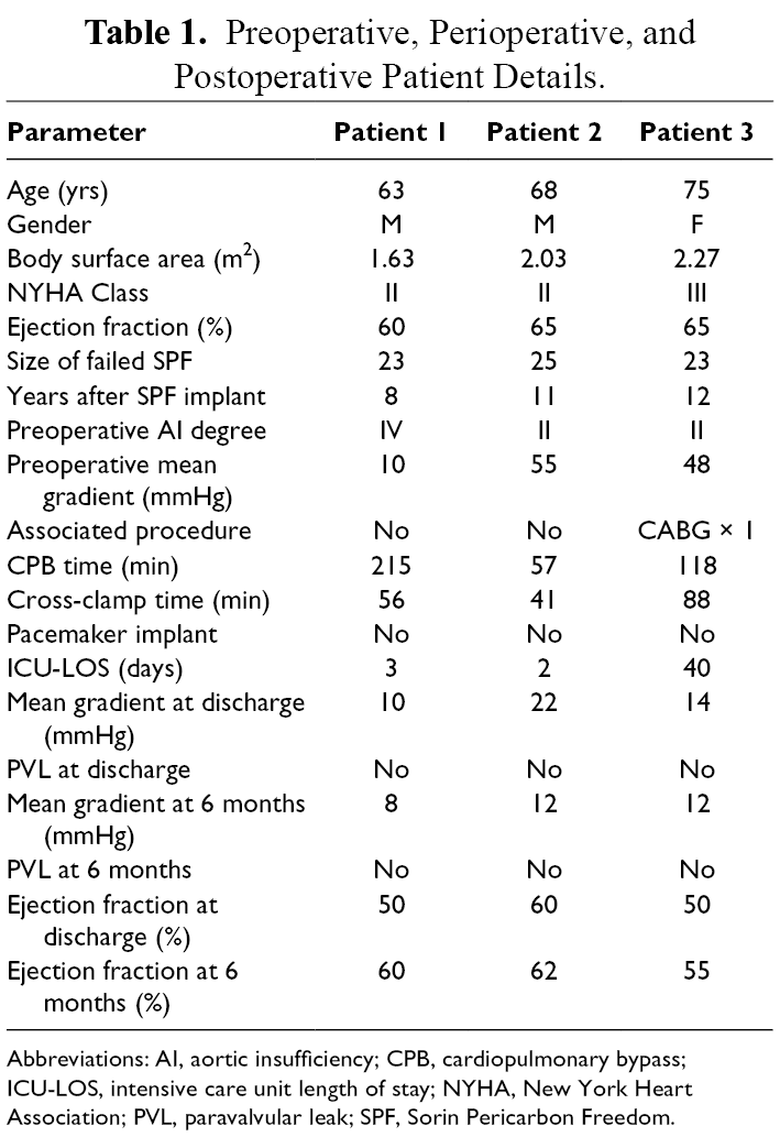
Abbreviations: AI, aortic insufficiency; CPB, cardiopulmonary bypass; ICU-LOS, intensive care unit length of stay; NYHA, New York Heart Association; PVL, paravalvular leak; SPF, Sorin Pericarbon Freedom.
Discussion
Reoperations on stentless aortic prostheses in cases of structural deterioration represent a surgical challenge. 2 This is particularly true in cases of valves requiring a subcoronary implantation, just like the SPF and SPF Solo. Despite the 2 prostheses, SPF and SPF-Solo, share the same valve design, the surgical technique differs, since the SPF is implanted by using a double-suture line and intraannular-subcoronary positioning, while the Solo is fixed in place by a single, supra-annular-subcoronary suture line. Since almost always patients implanted with these 2 devices develop, after several years, extensive calcifications involving the aortic annulus and the aortic root, the removal of the failed prosthesis and the implant of a new one becomes often impossible. The calcification process is further accelerated in the young and active patients, where these valves are often preferred due to their improved hemodynamic performances. 3 Therefore, more stentless valve reoperations will be required in the future. The surgical option in cases of widely calcified aortic root is the Bentall operation, quite often not a simple and straightforward procedure. High mortality rates, ranging from 5% to 12%, have been reported in many series of stentless prostheses reoperations. 4 We have a large experience with SPF stentless valve, with more than 322 implants. 5 Our experience with SPF, consisting entirely of Freedom valves, started in 2003; therefore, we began to see an increasing number of patients reaching the limits of the prosthesis durability. In our experience, the more frequent failure mode of the SPF is a calcified cusp tearing, usually the left, causing a sudden and severe aortic incompetence responsible for a clinical condition of acute heart failure (Fig. 4). In some cases of SPF reoperation it was possible, after the removal of the calcified leaflets (Fig. 5), to implant a new stented bioprosthesis inside the degenerated stentless valve. Those were cases of large-size SPF, usually 27 or 29 mm, with only partially calcified annulus. In smaller sizes, it was quite difficult to implant a stented prosthesis of an adequate size to avoid a severe patient/prosthesis mismatch. In these cases, a sutureless valve maybe very helpful. 6 Since 2016, the LPS prosthesis has become in our practice the ideal solution to treat patients with SPF structural degeneration. Preoperative precise evaluation by echocardiography (Fig. 6) and by computed tomographic (CT) scan (Fig. 7) is mandatory, in order to assess the size of the aortic annulus, aortic root, and sinotubular junction. Indeed, the LPS prosthesis implantation showed to be a low risk and straightforward procedure in most of these cases, allowing for appreciable hemodynamic results (Supplemental Video). Several experiences are reported in the literature of degenerated stentless valves or homografts treated with LPS AVR with excellent results, in the majority of cases. 7 -9 TAVI-ViV procedure has become more recently another surgical alternative for the treatment of degenerated bioprostheses, particularly in high-risk patients with associated severe comorbidities. 10,11 However, while the TAVI-VIV is nowadays a well-established procedure in cases of failed stented prostheses, the risk correlated to this technique in degenerated stentless valves, and particularly in SPF and SPF-Solo, is still elevated. The chance of coronary obstruction by the prosthetic leaflets represents the major drawback of TAVI-VIV in these patients. The global VIV registry reported a 50% risk of coronary obstruction associated with TAVI-VIV in degenerated SPF-Solo valves, much higher when compared to the 3.5% risk reported for overall VIV procedures. 12 Three-dimensional transesophageal echocardiography and multidetector CT scan maybe helpful in the assessment of the aortic root anatomy and of the prosthetic cusp height in relation to the coronary ostia. Based on these evaluations, patients with a failed SPF valve having poorly dilated aortic sinuses and coronary arteries originating very close to the aortic valve annulus should be considered at very high risk for a TAVI-VIV procedure. We have treated 3 patients with malfunctioning SPF valve by TAVI-VIV. Two patients received a successful transapical Edwards Sapien 3 implant, and were both patients with patent coronary grafts. The third patient, with anatomy favorable to TAVI, underwent an uneventful transfemoral implant of a Medtronic Evolut Pro.
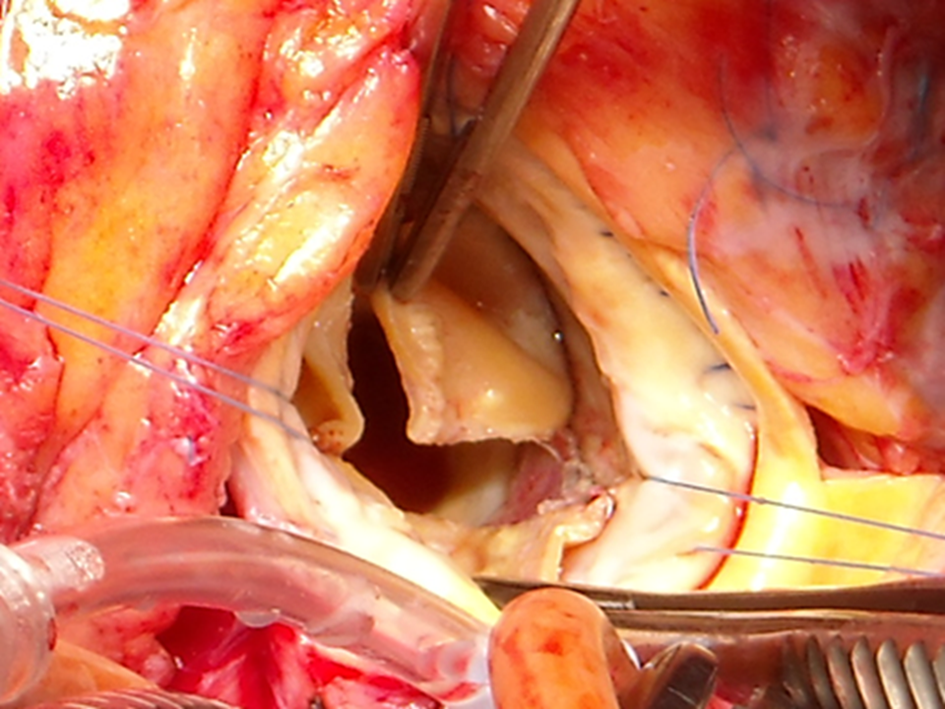
Failed Sorin Pericarbon Freedom stentless valve with detachment of noncoronary and left cusp.
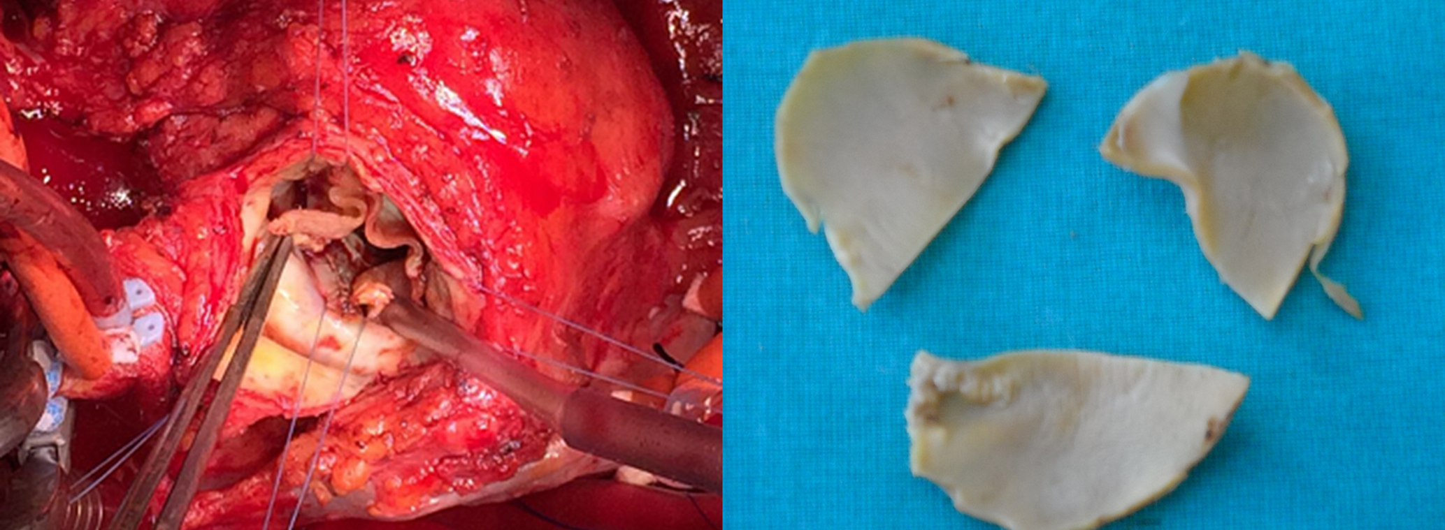
Removed Sorin Pericarbon Freedom leaflets.
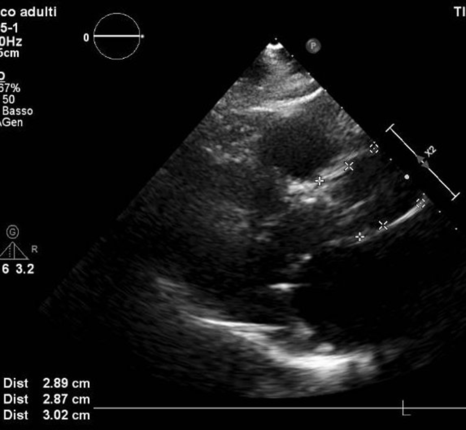
Preoperative echocardiography and sizing of valve annulus, sinuses of Valsalva, and sinotubular junction.
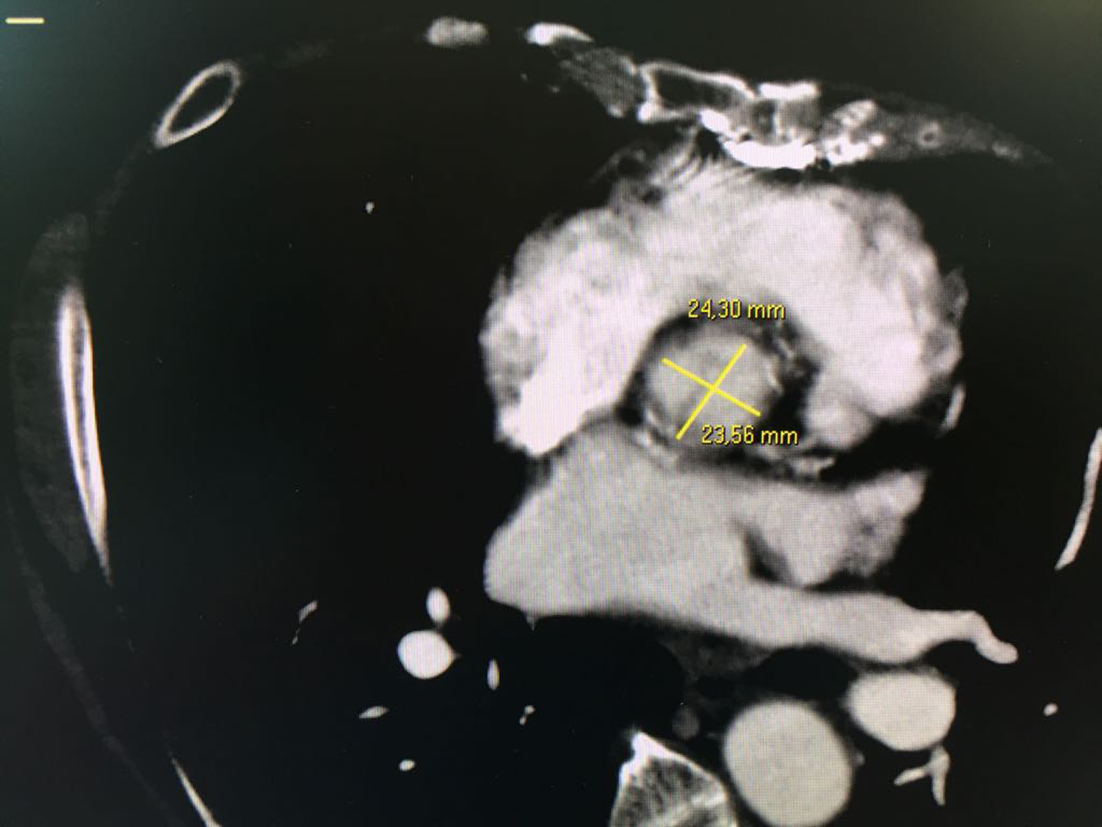
Preoperative computed tomography scan and sizing of the failed prosthetic annulus.
In conclusion, our experience endorses the surgical treatment of patients with degenerated stentless SPF and SPF-Solo valve by LPS prosthesis implantation. The procedure carries a low risk and it is fast and reproducible. The correct sizing of the aortic annulus is mandatory, to avoid inadequate results. TAVI-VIV represents, for this subset of patients, a promising surgical tool alternative to open-heart approach, but it is less easily reproducible, associated with a much higher risk, and requires high level of expertise of the operator.
Supplemental Material
Visual Abstract - Supplemental material for Successful Reoperation in 3 Cases of Failed Sorin Freedom Stentless Aortic Valve Using the LivaNova Perceval Sutureless Prosthesis
Supplemental material, Visual Abstract, for Successful Reoperation in 3 Cases of Failed Sorin Freedom Stentless Aortic Valve Using the LivaNova Perceval Sutureless Prosthesis by Guglielmo Stefanelli, Fabrizio Pirro, Vincenzo Smorto, Emilio Chiurlia and Luca Weltert in Innovations: Technology and Techniques in Cardiothoracic and Vascular Surgery
Footnotes
Declaration of Conflicting Interests
The author(s) declared no potential conflicts of interest with respect to the research, authorship, and/or publication of this article.
Funding
The author(s) received no financial support for the research, authorship, and/or publication of this article.
Supplemental Material
Supplemental material for this article is available online.
References
Supplementary Material
Please find the following supplemental material available below.
For Open Access articles published under a Creative Commons License, all supplemental material carries the same license as the article it is associated with.
For non-Open Access articles published, all supplemental material carries a non-exclusive license, and permission requests for re-use of supplemental material or any part of supplemental material shall be sent directly to the copyright owner as specified in the copyright notice associated with the article.
