
Abstract
Avascular necrosis (AVN), one of the most common therapy-related and debilitating side effects of antileukemic treatment, can adversely affect a patient’s long-term quality of life. Our case study presents a young woman with bilateral elbow AVN and hip AVN after acute lymphoblastic leukemia treatment, with a unique treatment strategy for her elbow pain. The treatment strategy included elbow joint denervation with arthroscopic debridement and distal humerus core decompression. The goal of this procedure was to improve symptomatic pain while retaining bone stock in the distal humerus hopefully allowing better function of the patient’s elbow. This treatment may not only improve the quality of life in a young patient but also delay the need for future surgery. Our patient had improved pain relief in her elbow postsurgery. This procedure may be used for pain control and may have beneficial future implications in this limited population.
Keywords
Introduction
Avascular necrosis (AVN), or osteonecrosis, is a disabling state of bone death and decay usually resulting from a lack of blood flow and ischemic injury. It is estimated that between 10 000 and 20 000 patients are diagnosed each year in the United States alone. 1 Avascular necrosis can range in severity and clinical presentation, from mild pain to complete disability with significant loss of function. Diagnosis is most commonly made by radiographic imaging. 2 Avascular necrosis has been linked to multiple conditions and factors, including chemotherapy, but most commonly is association with corticosteroid use. 1 Avascular necrosis is more common among adults, but has been shown to affect pediatric patients as well. Acute lymphoblastic leukemia (ALL) is a malignancy of hematopoietic stem cells that originates from B- and T-cell lineage lymphoid precursors and is driven by a spectrum of genetic aberrations. 3 Acute lymphoblastic leukemia is the most common pediatric cancer worldwide, and common frontline treatment includes multiagent chemotherapy regimens. While the prognostic values of these treatments are good in the pediatric population, these toxic agents also present with side effects that can be debilitating. Osteonecrosis remains 1 of the most common therapy-related and debilitating side effects of antileukemic treatment which can adversely affect a patient’s long-term quality of life. 4 We present an unusual case of a young woman with bilateral elbow AVN and hip AVN after ALL treatment, with a unique treatment strategy for her elbow pain. While treatment protocols vary, surgical innervations such as elbow joint denervation and core decompression can greatly improve a patient’s quality of life through pain relief. This is the first case reported using these surgical techniques in this unique patient population.
Methods
An 18-year-old Caucasian woman presented to the senior author’s clinic in January 2020. She has a history of ALL for which she was treated on the AALL1131 protocol. She completed her oncologic treatment in June 2018 and has since been in remission. On presentation, she had been diagnosed with AVN of her hips bilaterally, which was being treated conservatively by orthopedic colleagues. She was diagnosed with bilateral elbow osteonecrosis from longstanding chemotherapy and prednisone treatments. Her AVN involved the distal humerus and proximal radius on the left, and only the distal humerus on the right. Symptomatically, she reported pain with extension and forced flexion of her left elbow, with significantly less pain in her right. She was in a wheelchair secondary to her hip AVN and reported pain when trying to push herself up to her walker. The senior hand surgeon conducted a physical examination on initial consultation of her elbows. Her left elbow had a range of motion of 20° to 130° with pain on extension and flexion, no pain with medial elbow stress, and some pain with lateral stress. Her right elbow presented with a range of motion of 10° to 150° without pain.
Standard radiographs and elbow magnetic resonance imaging were reviewed by the hand surgery team. Imaging of the patient’s left elbow revealed AVN of the left distal humerus and left proximal radius, with some articular surface degeneration without subchondral collapse. Imaging of the patient’s right elbow revealed a small focal area of AVN in the distal humerus laterally without subarticular or articular bone changes.
Results
The hand surgery team presented the patient and her parents with treatment plan including left elbow joint denervation with distal humeral core decompression and elbow arthroscopic debridement. The hand surgery team believed the surgery treatment would help alleviate her pain.
In February 2020, the surgical team performed left elbow joint denervation, arthroscopic debridement, and distal humerus core decompression. After a medial incision was performed, the ulnar nerve was identified revealing 2 articular sensory branches which were neurotized. The nerve was decompressed across the elbow leaving it in situ (Figure 1). A lateral incision was made over the elbow. A small sensory articular branch of the median nerve between the brachialis muscle and brachioradialis muscle was identified and cauterized (Figure 2). Elbow arthroscopy was performed through open portals. Significant synovitis was visualized without arthritic cartilage changes. A partial synovectomy was performed. The core decompression was performed through medial and lateral open approaches. The necrotic tissue was debrided, and the medullary canal was packed with bone allograft. The incision was closed, and the patient was placed in a long-arm splint. She had a cast applied 1 week postoperatively for 4 weeks. Therapy was initiated after 5 weeks. At her latest follow-up, she reported 0/10 pain in the elbow but was starting to have left wrist pain. The current plan is for her to follow up in 6 months, while receiving conservative treatment for her other maladies.
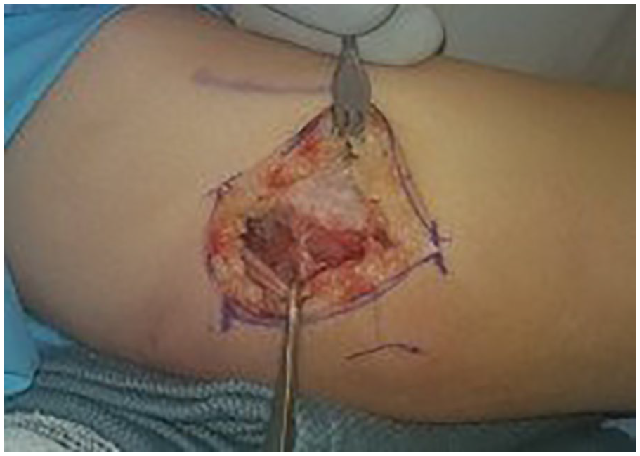
Dissection of the ulnar nerve.
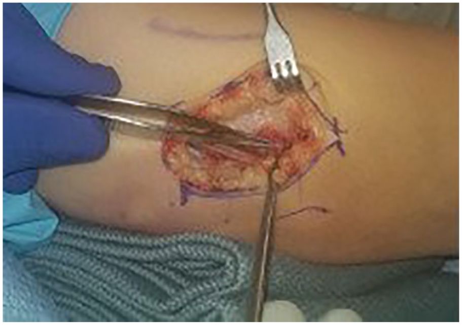
Dissection of capsular branches.
Conclusions
As leukemias are a very common group of malignancies and osteonecrosis is one of the most common side effects of antileukemic treatment, understanding how to both prevent and care for this complication is immensely important. Because AVN often presents asymptomatically or with mild symptoms, early detection is crucial, especially in the pediatric population where there is increased concern regarding lifetime compromise of joint function, adequate skeletal growth and development, and potentially multiple future surgeries. 5 Osteonecrosis therapy remains limited and many current practices primarily focus on its prevention to combat AVN. In adult patients, physical therapy and eventual joint replacement remain the most common therapies for symptomatic osteonecrosis.
Adolescents have been shown to be most at risk for AVN after ALL treatment, and current literature suggests that the underlying pathophysiology for AVN development in adolescents is likely due to age-specific factors ultimately affecting bone morphology, metabolism, and/or nourishment. 4 It is also hypothesized that this could be partially related to increased end-organ susceptibility caused by the increased growth rate and hormonal changes in this period of life. 6 Testosterone, estrogen, growth hormone, and insulin-like growth factor 1 have all been shown to be implicated in these processes. Coagulation is another link to osteonecrosis, and concentrations of procoagulant and anticoagulant factors are modulated and change crucially during growth and their levels differ significantly during adolescence than during adulthood. 1 Glucocorticoids have been heavily linked to osteonecrosis, and are often coadministered during chemotherapy. 7 It has been suggested that reducing steroid exposure during treatment may lower the risk of osteonecrosis and outweighs the cumulative dose as a risk factor for the development of treatment-related osteonecrosis. 8
Due to our patient’s young age and already debilitating medical history and conditions, a treatment was pursued allowing improvement of pain with a better quality of life for this young patient: elbow joint denervation along with core decompression and arthroscopic debridement as a means of pain control. Joint denervation in the setting for the treatment of chemotherapy-induced osteonecrosis has not been previously reported. Joint denervation has previously been reported as a tool for control of chronic elbow pain. Arthroscopic debridement has also been shown to be slightly effective in the management of reported pain from osteoarthritis of the elbow. 9 Core decompression has been used as a technique for AVN of the hips for pain and preservation of bone stock. The combination of these 3 procedures was performed to improve our young patient’s pain and quality of life.
Despite the rate of occurrence, there is no consensus on osteonecrosis management in pediatric patients with ALL. A recent group conducted a systematic review of the various treatments of osteonecrosis in pediatric patients with ALL and identified preventative strategies (such as glucocorticoid dosing and care), nonsurgical, and surgical interventions. 10 Nonsurgical interventions have often been suggested for conservative treatment of AVN, including physical therapy. Bisphosphonates have been used for nonsurgical treatment and data showed that a small number of patients reported symptomatic benefits, but no radiologic benefits. Other studies examined the use of hyperbaric oxygen chambers in the treatment of these patients and patients receiving this therapy reported symptomatic improvement. Surgical interventions described include bone or cartilage stimulating methods, containment-improvement or pressure relieving methods, and joint replacements. Some studies have investigated implantation of autologous osteogenic cells and osteochondral grafting in pediatric patients with ALL and reported favorable outcomes. More firsthand research is needed into these potential therapies for osteonecrosis in patients with ALL. Our patient with ALL reported symptomatic improvement in her elbow, and these surgical methods should be considered in the care of future patients. We believe this procedure should be considered for this unique patient because of its improved pain relief and low morbidity with retaining boney architecture for future options.
Footnotes
Ethical Approval
This study was approved by our institutional review board.
Statement of Human and Animal Rights
All procedures followed were in accordance with the ethical standards of the responsible committee on human experimentation (institutional and national) and with the Helsinki Declaration of 1975, as revised in 2008 (5).
Statement of Informed Consent
Informed consent was obtained from all individual participants included in the study.
Declaration of Conflicting Interests
The author(s) declared no potential conflicts of interest with respect to the research, authorship, and/or publication of this article.
Funding
The author(s) received no financial support for the research, authorship, and/or publication of this article.