





Abstract
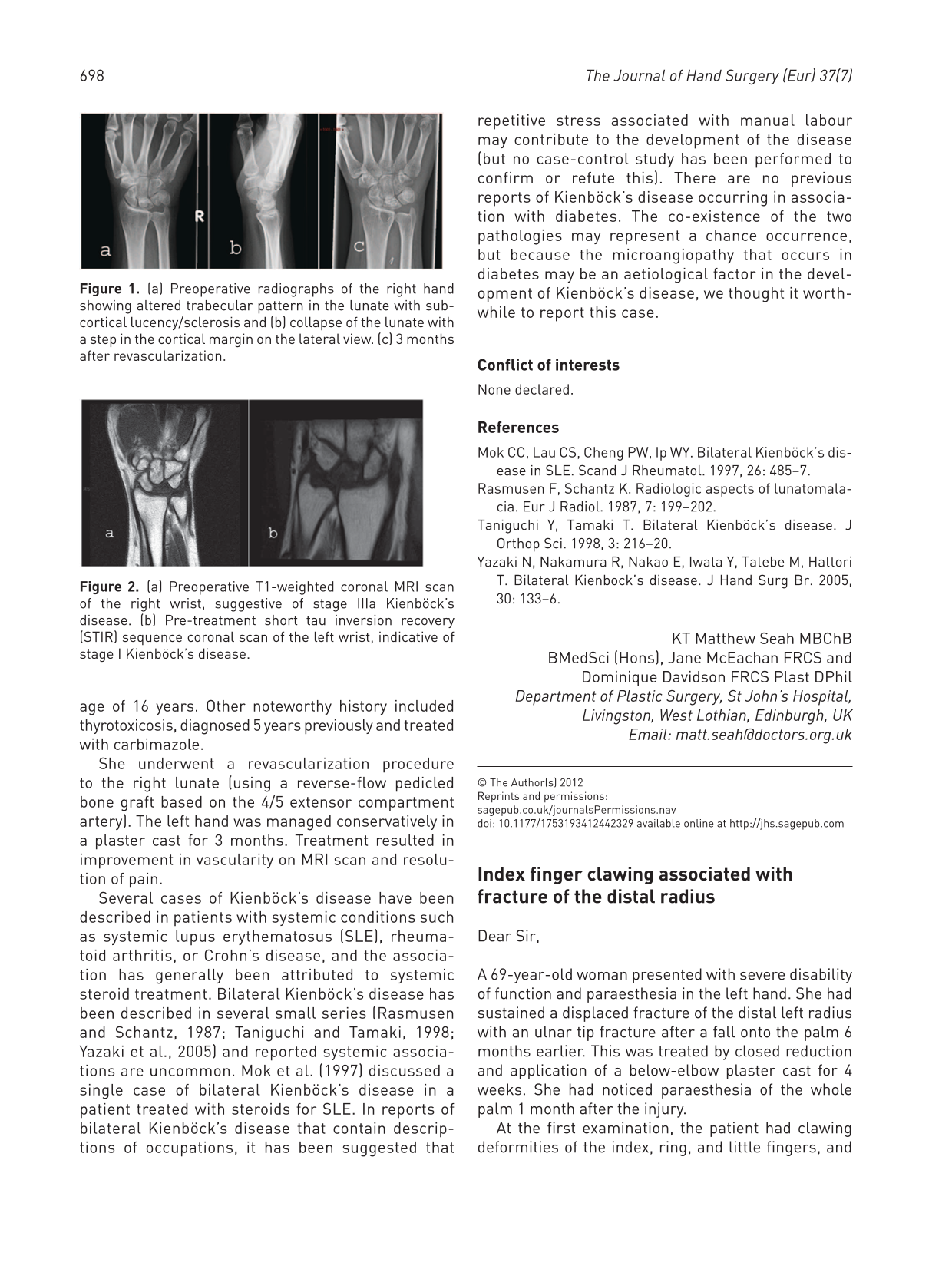
Dear Sir,
A 69-year-old woman presented with severe disability of function and paraesthesia in the left hand. She had sustained a displaced fracture of the distal left radius with an ulnar tip fracture after a fall onto the palm 6 months earlier. This was treated by closed reduction and application of a below-elbow plaster cast for 4 weeks. She had noticed paraesthesia of the whole palm 1 month after the injury.
At the first examination, the patient had clawing deformities of the index, ring, and little fingers, and atrophy of the entire intrinsic musculature (Figure 1a). Although there was a wrist deformity with shortening of the radius and protrusion of the ulnar head, a good range of motion was preserved without pain. All intrinsic muscles were graded less than 2 on the Medical Research Council scale. Tinel’s sign was strongly positive at the entrances of the carpal tunnel and Guyon’s canal. Sensory examination showed severe paraesthesia in the median and ulnar innervated areas. Radiographs showed a malunited deformity of the radius (Figure 1b), and short TI inversion recovery (STIR) -sequence MRI showed a marked change in intensity throughout the intrinsic muscles (Figure 1c).

(a) Severe clawing of the index finger. (b) Radiographs show the malunited deformity before operation. (c) Axial STIR-sequence MRI at the level of the base of the metacarpal bones shows the high intensity change throughout the intrinsic muscles.
We carried out nerve releases and reconstructive tendon transfers during the same operation. The median nerve was firmly bound at the entrance to the carpal tunnel. The ulnar nerve was surrounded by dense fibrous tissue around the distal end of the ulna and was severely stretched at the entrance of Guyon’s canal (Figure 2). We carried out the following multiple tendon transfers to achieve an earlier functional recovery of pinching ability: Camitz procedure using the palmaris longus for the opponensplasty; Neviaser procedure with the abductor pollicis longus elongated by a graft from the opposite palmaris longus tendon for radial deviation of the index finger; and an index flexor digitorum superficialis lasso tendon transfer for correction of the index-clawing deformity. The patient regained thumb opposition and strong pinching ability 6 weeks after the operation, in spite of there being no recovery during electrophysiological examination.
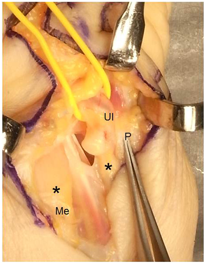
The median nerve (Me) was firmly bound at the proximal edge of the flexor retinaculum. The ulnar nerve (Ul) was severely stretched on the ulnar side at the entrance to Guyon’s canal. * = pseudoneuroma; P = pisiform bone.
A clawing deformity generally occurs in the ring and little fingers, and severe index finger clawing is quite rare. Sensory examination of both median and ulnar nerve-innervated areas, including the middle finger, showed severe paraesthesia in this case. In spite of severe palsies of both nerves, the middle finger did not show a claw deformity. We believe this was because neuronal damage caused by compression neuropathies after the distal radius fracture was not complete and the branch to the second lumbrical muscle might have been be spared, similar to some cases of severe carpal tunnel syndrome (Logigian et al., 1987).
The functional disability from combined median and ulnar nerve palsies is more severe than that of a single nerve palsy, although combined low median and ulnar nerve palsies is a rare complication in fractures of the distal radius (Rychak and Kalenak, 1977). This fracture mostly occurs in elderly people, and recovery from nerve injuries is uncertain in this age group (Matsuzaki et al., 2004). In order to obtain early functional recovery, we believe that combining tendon transfers with nerve decompression to act as internal splints is very useful for severe nerve palsy in elderly patients.
Footnotes
Conflict of interests
None declared.