






Abstract
The Kapandji test is a simple method to score thumb opposition; however, the position of the interphalangeal joint of the thumb during this test has not been described. We aimed to quantitatively examine the effect of the thumb interphalangeal joint position on movements of the trapeziometacarpal joint during thumb opposition using the Kapandji test. The Kapandji test was carried out in 20 healthy participants during thumb interphalangeal joint extension and flexion. Movements of the joints and the activity of thenar muscles were recorded using motion capture and electromyography, respectively. We found that interphalangeal joint extension increased the trapeziometacarpal joint movement and thenar muscle activity compared with interphalangeal joint flexion, which contributed to thumb opposition at Kapandji Positions 0–6. These findings suggest the position of the thumb interphalangeal joint affects the trapeziometacarpal joint during thumb opposition, and assessment of thumb opposition using the Kapandji test is best done with the thumb interphalangeal joint in extension.
Introduction
The functional assessment of the trapeziometacarpal (TM) joint includes the measurement of palmar and radial deviation angle in the thumb metacarpal and also thumb opposition. Thumb opposition is the movement of the thumb pulp to face the other digits for prehension, and the movement of the thumb metacarpal has an important role in thumb opposition (Napier, 1955). It is necessary to use an appropriate method to measure the TM joint movement during thumb opposition (Kuroiwa et al., 2019; Mende and Tonkin, 2021; Tonkin, 2020).
Thumb opposition can be assessed by various methods (Kapandji, 1992; Tonkin, 2020). The Kapandji test records successive positions of thumb opposition (from 1 to 10) and is widely used clinically because of its simplicity (Barakat et al., 2013; Dilokhuttakarn et al., 2017; Kuroiwa et al., 2019; Mende and Tonkin, 2021; Waitayawinyu et al., 2019). However, it has a limitation, in that the position of the thumb interphalangeal (IP) joint can affect the movements of the thumb TM joint (Tonkin, 2020). Although Tonkin (2020) has proposed carrying out the Kapandji test by extending the thumb IP joint, it is unclear whether this method resolves the limitation.
This study aimed to quantitatively examine the effect of the thumb IP joint position on thumb TM joint movements during thumb opposition by analysing movements of the thumb joints and electromyographic (EMG) signals of the thenar muscles.
Methods
Participants
Twenty healthy right-handed adults participated in this study. Their mean age was 23 years (SD 1.3). Ten were male and ten female. None of the participants had any neurological or orthopaedic disorders of the hands. The participants carried out the Kapandji test in the right hand.
The institutional review board (the Ethics Committee of Hiroshima University) approved the experimental protocol used in this study (approval number E604-3), and written informed consent was obtained from all participants. The study was performed in accordance with the guidelines laid down by the Declaration of Helsinki (1964) and its subsequent amendments.
Procedure
First, participants positioned their hand with the thumb in a neutral position and the TM joint was marked for reference (Figure 1) (Li et al., 2008; Lin et al., 2011). Next, they carried out the Kapandji test, from positions 0–10, with the thumb IP joint in extension and flexion. When the IP joint was extended, we instructed participants to touch each Kapandji position with the pulp of the thumb as far as possible across the palm, but not with a ‘crawling’ movement (Tonkin, 2020). When the IP joint was flexed participants were instructed to touch each Kapandji position using the tip of their thumb. These tests were done first with extension of the IP joint, followed by flexion. The wrist joint was in the neutral position throughout the Kapandji scoring procedure. Participants held the thumb on each Kapandji position for at least 3 seconds and performed three trials for each position.
Marker displacements and the reference position of the trapeziometacarpal joint of the thumb.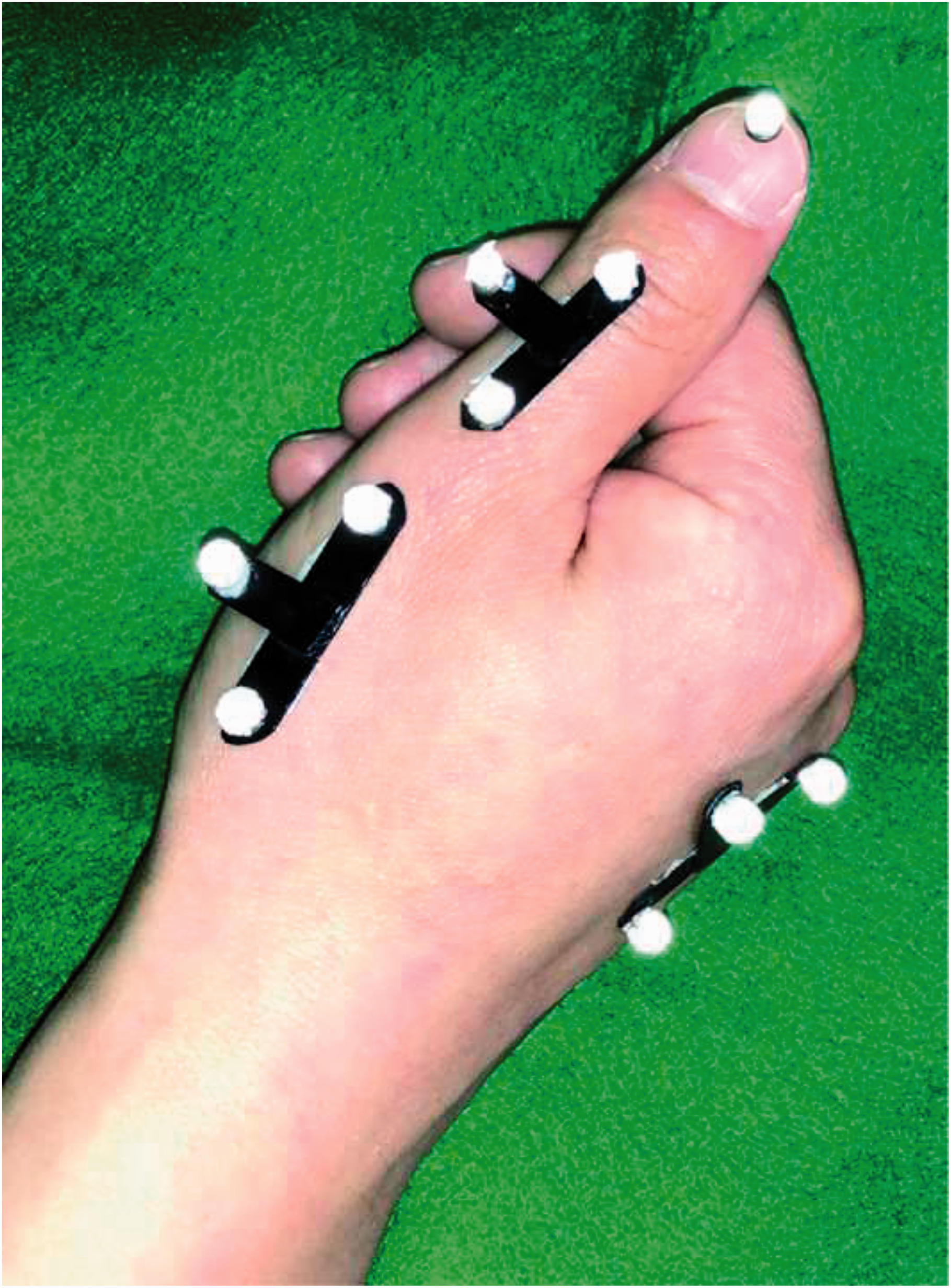
Motion capture
Three-dimensional displacements of the thumb were recorded during the Kapandji test using a motion capture system. We attached ten reflective markers (4 mm diameter) on the dorsal surface of the hand using adhesive tapes (Figure 1). One marker was attached to the tip of the thumb nail and the others were attached to the proximal phalanx, thumb metacarpal and middle metacarpal using T-shaped components that each had three markers. We used a three-dimensional motion capture system (Optitrack with 12 FLEX 3 cameras; NaturalPoint Inc., Corvallis, OR, USA) to obtain the marker displacements. The sampling frequency was set at 100 Hz.
To assess the thumb movements during the Kapandji test marking, we obtained TM, metacarpophalangeal (MP) and IP joint angles of the thumb from the three-dimensional displacements of the markers, which were exported for subsequent data processing and analysis using a custom-written program in Matlab 2021b (MathWorks Inc, Natick, MA, USA). First, all displacement data were filtered by a second-order Butterworth low-pass filter at a cut-off frequency of 5 Hz. Next, we computed TM, MP and IP joint angles of the thumb. We defined the local coordinate systems at the thumb proximal phalanx, thumb metacarpal and middle metacarpal (Li et al., 2008). TM and MP joint angles were calculated using the Euler angles of ZYX rotation sequence, where the rotation along the x-, y- and z-axes represented the flexion/extension, abduction/adduction and pronation/supination, respectively. TM joint angles were calculated from the reference position. The flexion/extension angle at the IP joint was calculated according to a previous report (Li et al., 2008). The angles of the TM and MP joints were thereby assessed in all three anatomical positions (flexion/extension, abduction/adduction and pronation/supination) and the angle of the IP joint was assessed in one anatomical direction (flexion/extension).
To estimate the thumb joint movements, we calculated TM, MP and IP joint angles in all directions at each Kapandji position. We recorded the mean joint angle for 1 second while the thumb touched and remained in each Kapandji position, and calculated the mean joint angles from three trials.
The motion capture system of this study was based on the accuracy that the root mean square (RMS) error was 0.14°, but we report the joint angles as integer values.
Electromyography
EMG signals were recorded from two thenar muscles, the flexor pollicis brevis (FPB) and abductor pollicis brevis (APB) using surface electrodes. The activities of these muscles were recorded according to the standard procedures for EMG electrode placement (Hermens et al., 2000). EMGs were recorded at 1 kHz with PowerLab16 (AD instruments Inc., Bella Vista, NSW, Australia) using 7-mm surface electrodes and electrically synchronized with the motion capture system.
First, the EMG signals were filtered using bandpass (20–500 Hz) and notch (60 Hz) filters. They were then were smoothed using a RMS algorithm with a 100 ms time window. The signals were then normalized for activity at maximum voluntary contraction (Tagliabue et al., 2015). We computed the mean EMG signal for 1 second when the thumb touched and remained in each Kapandji position and calculated the mean muscular activity obtained from three trials.
Because of the proximity of the APB muscle to the FPB muscle, we examined the EMG cross-talk to show that the two EMG signals were independently recorded. The cross-correlation coefficient was calculated to indicate the EMG cross-talk between two muscles using maximum voluntary contraction data.
Statistical analysis
To analyse the effect of the two positions of the thumb IP joint on the thumb TM joint movement (joint angles), activity of the thenar muscles, the amount of thumb MP and IP joint flexion at all Kapandji positions and to identify the Kapandji position reflecting thumb opposition, we used a repeated-measures two-way analysis of variance (ANOVA) with joint position (thumb IP extension and flexion) and Kapandji position (from 0 to 10) as factors. If the ANOVA showed a significant interaction or main effect, a pairwise comparison was done using a paired t-test with Bonferroni correction. In the pairwise comparison of the Kapandji positions, the differences between adjacent Kapandji positions were analysed. The significance level was set at p < 0.05.
Results
Main effects and interactions in analysis of variance.
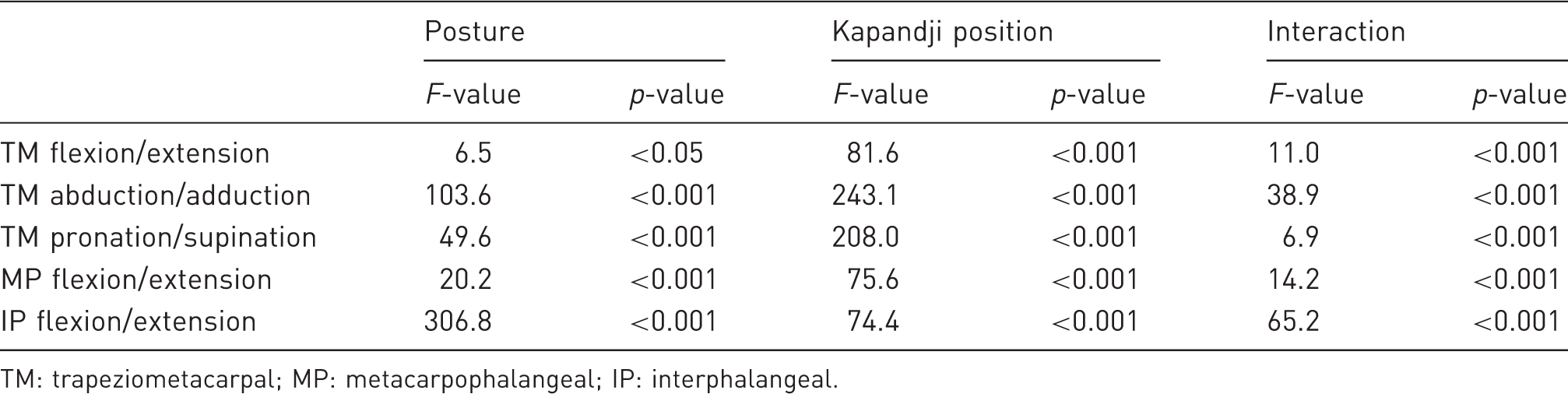
TM: trapeziometacarpal; MP: metacarpophalangeal; IP: interphalangeal.
Trapeziometacarpal joint angles with flexion and extension of the interphalangeal joint at each Kapandji position. Results in degrees are expressed as mean (standard deviation).
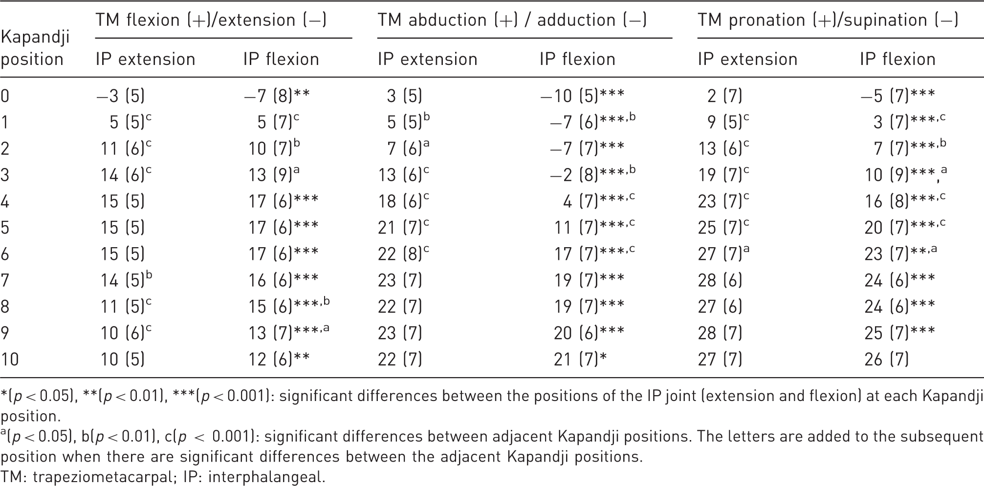
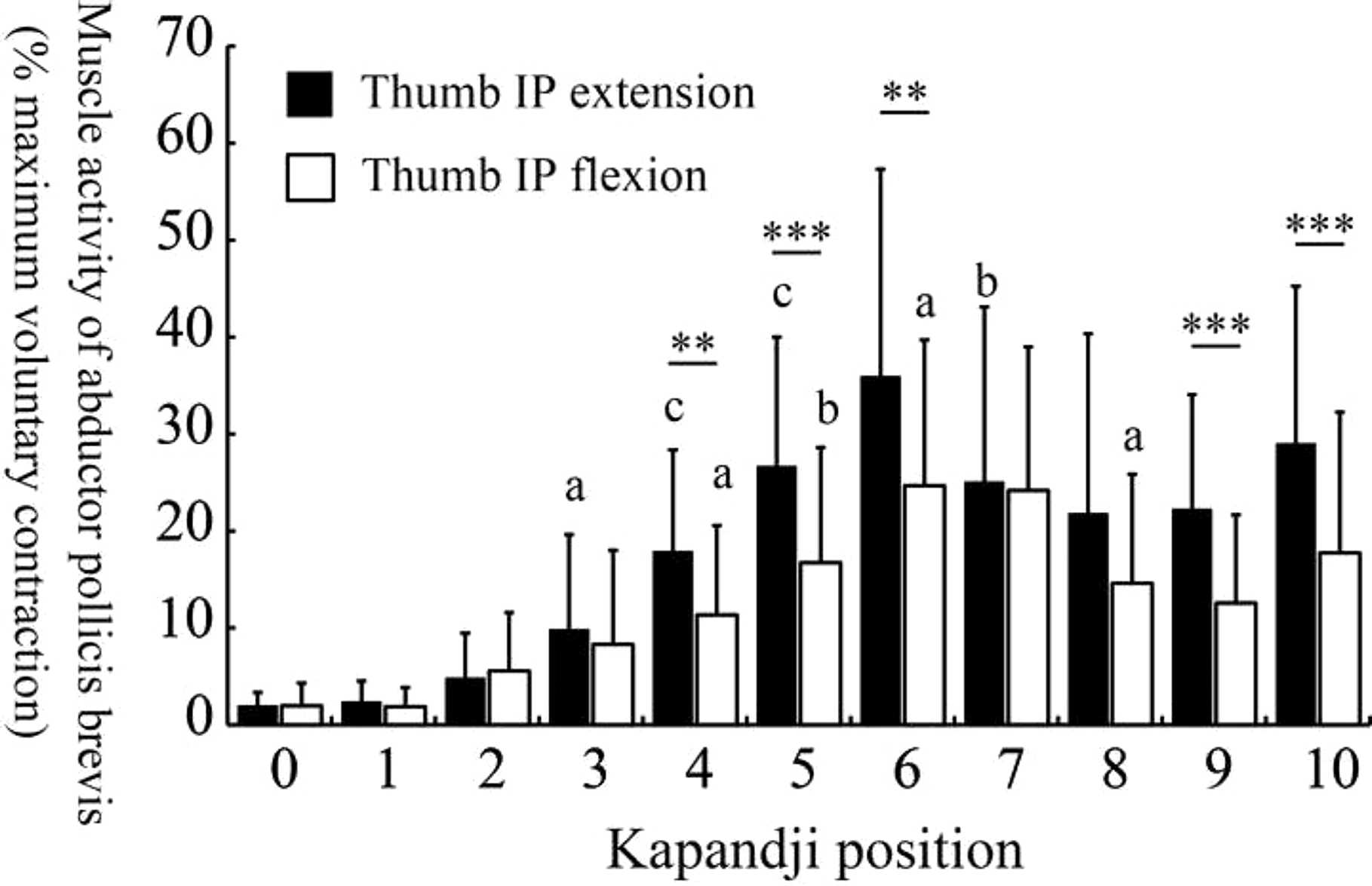
(p < 0.05), **(p < 0.01), ***(p < 0.001): significant differences between the positions of the IP joint (extension and flexion) at each Kapandji position.
(p < 0.05), b(p < 0.01), c(p < 0.001): significant differences between adjacent Kapandji positions. The letters are added to the subsequent position when there are significant differences between the adjacent Kapandji positions.
TM: trapeziometacarpal; IP: interphalangeal.
Metacarpophalangeal and interphalangeal joint angles with flexion and extension of the interphalangeal joint at each Kapandji position. Results in degrees are expressed as mean (standard deviation).
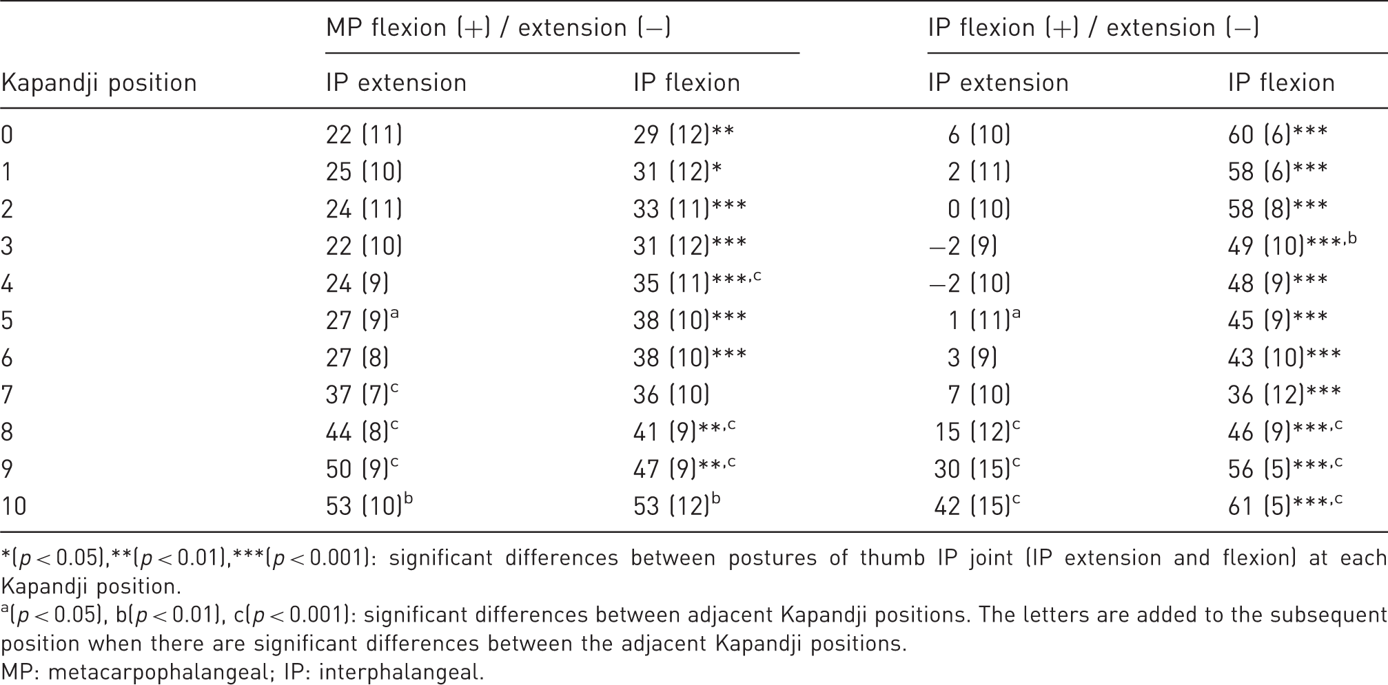
(p < 0.05),**(p < 0.01),***(p < 0.001): significant differences between postures of thumb IP joint (IP extension and flexion) at each Kapandji position.
(p < 0.05), b(p < 0.01), c(p < 0.001): significant differences between adjacent Kapandji positions. The letters are added to the subsequent position when there are significant differences between the adjacent Kapandji positions.
MP: metacarpophalangeal; IP: interphalangeal.
One participant was excluded from the EMG analysis because of high noise owing to sweating of the hand. Both the joints positions and Kapandji positions had significant main effects on activities of the FPB and APB muscles (FPB: F = 22.4, p < 0.001 and F = 23.6, p < 0.001, respectively; APB: F = 12.4, p < 0.01 and F = 27.8, p < 0.001, respectively) and displayed significant interaction (FPB: F = 4.7, p < 0.001; APB: F = 7.4, p < 0.001). Thumb IP joint positions significantly affected the activity of the FPB muscle at all Kapandji positions (Figure 2) and also had a significant effect on the activity of the APB muscle (Figure 3). Activities of the FPB and APB muscles gradually increased from Kapandji Position 3 to 6. However, after Kapandji Position 7, the activity of the FPB muscle reached a plateau, whereas that of the APB muscle decreased significantly (Figures 2 and 3).
Muscle activity in the flexor pollicis brevis at each Kapandji position. Muscle activity of the abductor pollicis brevis at each Kapandji position.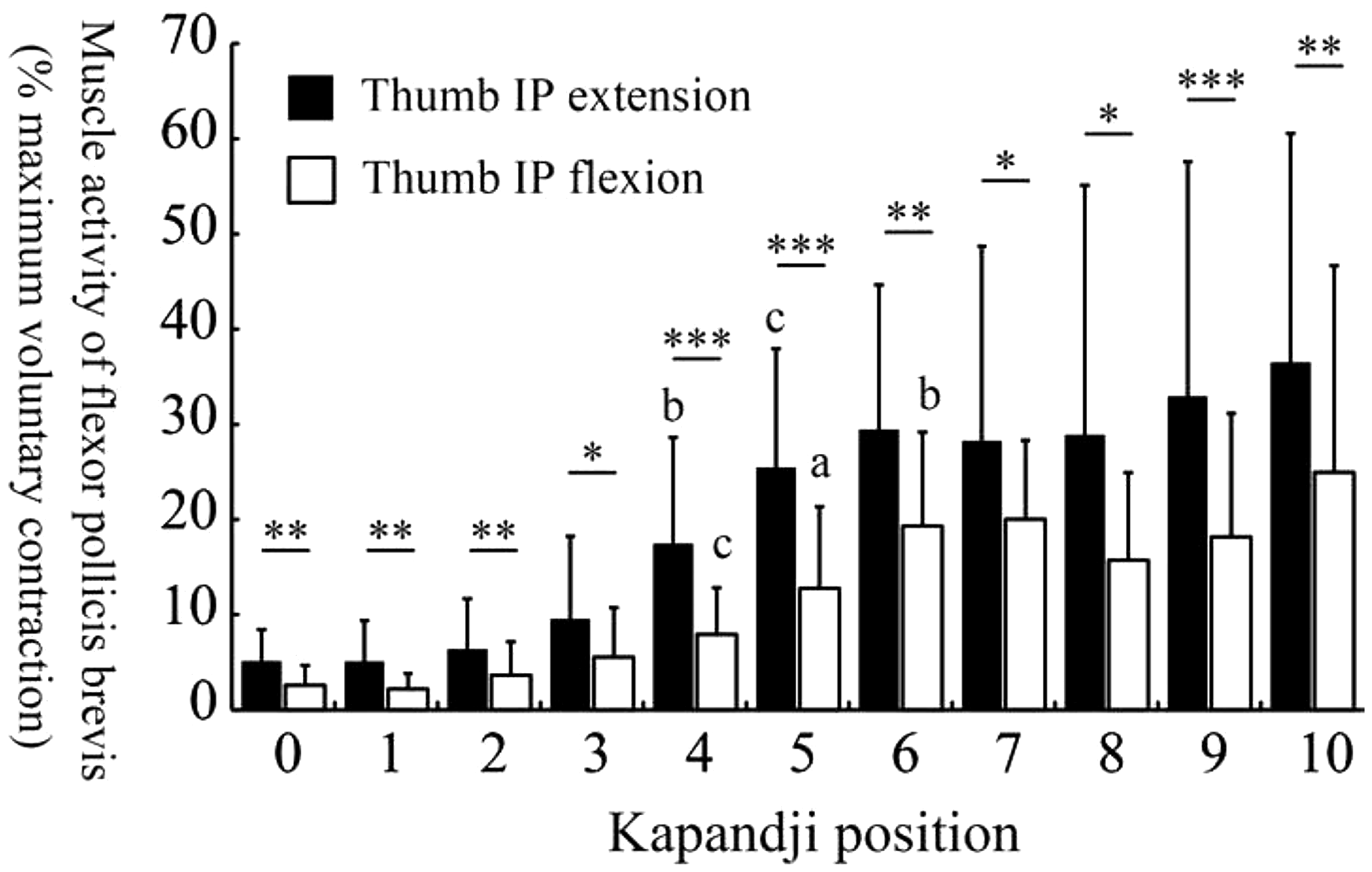
The mean cross-correlation coefficient was 0.28 (SD 0.15), indicating that there might be little cross-talk between the two recording sites and therefore that independent signals were recorded from the two muscles (Talib et al., 2019).
Discussion
The main result of this study was that the position of the thumb IP joint affected the movements of the thumb TM joint and activity of the thenar muscles during thumb opposition. Thumb IP extension increased TM joint movements, particularly abduction and pronation, more than thumb IP flexion; however, the MP and IP joint angles of the thumb were larger during thumb IP flexion than during thumb IP extension, which could be due to the action of the flexor pollicis longus. This means that flexion of the thumb MP and IP joints, which occur in thumb flexion, may compensate for the movements of the thumb TM joint during opposition with IP flexion. To support this hypothesis, the activity of the FPB and APB muscles involved in the movements of the thumb TM joint (Brand and Hollister, 1999; Kaufman et al., 1999) is less during thumb IP flexion than during thumb IP extension. In addition, the result for the extension of the IP joint involving the FPB and APB muscles (Brand and Hollister, 1999; Gupta and Tamai, 2021) is consistent with the results for the FPB and APB muscles, both of which are greater in thumb IP extension than in thumb IP flexion. As pronation of the thumb TM joint occurs by the contraction of the thumb intrinsic muscles (Li et al., 2008), compensatory flexion of the thumb IP joint would affect not only movements of the thumb TM joint but also the activity of the thenar muscles.
The Kapandji test appears to reflect thumb opposition from Positions 0 to 6. The joint angles during abduction and pronation of the TM joint and activity of thenar muscles showed a gradual increase from Kapandji Positions 0 to 6. These findings are similar to those of a previous study in which movements of the thumb TM joint occurred from Kapandji Position 0 to 6 (Kuroiwa et al., 2019). In contrast, after Kapandji Position 7, the thumb gradually flexed and movements of the TM joint gradually decreased or reached a plateau, even with IP extension. Additionally, after Kapandji Position 7, although the activity of the FPB muscle reached a plateau, that of the APB muscle, involved in the abduction of the TM joint, decreased in both IP joint positions. This means that the Kapandji positions above 7 may reflect flexion of the thumb rather than opposition (Tonkin, 2020).
Previous studies have reported that patients with carpal tunnel syndrome (CTS) can perform thumb opposition up to the Kapandji Position 10, despite atrophy of the thenar muscles (Dilokhuttakarn et al., 2017; Kuroiwa et al., 2019). A previous study indicated that in patients with CTS, flexion of the thumb MP and IP joints compensated for thumb opposition (Marquardt et al., 2014).
This study has some limitations. First, the surface markers and T-shaped components were attached to the skin surface and as a result, skin movements might have influenced joint angles. Second, it is unclear how the extrinsic muscles of the thumb, such as the flexor pollicis longus, contribute to the positions of the thumb IP joint. Finally, all data were obtained from healthy participants. The effect of the position of the IP joint on movements of the TM joint or thumb opposition should be studied in patients with neurological disorders.
In conclusion, we found that the position of the thumb IP joint did indeed affect movements of the TM joint and the activities of the thenar muscles during opposition. We also demonstrated that Kapandji positions from 0 to 6 reflected thumb opposition, whereas positions beyond 7 reflected compensatory flexion of the MP and IP joints. Therefore, the Kapandji test should be done with the IP joint in extension when examining the TM joint movement and thumb opposition.
Footnotes
Acknowledgements
We thank Professor Masayuki Kakehashi (Hiroshima University) for providing statistical advice.
Declaration of conflicting interests
The authors declare no potential conflicts of interest with respect to the research, authorship, and/or publication of this article.
Funding
The authors disclosed receipt of the following financial support for the research, authorship, and/or publication of this article: JSPS KAKENHI Grant Number JP19K11416 supported this work.
Ethical approval
Ethical approval to report this case was obtained from the Ethics Committee of Hiroshima University (No. E604-3).
Informed consent
Written informed consent was obtained from the participants for their anonymized information to be published in this article.
Author contributions
All authors contributed to the study's conception and design. Material preparation and data collection were performed by HK, KK and SD. HK, YS and TS performed data analysis. HK wrote the first draft of the manuscript. All authors reviewed and edited the manuscript and approved the final version of the manuscript.
