






Abstract
Cerebral cavernous malformation is a rare but important cause of cerebral hemorrhage in pregnancy and puerperium. In pregnancy, cavernomas can more easily bleed as a result of increased female hormones and growth factors such as vascular endothelial growth factor. We present the case of a pregnant woman who had been diagnosed with a cerebral cavernoma about ten years previously, after repeated headache episodes; at the 28th week of pregnancy the woman was hospitalized for epileptic seizures and active bleeding from the anterior cerebral artery. We describe the management of the case, the decision for a preterm delivery and for a resolutive neurosurgical procedure.
Introduction
Cerebral cavernous malformations (CCMs) are found in 0.4–0.8% of the general population. 1 Diagnosis is often made by magnetic resonance imaging (MRI) as an incidental finding or after severe symptoms such as headache, seizures, focal neurological symptoms. Symptoms may be absent but seizures are the most frequent finding. Complications are not uncommon: the annual incidence of hemorrhage is found from 0.7% to 6% per year and re-bleeding in the percentage of 4.5 per year.2‐3
Cavernomatosis in pregnancy is a rare presence and there are currently no guidelines on management in pregnancy and the puerperium for women with CCM.
Here we describe a case of a pregnant woman known to have a CCM, admitted to hospital for cavernoma haemorrhage in the third trimester of pregnancy, highlighting the need for evidence-based care planning by a multidisciplinary team.
Case
A Caucasian 40-year old woman in her first pregnancy, presented at 28 weeks and 2 days and was hospitalized for seizures. She reported a history of CCMs, diagnosed at the age of 30 after performing MRI due to persistent headache. The patient, although aware of her vascular malformation, had not undergone a gynecological examination before pregnancy or preconception counseling. She had had no previous seizures. Pregnancy history was unremarkable, with normal first and second trimester screenings, normal estimated birth weight and Doppler velocimetry.
At the time of her presentation to the emergency department, the patient complained of headache and reported seizures within the previous twenty-four hours. She had no features suggestive of a hypertensive disorder of pregnancy; the arterial pressure was less than 140/90 mmHg; there were no signs of edema, the patient did not complain of abdominal pain, the hepatic profile and the renal profile were normal on blood tests and there was no proteinuria at a rapid evaluation of the urine or after the twenty-four hour collection.
A cerebral MRI was performed and showed bleeding from the cavernoma present in the territory of the anterior cerebral artery and the middle cerebral artery was performed (Figures 1 and 2).
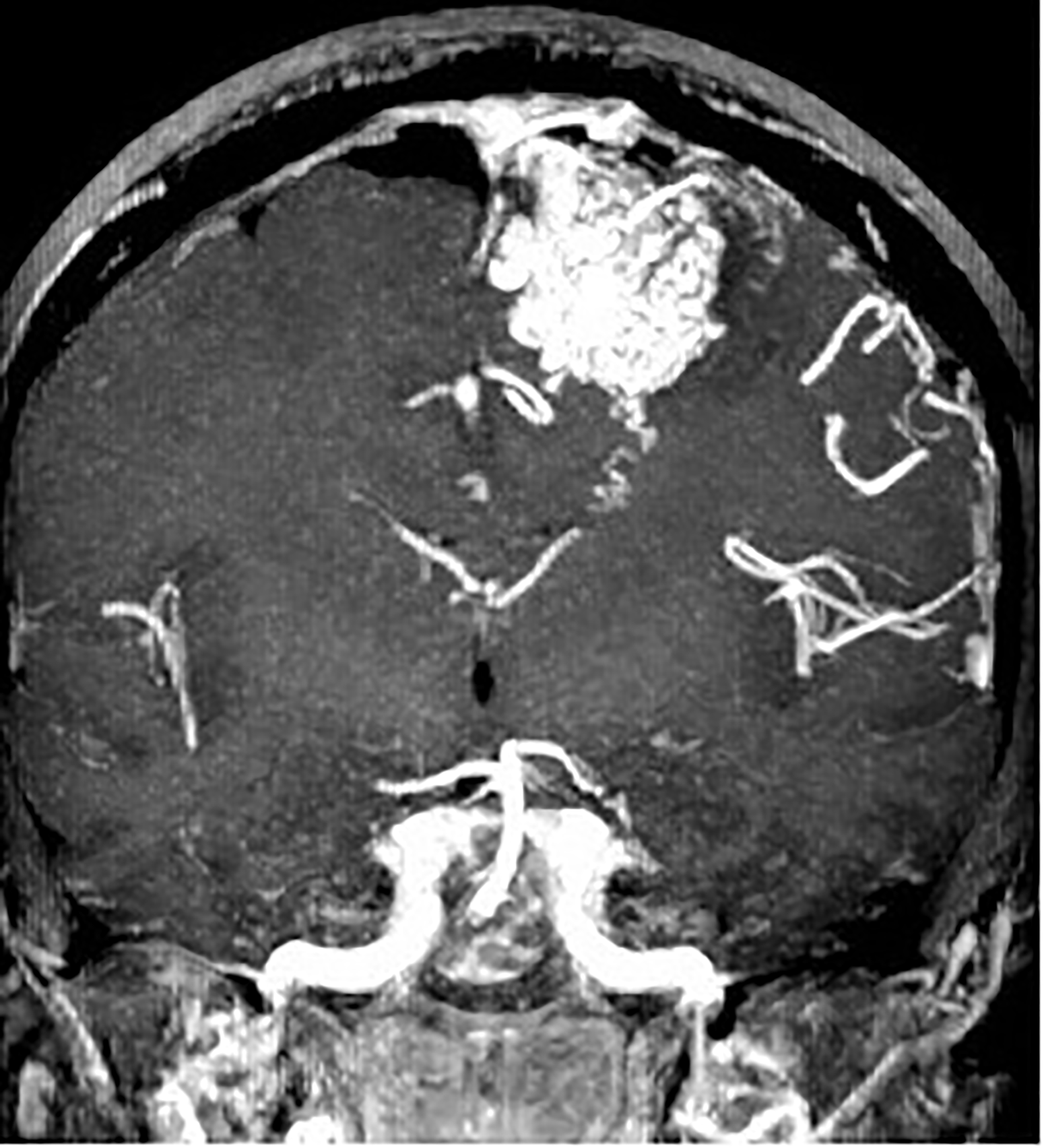
MRI of the cavernoma.
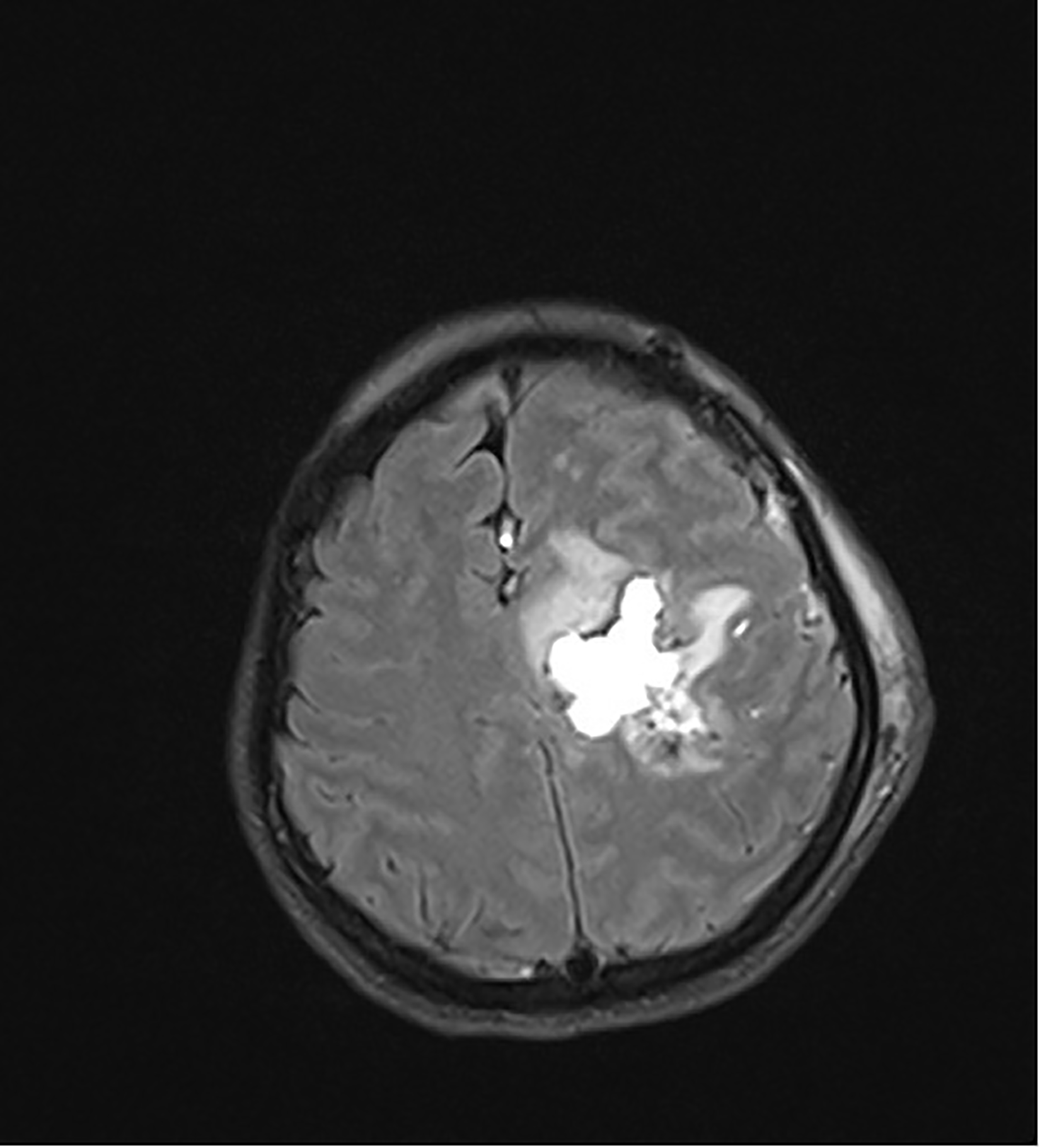
MRI showing the hemorrhagic lesion with periwound edema.
After advice from neuroradiology and neurosugery, it was felt there was no indication for emergency surgery. After careful neurological consultation, electroencephalogram (EEG), that result was normal, was performed and therapy was started, Levetiracetam 500 mg twice a day.
After performing cardiotocography and ultrasound of the fetus, the gynecologist advised corticosteroid prophylaxis for fetal lung maturation.
In accordance with the limited literature available, in case of bleeding of a CCM in the third trimester of pregnancy there is an indication to perform a cesarean section in order to allow a subsequent neurosurgical intervention and minimize the consequences of a possible massive bleed. In fact, in consideration of the risks for the mother and the fetus related to a neurosurgery during pregnancy, after 28 weeks it is often preferable to deliver the baby and then the neurosurgery. Therefore, careful counseling with the patient about maternal-fetal risks related to the continuation of pregnancy was carried out. After discussion with the obstetric team, the patient and other specialists, a decision was made to perform a cesarean section at 30 weeks. Before the caesarean section, and neuroprotection with magnesium sulfate.
Elective cesarean section was performed under general anaesthetic using propofol 100 mg and rocuronium 100 mg. Opioids were used only after the baby extraction. Anesthesia was maintained with propofol in continuous infusion 4 mg/kg/hour. Neuroprotection adopted was propofol 4 mg/kg/hour. 0.9% sodium chloride was administered intravenously during the procedure. NaCl 0,9% is preferable to glucose solution or lactated Ringer's because crystalloid is associated with less accumulation of fluid in the brain.
After surgery the patient was transferred for 24 h of observation in the Post-Operative Intensive Care Unit.
The patient was aware of the need for further investigations with contrast MRI as well as neurosurgical intervention and expressed the desire not to undertake breastfeeding. Therefore, cabergoline 1 mg was administered.
The female infant was born in good condition, with APGAR score of 8 and 9 at 1 and 5 minutes respectively, birthweight 1900g and cord *** pH 7.27. She remained in neonatal intensive care for six weeks.
Three days after delivery, the patient reported an intense headache with reduced mobility of the right side of her body. In view of her history and suspicion of recurrence of cerebral hemorrhage, urgent head computed tomography (CT) was performed. CT did not show any areas of new bleeding or infarction. The patient presented on CT an inhomogeneous area of altered density in the subcortical white matter of the left semioval center due to previous bleeding outcomes with modest peripheral edema from arteriovenous vascular malformation. Due to the progressive worsening of symptoms, in line with existing literature,4‐5 neurosurgical intervention for CCM removal was planned.
The surgery was performed under general anesthesia; a craniotomy was performed with subsequent removal of the vascular malformation. The surgery lasted two hours and thirty minutes. The patient had an uncomplicated seven-day post-operative hospital stay. She had a neurosurgical control one month after the surgery and a MRI with contrast which was negative three and six months after the surgical procedure.
After this admission the patient was counselled about the need for planning a possible second pregnancy and hospital management of this.
Discussion
The scientific literature is controversial regarding the course of CCMs in pregnancy. 4 According to older literature, pregnancy predisposes women to hemorrhage from CCMs,2‐6 while according to more recent studies the risk of intracranial hemorrhage remains almost unchanged during pregnancy.7–8
The indications for neurosurgery in non-pregnant patients with CCM are the presence of seizures unresponsive to medical treatment, acute bleeding and re-bleeding.9–11 Surgical removal can be considered prophylactic in single asymptomatic brain cavernomas located in surgically accessible brain areas.12–14
The need for neurosurgery during pregnancy is rare but, if necessary, due to the rapid progression and the precarious haemodynamic balance of the pregnant woman, it must be performed immediately.
There are currently no guidelines for pregnancy management in patients with cerebral cavernomatosis. In 2012, Burkhardt et al. outlined a system for the management of these patients: in the case of asymptomatic cavernomas maintain a careful follow-up, in the case of mild symptoms consider a re-evaluation by MRI and continue a careful clinical evaluation, in case of therapy-resistant seizures or rapidly progressing neurological disorders the management depends on the trimester of pregnancy. In the first and second trimester there is an indication to perform neurosurgery, in the third trimester to perform childbirth and then surgery. 5 In this case, vaginal delivery is not contraindicated in patients with cerebral cavernomatosis7–8 but a cesarean section is generally preferred to avoid the risk of bleeding due to an excessive increase in blood pressure during vaginal delivery. 15
Neurosurgery in pregnancy should be considered only in severe cases. The use of gadolinium is contraindicated in pregnancy and consequently the preoperative evaluation often remains poor. Some centres advocate a first surgery in pregnancy followed by further surgery in the puerperium, after MRI with gadolinium. 16 Furthermore often neurosurgery requires general, hypotensive and hypothermic anesthesia which can potentially reduce placental blood flow and cause adverse pregnancy events. 15 For all these reasons it is necessary to perform neurosurgery in highly specialized centers.
In conclusion, pregnancy is not contraindicated in patients with CCMs but these patients must be followed by a multidisciplinary team, in a specialized center, to minimize the increased risk of adverse events in pregnancy. Even the puerperium is recognized by the literature as a critical moment especially due to the possible onset of cerebral hemorrhages. For this reason, a close follow-up of these patients is also necessary. 15
Footnotes
Acknowledgments
None.
Declaration of conflicting interests
The author(s) declared no potential conflicts of interest with respect to the research, authorship, and/or publication of this article.
Funding
The author(s) received no financial support for the research, authorship, and/or publication of this article.
Ethical approval
Not applicable.
Informed consent
The patient provided written informed consent for the publication of this case report.
Guarantor
Federica Del Prete.
Contributorship
Lucia Merlino: conceptualization, methodology. Federica Del Prete: conceptualization, methodology, data curation, writing. Luca Titi: visualization, supervision. Maria Grazia Piccioni: visualization, supervision. All authors reviewed and edited the manuscript and approved the final version of the manuscript.
