






Abstract
It has been documented that exogenously administered irisin (1010 fibronectin-type III domain-containing 5 [FNDC5]), which is a new polypeptide hormone, induces the browning of subcutaneous fat and thermogenesis. In this study, effects of physical activity and exogenous administration of irisin were investigated on parameters related with reproduction and metabolism in the high-fat diet-induced obesity model of the female C57BL/6J mice. Sixty mice were gathered at age approximately 5 to 6weeks and were divided into 3 groups. Control group remained sedentary. Irisin group remained also sedentary but intravenously received 1010 FNDC5-expressing adenovirus after 20 weeks. Exercise group performed treadmill after 6 weeks. All mice were sacrificed 22 to 23 weeks after the start of the study. There was a significantly greater Δ weight in the controls compared with the irisin and exercise groups (P < .05). Glucose and insulin levels were significantly higher in the controls (P < .05). The serum irisin level was significantly higher in the exercise group (P < .05). Serum luteinizing hormone levels were significantly increased in the irisin group (P < .05). Serum anti-Müllerian hormone levels were significantly higher in irisin and exercise groups (P < .05). There were significant negative correlations between serum irisin levels and Δ weight and homeostatic model assessment of insulin resistance (r = −0.327, r = −0.297, respectively; P < .05 for both). The numbers of primordial follicles per ovary were similar (P > .05), whereas primary and secondary follicles per ovary were higher in the irisin and exercise groups compared with controls (P < .05). Pharmacologic introduction of irisin may improve metabolic factors such as insulin sensitivity and obesity by promoting weight loss and consequently improving the reproductive potential.
Keywords
Introduction
Exercise is important to achieve a healthy systemic metabolism and may protect against metabolic disorders such as obesity, but the mechanism is poorly understood. Notably, exercise increases the energy expenditure of the whole body beyond the calories spent in the actual performed work. 1 On the other hand, how adipose tissues contribute to this phenomenon is still unclear. Bostrom et al 2 tried to identify factors secreted from muscle that may increase whole-body energy expenditure because transgenic mice that selectively expressed PGC1-α in muscle presented a notable resistance to diabetes and obesity. The authors described a remarkable new hormone, namely irisin, which is regulated by PGC1-α.
Irisin is a recently discovered polypeptide hormone. 2,3 After being released from muscle following exercise, circulating irisin exerts its function as a hormone that stimulates the browning of white adipose tissue, which translates into burning more calories and therefore increasing oxygen consumption and thermogenesis in adipocytes. The therapeutic potential of irisin is intriguing. Exogenously administered irisin induced thermogenesis, and it could theoretically be administered as an injectable polypeptide as a therapeutic alternative in obese humans.
The world is dealing with a public health emergency due to the increasing rate of obesity and obesity-related disorders. 4 Obesity may affect several aspects of reproduction in both females and males. The association between obesity and alterations in female reproductive function has long been recognized 5,6 and was confirmed more recently. 7 The Nurses’ Health Study, 8 which was carried out in the United States, reported that the risk of ovulatory infertility increased in women who had increased body mass index (BMI) values. Numerous prospective and cross-sectional studies documented similar findings (reviewed in the study by Linne 9 ). The authors of an observational study performed in the United Kingdom 4 found that BMI had a significant negative impact on fertility. Obesity might affect the metabolism and apoptosis rate of ovarian follicles, which can be evaluated using a number of markers such as Forkhead boxO (FOXO), nitric oxide (NO), and anti-Müllerian hormone (AMH).
Forkhead boxO transcription factors are potential markers expressed in various cells including ovarian follicles and control several cellular process including metabolism and apoptosis. Anti-Müllerian hormone is a substance produced by granulosa cells in ovarian follicles. It is first made in primary follicles that advance from the primordial follicle stage, indicating its significance on ovarian reserve. Nitric oxide is another vital molecule that is involved in different biologic functions. These markers may be related with obesity.
The aim of this study was to investigate and compare the effects of exogenous administration of irisin and physical activity on several parameters related with metabolism, reproduction, and immunologic markers on ovary in a high-fat diet-induced obesity model of female C57BL/6J mice. We hypothesized that exogenous administration of irisin would have similar effects to physical activity on these parameters.
Material and Methods
Experimental Animals
Sixty female C57BL/6J mice were gathered in Institute of Experimental Medicine, Istanbul University (Istanbul, Turkey), aged approximately 5 to 6 weeks. The animals were housed 5 to a cage, maintained on a 14-hour light/10-hour dark cycle, and allowed access to food and water ad libitum during the entire experiment. The mice received a 60% kcal high-fat diet (cat#58Y1, TestDiet, St. Louis, Missouri). All mice remained on the same diet until sacrificed. The protocols were conducted in accordance with the Turkish Legislation for the Use and Care of Laboratory Animals and were approved by the Experimental Animal Use Ethics Committee of Istanbul University.
Mice were randomly assigned to 3 groups: irisin (n = 20), exercise (n = 20), and controls (n = 20), as presented in Figure 1. Mice in the irisin group remained sedentary and were intravenously injected with 1010 fibronectin-type III domain-containing 5 (FNDC5)-expressing adenovirus (cat# 041429A, Applied Biological Materials Inc, Richmond, British Columbia, Canada) 20 weeks after the start of the study. The age of the mice when first gathered and dose and timing of adenovirus were selected in line with the study of Bostrom et al. 2 Six weeks after the start of the study, approximately 12-week-old mice in the exercise group were exercised using freewheel running as described previously until sacrificed. 10 The serum levels of irisin after 10 weeks of regular exercise were twice as high as baseline levels in the study of Bostrom et al. 2 Hence, adenovirus administration was after the initiation of exercise in mice. The controls remained sedentary until sacrificed. One mouse in the exercise group died of an unknown cause.
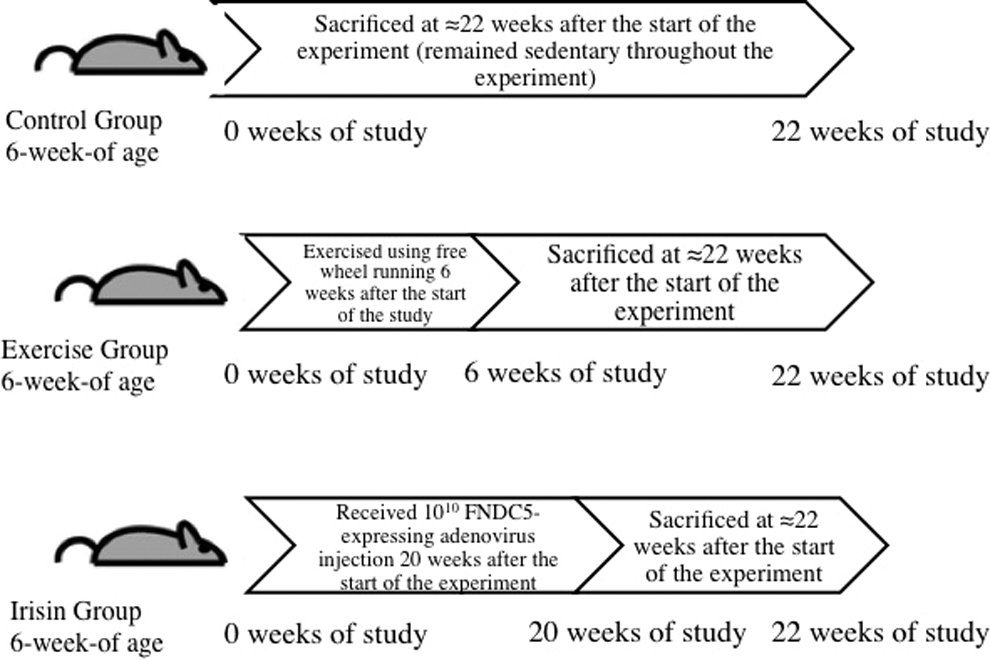
Experimental model.
Ovarian Histology and Quantification of Follicles
All mice were sacrificed 22 to 23 weeks after the start of the study (depending on the time of their estrous phase) by CO2 asphyxiation after fasting for 5 hours, and their ovaries were immediately excised and fixed in 4% paraformaldehyde. Paraffin tissue sections were stained with hematoxylin–eosin (H&E) for histologic evaluation. The estrous cycle was evaluated 2 to 3 times a week in all mice starting after the administration of FNDC5-expressing adenovirus by assessing the vaginal opening every morning. 11
For histologic examination of ovarian morphology and follicle counts, ovaries were fixed in 10% neutral-buffered formalin for 24 hours, dehydrated in alcohol, and embedded in paraffin using standard protocols. Sections of 5 µm thickness were mounted on glass slides and stained with H&E for general tissue morphology. All analyses were observed and photographed using an Olympus BX53 (Olympus, Hamburg, Germany).
Nuclei were used as markers to count follicles, and the stage of follicular development was determined in line with previous literature. 12 In short, follicles were categorized according to the number and shape of somatic cells that surrounded the oocyte: primordial follicles with flattened cells, primary follicles with 1 layer of cuboidal cells, and secondary follicles with 2 partial layers of cells.
Hormone Assays
Mouse blood was obtained through cardiac puncture promptly after death. The blood was centrifuged, and serum was placed into vials and stored at −80°C until required for evaluation. Mouse serum glucose, insulin, irisin, estradiol (E2), luteinizing hormone (LH), follicle-stimulating hormone (FSH), and AMH levels were quantified using commercially available enzyme-linked immunosorbent assay kits in accordance with the manufacturer’s instructions, and all interassay and intraassay coefficients of variation were less than 10% (Merck Millipore, Darmstadt, Germany).
Immunohistochemical Analysis
For immunohistochemical analysis, paraffin-embedded ovaries were cut into 8-μm sections and analyzed using standard procedures as previously described. Briefly, after deparaffinization, antigen retrieval, endogen peroxidase, and protein-blocking procedures, all sections were incubated with FOXO1 (cat#C29H4; Cell Signaling Technology, Danvers, Massachusetts), FOXO3 (cat#D19A7 Cell Signaling Technology), endothelial NO synthase (eNOS; cat#RB-9279-P; Thermo Fisher Scientific Inc, Waltham, Massachusetts), inducible NO synthase (iNOS; cat#RB-1605-P, Thermo Fisher Scientific Inc), and AMH (cat#sc-6886; Santa Cruz Biotechnology, Inc, Dallas, Texas) antibodies for 1 hour at room temperature. All antibodies were diluted with UltrAb Diluent (cat# TA-125-UD Lab Vision Antibody Diluent OP Quanto; Thermo Fisher Scientific Inc) and then treated with a commercially available secondary antibody kit (Thermo Fisher Scientific Inc) and stained with 3-amino-9-ethyl carbazole chromogen (cat#TA-004-HAC; Thermo Fisher Scientific Inc). Finally, all sections were counterstained with Mayer hematoxylin to identify ovarian cell types and components. Two independent observers evaluated the immunostaining levels between the 3 groups. Results contain observations of consistent immunostaining patterns.
Apoptotic Index and HSCORE
Apoptosis in tissue samples was detected using the commercially available Terminal Deoxynucleotidyl Transferase dUTP Nick End Labeling (TUNEL) assay (ApopTag Plus in situ apoptosis detection kit; Chemicon International, Germany). The TUNEL-positive cells were used to evaluate the apoptotic index (percentage of TUNEL-positive cells in 1000 cells). 13
The evaluation of the FOXO1, FOXO3, eNOS, iNOS, and AMH immunohistochemical staining was performed using histological score (HSCORE). 14 This approach provides a value for the overall staining intensity and also the percentage of cells stained. Positively stained cells were counted and graded by 2 independent observers depending on the staining intensity. Staining intensity grading scores were defined as follows: 0 = no staining, 1 = weak but detectable above control, 2 = moderate, and 3 = strong staining.
For each tissue sample, the HSCORE value was calculated with the following equation:

where “i” is the staining intensity grading score and “pi” the corresponding percentage of stained cells for each intensity grading.
Characteristics of Obesity
The mice were weighed at 2-week intervals, and their weights were recorded. The homeostatic model assessment (HOMA), an index of insulin resistance, was determined at the end of the study using the following equation: (fasting insulin [mIU/L] × fasting glucose (nmol/L)/22.5. 15
Statistical Analysis
Continuous variables are presented as mean (standard deviation). According to the histograms, all variables were distributed normally. Student t test was used in 2-group comparisons. One-way analysis of variance was used to compare the 3 groups, followed by post hoc Tukey tests. Pairwise correlation of the hormonal and selected mediators was achieved using Pearson correlation coefficient. Analyses were performed using the version 20.0 of Statistical Package for the Social Sciences software (SPSS, Inc, Chicago, Illinois) and version 6 of GraphPad Prism software (GraphPad Software, Inc, La Jolla, California). A P value of <.05 was considered significant.
Results
Table 1 presents the characteristics of the mice. There was no significant difference between the initial weights of mice between the groups (irisin group: 14.52 ± 0.93 g vs exercise group: 14.60 ± 0.87 g vs controls: 14.71 ± 0.79 g; P > .05). When Δ weight was calculated, there was no significant difference between the irisin and exercise groups (9.47 ± 0.89 g vs 7.75 ± 0.86 g; P > .05; Figure 2). There was a significantly greater weight difference in the controls compared with the irisin and exercise groups (controls: 11.66 ± 1.19 g; P < .05). Glucose and insulin levels were significantly higher in the controls compared with the irisin and exercise groups (P < .05); the difference between the irisin and exercise groups was not significant (P > .05). The HOMA-IR was again similar between the irisin and exercise groups; however, it was significantly lower when compared with the control group (irisin: 7.54 ± 0.77 vs exercise: 6.65 ± 0.67 vs controls: 9.31 ± 1.23; P < .05).
Characteristics of the High-Fat Diet-Induced Obese Female C57BL/6J Mice.a

Abbreviations: AMH, anti-Müllerian hormone; E2, estradiol; FSH, follicle-stimulating hormone; HOMA-IR, homeostatic model assessment of insulin resistance; LH, luteinizing hormone; NS, not significant at P > .05.
aSimilar superscripts (b, c, and d) indicate a statistically significant difference (P < .05).
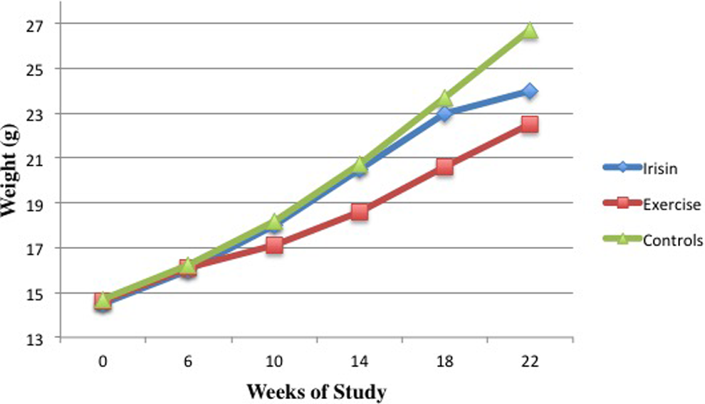
Body weight changes with time.
Serum E2 levels were slightly higher in the controls compared with the irisin and exercise groups, but the difference did not reach statistical significance (P > .05). Serum FSH levels were slightly lower in the controls compared with the irisin and exercise groups, but the difference did not reach statistical significance (P > .05). The serum irisin level was significantly higher in the exercise group compared with controls (exercise: 830.96 ± 303.56 µg/mL vs controls: 631.95 ± 201.99 µg/mL; P < .05). The difference in serum irisin levels between the irisin and exercise groups did not reach statistical significance, although it was higher in the exercise group (irisin: 742.02 ± 282.07 µg/mL vs exercise: 830.96 ± 303.56 µg/mL; P > .05). Serum LH levels were significantly increased in the irisin group compared with controls (irisin: 85.00 ± 32.21 IU/L vs controls: 59.78 ± 32.73 IU/L; P < .05). The difference in serum LH levels between the exercise group and controls did not reach significant difference (exercise: 70.95 ± 9.46 IU/L vs controls: 59.78 ± 32.73 IU/L; P > .05). Serum AMH levels were significantly higher in irisin and exercise groups compared with controls (irisin: 61.63 ± 22.78 ng/mL vs exercise: 59.35 ± 14.23 ng/mL vs controls: 42.48 ± 17.03 ng/mL; P <.05).
As presented in Table 2, the numbers of primordial follicles per ovary were similar (P > .05), whereas primary and secondary follicles per ovary were higher in the irisin and exercise groups compared with controls (P < .05). The exercise group had slightly higher numbers of follicles compared with the irisin group overall, but the differences were not statistically significant (P > .05). There were significant negative correlations between serum irisin levels and Δ weight and HOMA-IR (r = −0.327, r = −0.297, respectively; P < .05 for both).
Numbers of Follicles per Ovary of the High-Fat Diet-Induced Obese Female C57BL/6J Mice in the Estrous Phase on the Day of Sacrifice.

Abbreviation: NS, not significant at P > .05.
Immunohistochemical Analyses
Immunohistochemical localization of FOXO1 showed intense staining in the granulosa cells of the preantral and antral follicles and weak staining in the granulosa cells of follicles in earlier stages. No immunohistochemical localization was observed in the developing corpus luteum. The HSCORE for FOXO1 staining for each group is presented in Figure 3. The comparison of immunostaining assessed in each group revealed a similar HSCORE between the irisin and exercise groups (P > .05), whereas there was a significantly decreased HSCORE in the irisin group compared with controls and exercise group compared with controls (P < .05 for both).
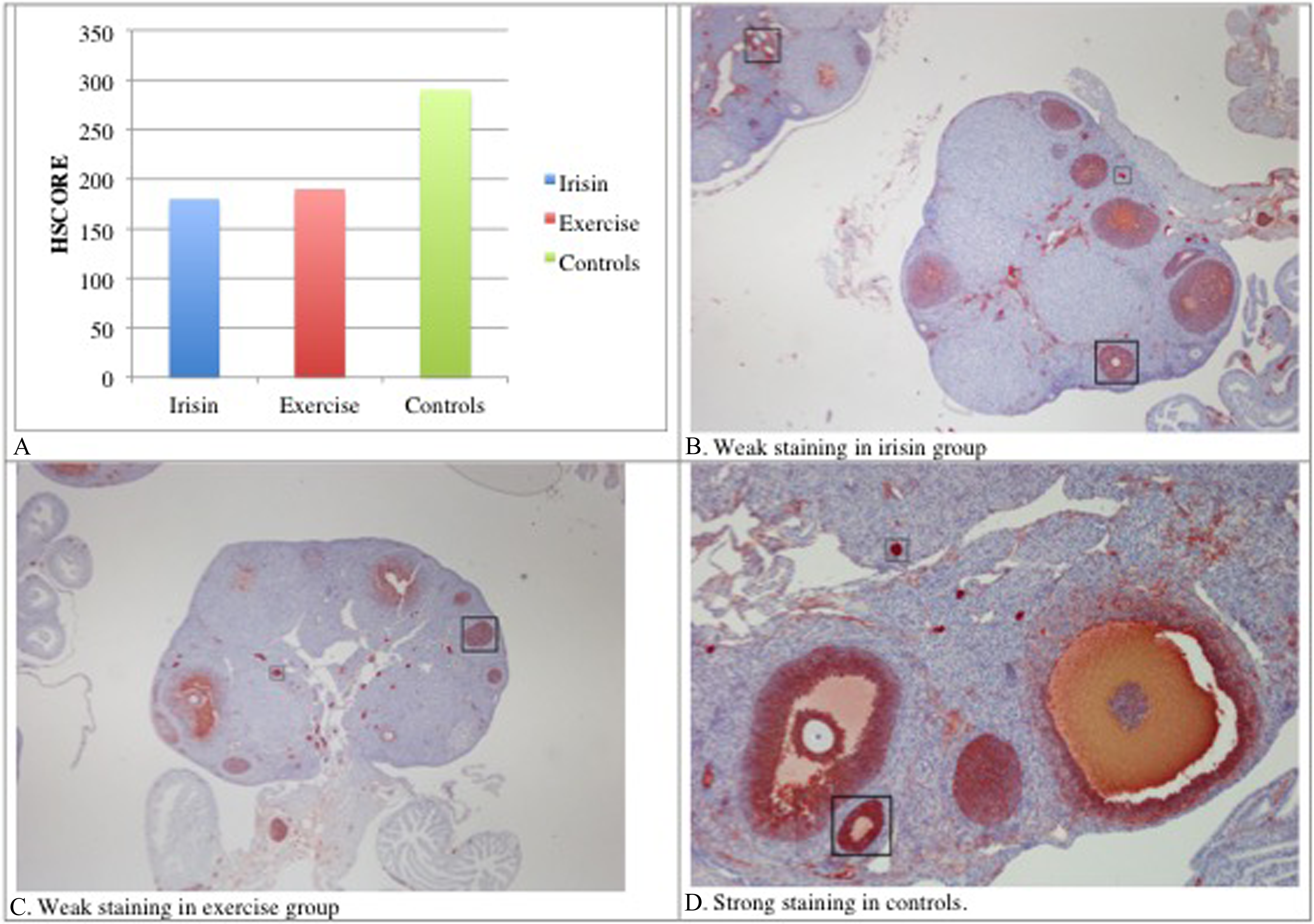
A, HSCORE for FOXO1. B-D, Representative images of weak versus strong immunohistochemical staining for FOXO1 for irisin, exercise, and control groups, respectively. Red color represents localization. Scale bar = 50 µm. FOXO indicates Forkhead boxO.
Immunohistochemical localization patterns of FOXO3 were similar to FOXO1. The HSCORE for FOXO3 staining for each group is presented in Figure 4. The comparison of immunostaining assessed in each group revealed a similar HSCORE between the irisin and exercise groups (P > .05) and a significantly decreased HSCORE in the irisin group compared with controls and the exercise group compared with controls (both P < .001).
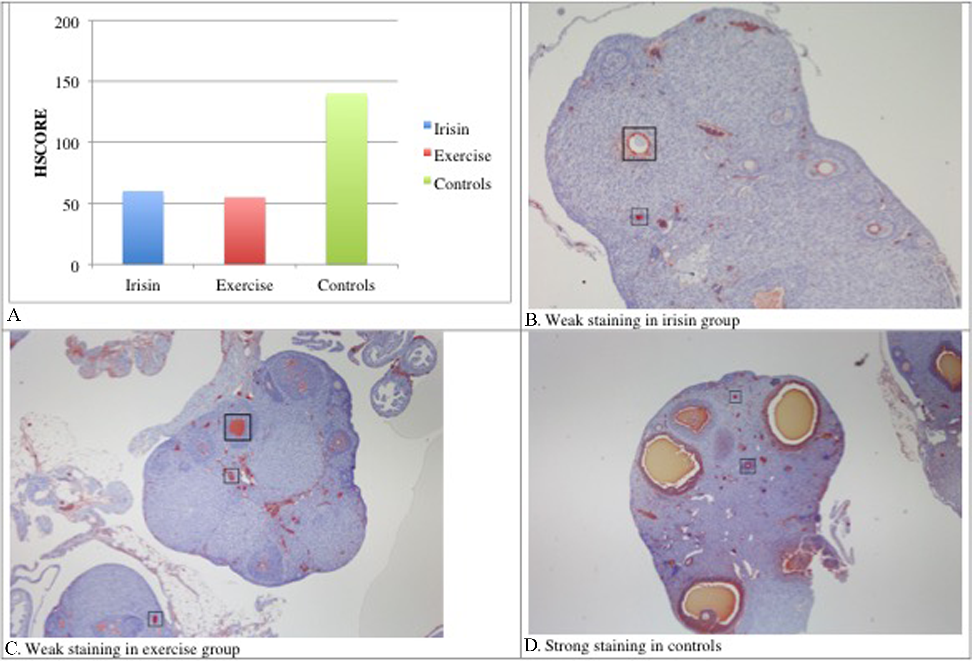
A, HSCORE for FOXO3. B-D, Representative images of weak versus strong immunohistochemical staining for FOXO3 for irisin, exercise, and control groups, respectively. Red color represents localization. Scale bar = 50 µm. FOXO indicates Forkhead boxO.
Immunohistochemical localization of AMH showed intense staining in the granulosa cells of the primary, secondary preantral, and antral follicles. The HSCORE for AMH staining for each group is presented in Figure 5. The comparison of immunostaining assessed in each group revealed similar AMH between the irisin and exercise groups (P > .05), whereas there was significantly increased AMH in the irisin group compared with controls and exercise group compared with controls (P < .001 for both).
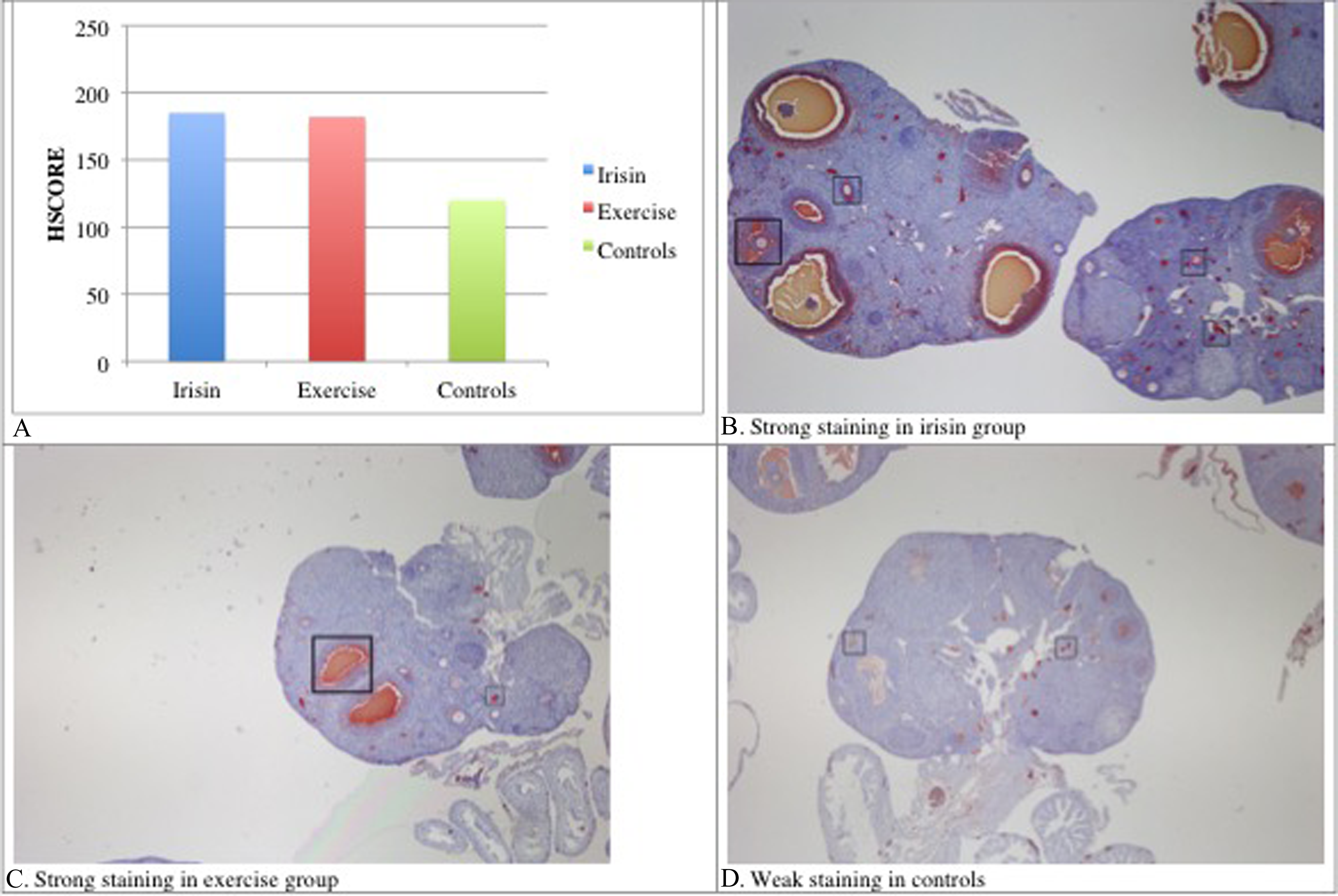
A, HSCORE for AMH. B-D, Representative images of weak versus strong immunohistochemical staining for AMH for irisin, exercise, and control groups, respectively. Red color represents localization. Scale bar = 50 µm. AMH indicates anti-Müllerian hormone.
Immunohistochemical localization of eNOS showed staining in the zones pellucida, theca interna cells, and blood vessels. The HSCORE for eNOS staining for each group is presented in Figure 6. The comparison of immunostaining assessed in each group revealed a similar HSCORE between the irisin and exercise groups (P > .05), whereas there was a significantly increased HSCORE in the irisin group compared with controls and the exercise group compared with controls (P < .001 for both).

A, HSCORE for eNOS. B-D, Representative images of weak versus strong immunohistochemical staining for eNOS for irisin, exercise, and control groups, respectively. Red color represents localization. Scale bar = 50 µm. eNOS indicates endothelial NOS.
Immunohistochemical localization of iNOS showed staining in the zona pellucida, antrum, and granulosa cells. The HSCORE for iNOS staining for each group is presented in Figure 7. The comparison of immunostaining assessed in each group revealed a similar HSCORE between the groups (P > .05).

A, HSCORE for iNOS. B-D, Representative images of weak versus strong immunohistochemical staining for iNOS for irisin, exercise, and control groups, respectively. Red color represents localization. Scale bar = 50 µm. iNOS indicates inducible NOS.
As presented in Figure 8, the introduction of exercise in the exercise group or administration of FNDC5-expressing adenovirus in the irisin group decreased apoptotic indices similarly in comparison with the controls (P < .001).

A, Apoptotic index (%) of each group. B-D, Arrows show TUNEL-stained cells in brown color, indicative of apoptosis. Scale bar = 100 µm. TUNEL indicates Terminal Deoxynucleotidyl Transferase dUTP Nick End Labeling.
Discussion
The data of the present study confirm that exogenous administration of irisin acts similarly to exercise in diet-induced obese female mice: Weight gain was less compared with sedentary mice, and insulin resistance and levels of reproductive hormones were improved. In addition, the number and quality of follicles improved, and various factors involved in ovarian steroidogenesis, ovarian follicular growth, and apoptosis were positively affected, which have positive implications for reproductive outcomes.
Irisin is a recently discovered polypeptide hormone that has received significant attention since its discovery. 2,3 It is known as a proteolytic cleavage product of FNDC5. After being released from muscle following exercise, circulating irisin exerts its function as a hormone that stimulates the browning of white adipose tissue, which translates into burning more calories and therefore increasing oxygen consumption and thermogenesis in adipocytes. Studies showed that it was positively correlated with BMI and muscle mass. 2,16 –18
In the initial study by Bostrom et al, mice that overexpressed irisin had slightly more weight loss compared with controls. 2 In later studies, increased serum levels of irisin have been documented in male athletes compared with obese women; muscle mass acted as the sole predictor of serum irisin levels. 19 In the present study, weight difference was comparably higher in the irisin and exercise groups compared with obese mice, although it was the highest in the exercise groups. Maybe higher or multiple doses of exogenous injections would have resulted in higher weight loss. Hence, obese individuals who cannot lose weight with or without exercise due to various conditions may potentially benefit from this exogenous administration of irisin.
Other studies have shown that serum irisin levels are negatively associated with insulin resistance and sensitivity. 17,20 In the present study, exercise and exogenous administration of irisin increased serum irisin levels. Serum levels were higher in the exercise group compared with the exogenous irisin-injected group, but the difference did not reach statistical significance. The long-term effects should be evaluated in future studies. The available literature suggests that it has beneficial effects on metabolism similar to exercise because of the insulin activation action. 21 Interestingly, in our study, serum levels of insulin and HOMA-IR were remarkably low in groups that exercised or received irisin exogenously compared with the obese controls. In the short term, exogenous administration of irisin may have improved insulin resistance by slowing weight gain, even in mice that were kept on the high-fat diet.
There were similar improvements in AMH levels in mice that exercised and received irisin exogenously compared with obese controls. Some studies documented a negative correlation between serum AMH levels and obesity. 22,23 In a study that included Caucasian, African, Hispanic, and Asian women, Moy et al presented that serum AMH levels were negatively correlated with BMI in Caucasian women only. 24 In the present study, both serum AMH levels and expression of AMH in ovarian follicles were significantly improved in mice that exercised or received exogenous irisin compared with obese controls. On the other hand, LH levels were high in the irisin group in comparison to exercise group and obese controls. Such an LH increase may itself induce secondarily a polycystic phenotype. This unfavorable hormonal alteration may be due to the short-term administration of exogenous irisin. Effects of long-term exposure to irisin injection or higher doses of exogenous injections on LH levels should be evaluated in future studies.
In humans, one of the less discussed complications of obesity is female infertility. Understanding the potential effects of obesity on the female reproductive system and fertility has the utmost importance because obesity is prevalent in younger women. 25 Nutritional and hormonal signals control oocyte development and ovulation timing. These signals also enable oocytes to become embryos in the event of fertilization. There is growing evidence to suggest that obesity negatively affects these processes in the ovary and consequently causes a decrease in reproduction potential. 26
The impact of obesity on oocyte quality and development has been studied in patients who undergo in vitro fertilization (IVF) treatment because oocytes can be analyzed in vitro. In such studies, decreased oocyte maturity was recorded in obese patients undergoing IVF. 27 –29 Xie et al 30 reported that chronic inflammation caused by obesity led to increased messenger RNA in the ovaries and reduced oocyte development.
It seems that diet-induced obesity negatively affects ovarian processes in women, female mice, and rats. High-fat diet female mice models provide researchers a chance to conduct in-depth studies. Such studies have clearly documented that diet-induced obesity leads to detrimental effects on oocytes. Jungheim et al documented delayed maturation of oocytes and an increased number of apoptotic ovarian follicles. 31 Wu et al recorded decreased rates of IVF and slower blastocyst development. 32 Caillon et al 33 identified that ovarian reserve marker levels differed between obese and normal weight mice, which led to a need for higher doses of gonadotropin for ovarian stimulation in obese mice. Several other researchers documented poor oocyte quality, decreased rates of blastocyst survival, and abnormal development. 31,34,35 Bermejo-Alvarez et al 36 identified anovulatory ovaries in mice that were fed with a diet high in fat. Bazzano et al 37 reported that obesity caused by a cafeteria diet in rat models had a negative impact on fertility by causing a prolonged diestrus phase, an increased number of antral follicles, decreased serum E2 levels, and induced follicular cysts. In the present study, ovarian primary and secondary follicle counts were markedly decreased in obese mice compared with mice that exercised or received exogenous irisin. Although the number of primordial follicles was similar between the 3 groups, we identified that with the administration of irisin, the number of primary and secondary follicles was comparable to that of the exercise group. Hence, even when the diet is high fat, the number of developing follicles may improve with exercise and/or administration exogenous irisin.
We tried to identify the potential effects of exercise and exogenous irisin on reproductive potential by evaluating various factors involved in ovarian steroidogenesis, ovarian follicular growth, and apoptosis at a histologic level. These factors were FOXO1, FOXO3, eNOS, iNOS, and rate of apoptosis.
A number of studies presented that the FOXO family of forkhead transcription factors FOXO1/O3 was expressed in granulosa cells and suggested potential roles for these transcription factors in controlling follicular development, atresia, and luteinization. 38 –42 A study by Cunningham et al on a porcine model has shown that FOXO1 regulates cell cycle progression. 43 FOXO3 has the most important role in the activation of primordial follicles. 44 Primordial follicles are in a state of developmental arrest and role of FOXO3 is to maintain this state of arrest. 45 Follicles grow and this process is irreversible. Growth is either completed, namely ovulation, or atresia takes place, and FOXO3 seems to manage this process. 46 A study on female mice documented that FOXO3-null mice have abnormally developed ovarian follicles that display degeneration and consequently infertility. 47
Depletion of FOXO1/O3 changes the expression of genes related to follicle growth versus apoptosis by interrupting interactions of FOXO1/3, which regulate follicle growth or death by modulating pituitary FSH production. 42 In the present study, we demonstrated that exercise and administration of exogenous irisin decreased FOXO1/3 expressions in the ovarian follicles. This decrease in expressions of FOXO1/3 may have a role in oocyte number and quality.
Angiogenesis is vital for folliculogenesis because it has a pivotal role in the growth of follicles. 48 Adequate blood supply as a result of angiogenesis is essential for the induction of good quality oocytes. Nitric oxide is regarded as an important angiogenic factor and has important roles in ovarian angiogenesis during folliculogenesis, 49 ovulation, 50 –52 and follicular apoptosis. 53,54 Nitric oxide is produced by NOS and has 3 isoforms, iNOS, eNOS, and brain NOS. Inducible NOS and eNOS are localized in oocytes and theca cells of immature mice. 55 In the present study, eNOS expression increased and iNOS expression remained similar in mice that exercised or received exogenous irisin compared with obese controls. The potential role of NO in controlling follicular atresia through apoptosis is unclear. 56 It seems to be an important factor of the microenvironment of the oocyte by having a physiologic role during the development of the oocyte. Thus, if the NO system may be manipulated, some of the dynamics of ovulation and follicular growth can potentially be controlled.
In the present study, apoptosis in the ovary was very high in obese controls compared with mice that exercised or received exogenous irisin. Apoptosis is usually needed for the control of inflammatory diseases of the ovary to suppress the inflammatory response. Therefore, we can argue that inflammation might have occurred in the ovaries of the obese mice.
This study is the first to document a similar improvement in the number and development of follicles and serum reproductive hormone levels in diet-induced obese mice that exercised or received exogenous irisin compared with sedentary obese mice. However, it is important to note that although mice are mammals, there are several differences between mice and humans. Consequently, further studies with animal models closer to humans will need to be conducted before our promising findings may be validated. In such studies, serum levels of irisin and its effect on obesity should be evaluated both in short- and long-term experiments.
In conclusion, altered fertility may be the price to pay for obese women. This issue has received more attention recently because women have progressively delayed childbearing, especially in Western countries; however, the onset of obesity may start even during adolescent ages in such countries. The negative effect of obesity on reproductive potential was something to be expected. The exact mechanism as to how obesity affects reproduction processes is still not clearly understood and requires further investigation.
Obese women who face decreased reproduction potential and are not able to exercise regularly or unable to exercise due to cardiovascular and/or musculoskeletal conditions may potentially benefit from exogenous administration of irisin because it has been shown to have a negative association with obesity and insulin resistance. Pharmacologic introduction of irisin may improve metabolic factors such as insulin sensitivity and obesity by promoting weight loss and consequently improving the reproductive potential.
Footnotes
Acknowledgment
The authors would like to thank David F. Chapman, BSc, for editing the language of the manuscript.
Declaration of Conflicting Interests
The author(s) declared no potential conflicts of interest with respect to the research, authorship, and/or publication of this article.
Funding
The author(s) disclosed receipt of the following financial support for the research, authorship, and/or publication of this article: This research received funding from the Scientific Research Projects Coordination Unit of Istanbul University (grant no 27395).
