
Abstract
Introduction
Camellia L., belonging to the Theaceae family, is a diverse group of evergreen shrubs and small trees known for their captivating flowers and economic importance. Species in this genus, are native to various regions across Asia, with China being a significant center of diversity. One of the most well-known species is Camellia sinensis (L.) Kuntze, which is cultivated worldwide for its significant role in tea production. Camellia species have been highly valued for their bioactivities of importance to human health, including antioxidant, anti-inflammatory, antidiabetic, antiproliferative, and antitumor activities.1,2 For example, extracts from Camellia impressinervis, Camellia euphlebia, Camellia microcarpa, Camellia tunghinensis, and Camellia chrysantha leaves showed a strong capacity to inhibit the formation of free radicals. 3 An extract of C. sinensis leaves was shown to possess an in vitro anti-inflammatory activity in 5-lipoxygenase inhibition assay. 4 It also exhibited a protective effect against oxidative stress stimulated by CCl4 intoxication in a rat model.
The present study was focused on the chemical composition and bioactivities of three Camellia species, including Camellia quephongensis Hakoda et Ninh, Camellia yokdonensis Dung bis & Hakoda, and Camellia longii Orel & Luu. Among them, C. quephongensis was discovered and taxonomically classified by Hakoda and Tran Ninh during the period from 2012 to 2013. This species is shade-tolerant and typically thrives under the canopy of secondary forests, commonly found along stream banks. 5 C. longii was discovered and taxonomically classified by Orel et al in 2014. 6 This species is solely documented from its type locality, located within the northern part of Cat Tien National Park (Cat Loc) in Lam Dong province, Vietnam. C. longii occurs sporadically in small groups of mature plants, or as solitary specimens throughout the understory of the ever-green, mixed (broad leaf and bamboo), tropical forest. C. longii thrives in relatively rich, wet, but well-drained soils and low-light conditions. 6 C. yokdonensis was discovered and taxonomically classified by Tran Ninh and Ngo Tien Dung in 2005. 7 This is the first endemic plant species in the world discovered in Yok Don National Park, Central Vietnam, in the evergreen forest on the east-facing slope of a mountain, altitude of 290 to 370 m.
There is a considerable body of evidence to show that phenolic compounds, including flavonoids and phenolic acids, are widely distributed in Camellia plants and contribute to various aspects of their bioactivities, particularly antioxidant, antidiabetic, and anti-inflammatory activities.2,8,9 For instance, rutin reportedly contributed to an inhibitory effect of Camellia japonica leaf extract on xanthine oxidase and had an antigout potential in a mouse model. 10 In another study, rutin and isoquercitrin isolated and characterized in C. sinensis leaves were shown to stimulate insulin release and improve glucose tolerance. 11 Besides phenolics, chlorophyll and carotenoids are two important pigments in Camellia plants that have several potential health benefits for humans. Research has shown that the consumption of diets rich in these constituents, such as green tea and vegetables, could be linked to reduced risks for life-style-related diseases and certain cancers.12,13 To our knowledge, no data about bioactivities, pigments, and phenolics in the leaves of these three Camellia species are available.
The aim of the present study was to compare chlorophyll, carotenoid contents, and phenolics in the leaves of the Camellia species. Besides, antioxidant activity and inhibitory effects of the plants’ extracts on α-amylase and albumin denaturation were determined. The findings of the study will provide valuable insights into the biochemical composition and bioactivities of these plants, which can have significant implications for various fields, including pharmacology, nutrition, and agriculture.
Results and Discussion
Chlorophyll and Carotenoid Contents
The results showed that the chlorophyll levels and TCC varied significantly among the examined Camellia species (Table 1). Among the samples, C. quephongensis had the highest concentration of chlorophyll a (1333.32 ± 0.66 μg/g), followed by C. yokdonensis. Conversely, C. longii showed the lowest amount of chlorophyll a but displayed the highest concentration of chlorophyll b (863.14 ± 7.81 μg/g). It also contained the lowest total chlorophyll content. No significant difference in the total amount of chlorophyll between C. quephongensis and C. yokdonensis was noted. As for carotenoids, C. longii demonstrated the greatest TCC (166.86 ± 5.01 μg/g), approximately twice as much as that observed in C. quephongensis. Previous research reported levels of chlorophyll a and b in dried C. sinensis leaves were 770 and 560 μg/g, respectively. 14 Furthermore, the ratio between the two compounds (1.4) is comparable with those calculated in the present study (ie, 1.4-1.7). One study showed that chlorophyll a and b in C. sinensis fresh leaves were as high as 4720 and 1680 μg/g, respectively. 15 It should be noted that concentrations of these pigments in dried leaves can be much lower compared to fresh leaves because their degradation may occur during drying process. 16
Chlorophyll and Carotenoid Contents in the Leaves of Camellia Species.

Data are presented as mean ± standard deviation of three independent replicates. Means with different lowercase letters (a, b, c) were significantly different at P < .05 within the row.
Pigments, such as chlorophyll and carotenoids, are potent antioxidants capable of scavenging free radicals, providing protection against carcinogens, and retarding the aging process within the body. The findings of this work will contribute to the understanding of the pigments found in Camellia.
Phenolic Compounds
The results indicated that all the monitored phenolics were found in the studied samples, with catechins identified as major constituents (Table 2). The concentration of catechin in C. quephongensis (640.59 ± 101.70 μg/g) was significantly higher than those in the other samples. It was 30 times higher as compared to C. longii, which contained the lowest amount of catechin among the Camellia species. Similarly, epigallocatechin gallate (EGCG) was found at the greatest level in C. quephongensis (695.70 ± 97.62 μg/g), 12 times as much as that in C. yokdonensis. Unlike catechin and EGCG, epicatechin presented a far higher concentration in C. yokdonensis (26 011.07 ± 417.82 μg/g) than in the other samples. Previous studies showed that approximately 3.0 to 12.2 mg of catechin, 4.4-20.1 mg of epicatechin, and 38.2-102.6 mg of EGCG per gram of dried tea leaves (C. sinensis) were detected.17,18 The differences in the catechin levels among the studies are attributable to multiple factors, such as growing conditions, sampling location, drying process, and extraction methods. It is widely known that leaves of Camellia species, particularly C. sinensis, are a rich source of catechins.2,19 It has been suggested that catechins in Camellia play their roles in defense against herbivores and pathogens 20 and contribute to cold tolerance. 21
Phenolic Content (μg/g dry Weight) in Leaves of the Camellia Species.

Data are shown as mean ± standard deviation of three independent replicates. Means with different lowercase letters (a, b, c) were significantly different at P < .05 within the row. n.d.: not detected.
The results also yielded evidence that C. quephongensis contained significantly higher levels of chlorogenic acid (9.10 ± 0.87 μg/g), ferulic acid (143.83 ± 6.41 μg/g), and rutin (34.63 ± 0.36 μg/g) in comparison with the other species (Table 2). Chlorogenic acid was the only compound that was not detected in C. longii. While C. yokdonensis had the lowest concentration of rutin (25.82 ± 0.57 μg/g), it comprised the greatest amounts of quercetin (10.02 ± 0.15 μg/g) and kaempferol (11.56 ± 0.16 μg/g) among the samples. Previously, various flavonoids along with catechins were reported to be abundantly present in the leaves of Camellia.22–24 They play crucial roles in the plant's survival, reproduction, and interactions with the environment, making them vital components of the plant's biochemistry and ecology.
Free Radical Scavenging Activity
In the present study, ABTS and DPPH radical scavenging assays were employed to estimate the antioxidant activity of the extracts. As shown in Figure 1, the capacity to trap ABTS free radicals varied significantly among the Camellia extracts, with C. quephongensis exhibiting the strongest activity (IC50 = 63.39 ± 2.09 μg/mL), followed by C. yokdonensis (IC50 = 244.44 ± 6.21 μg/mL). With the greatest IC50 (420.79 ± 6.91 μg/mL), C. longii could exert the lowest activity. Notably, IC50 of the C. quephongensis extract was equivalent to that of ascorbic acid (60.52 ± 1.22 μg/mL), indicating that its ABTS radical scavenging activity was as potent as that of ascorbic acid. Previously, we revealed that an ethanolic extract of C. quephongensis leaves exhibited a powerful action on ABTS radical scavenging, which was comparable with ascorbic acid. 25 The results also showed C. quephongensis displayed the strongest antioxidant efficacy determined by the DPPH test (65.00 ± 0.77 μg/mL) (Figure 2). With an IC50 value of 78.95 ± 0.64 μg/mL, C. yokdonensis may present a comparable activity. Perhaps, C. longii could be less effective in quenching DPPH radicals compared to the other species examined in the study due to its much higher IC50 (151.12 ± 3.34 μg/mL). In comparison with ascorbic acid (IC50 = 5.82 ± 0.07 μg/mL), the three Camellia extracts may possess lower DPPH scavenging activity. This corroborates the previous findings, showing that C. longii and C. quephongensis extracts were not as strong as ascorbic acid with respect to neutralizing DPPH radicals.9,25 For both tests, increasing concentrations of the extracts considerably scavenged the radicals in a dose-dependent manner. It is reported that the ABTS assay relies on the release of ABTS•+ and is commonly employed to assess systems comprising both hydrophilic and lipophilic antioxidants. 26 In contrast, the DPPH test is better suited for evaluating hydrophobic constituents. Consequently, acetonic extracts are more inclined to exhibit a strong ability to counteract ABTS radicals, and they may not be as abundant in DPPH scavenging agents. 27
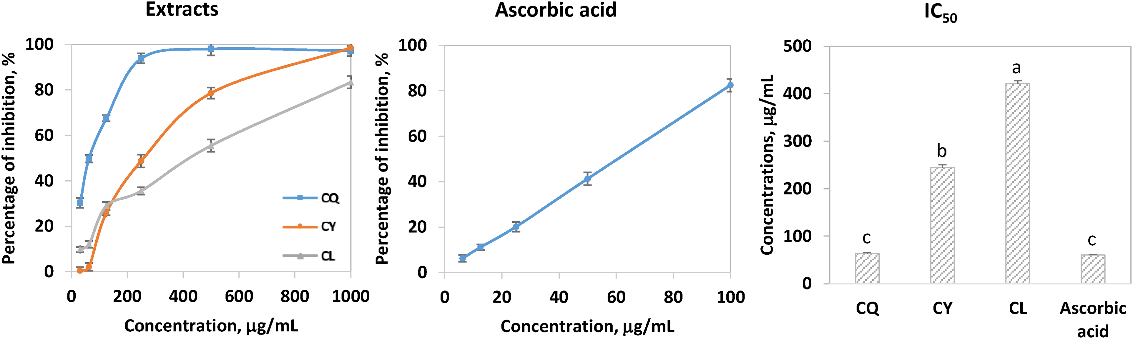
ABTS radical scavenging activity of the leaf extracts of the Camellia species. CQ, CY, and CL represent Camellia quephongensis, Camellia yokdonensis, and Camellia longii. Error bars indicate standard deviation of the means. Different letters (a, b, c) indicate significant differences (P < .05) in the activity among the extracts and ascorbic acid.
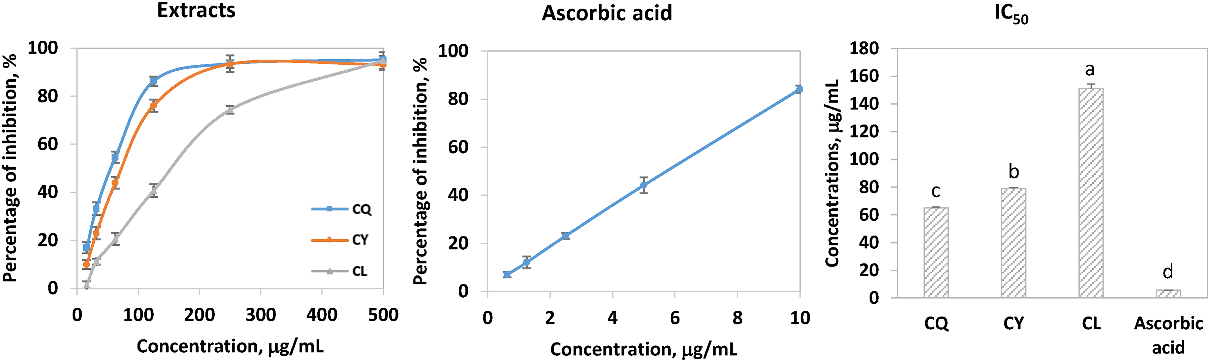
DPPH radical scavenging activity of the leaf extracts of the Camellia species. CQ, CY, and CL represent Camellia quephongensis, Camellia yokdonensis, and Camellia longii. Error bars indicate standard deviation of the means. Different letters (a, b, c, d) show significant differences (P < .05) in the activity among the extracts and ascorbic acid.
As stated earlier, extracts from Camellia leaves generally possess strong antioxidant effects. For instance, one study reported extracts from C. sinensis mature leaves growing in Malaysia exerted strong ABTS and DPPH scavenging capacities (IC50 = 180 and 40 μg/mL, respectively). 28 The figures for extracts of Camellia fascicularis leaves collected in China were 432.72 and 14.07 μg/mL. 29 The C. sinensis and C. fascicularis could have a lower capacity in neutralizing ABTS radicals when compared to C. quephongensis, but they all showed higher potency in removing DPPH radicals. This can be partially explained as discussed above on acetonic extracts. Research has indicated catechins are among the main contributors to antioxidant activity of Camellia plants. For example, levels of catechins in various types of tea made from C. sinensis leaves collected in China were shown to have a strong relationship with free scavenging potential. 30 Moreover, these compounds demonstrated significant efficacy in inhibiting the formation of free radicals, especially EGCG.30,31 Similarly, our previous study showed that EGCG and epicatechin had strong positive correlations with the potential of C. quephongensis leaf extracts to remove free radicals. 25 In the present study, catechin, epicatechin, and EGCG were the major phenolics in all the examined samples. Perhaps, the highest concentration of EGCG in C. quephongensis accounted for its superior ABTS and DPPH activities.
Reducing Power Capacity
Along with the free radical scavenging assays, a reducing power assay was used to predict the antioxidant activity of the Camellia extracts. As depicted in Figure 3, C. quephongensis and C. longii presented a significantly higher ability to reduce Fe3+ to Fe2+ compared to C. yokdonensis. This could be because C. quephongensis and C. longii may contain potent antioxidants, which either have high concentrations or are chemically structured with electron-donating groups. The reducing power of a molecule has a relationship with its electron transfer capacity and therefore correlates with antioxidant activity. 32 Camellia plants are known to be rich in flavonoids whose molecules are characterized by the presence of hydroxyl groups attached to aromatic rings. These phytochemicals have been shown to function as potent reducing agents and antioxidants. 33 Research has suggested o-catechol group in the B-ring of flavonoid molecules plays an important role in antioxidant capacity. 34 High levels of flavonoids in plant samples are often indicative of powerful antioxidant activities. 35

Reducing power capacity of the leaf extracts of the Camellia species at 250 μg/mL. CQ, CY, and CL represent Camellia quephongensis, Camellia yokdonensis, and Camellia longii. Error bars show standard deviation of the means.
Inhibition of Albumin Denaturation
Exposure of proteins to heat can lead to denaturation, causing alterations in their molecular structure and subsequent loss of their normal physiological functions. Denaturation of tissue proteins is closely associated with inflammatory diseases. 36 In this work, the Camellia extracts were assayed for their ability to inhibit bovine albumin denaturation. In other words, the in vitro anti-inflammatory activity of the extracts was predicted. As shown in Figure 4, at each of the examined concentrations (250, 500, and 1000 μg/mL), the C. quephongensis extract presented the highest percentage of inhibition, followed by the C. yokdonensis extract. The activity appeared to show a dose-dependent increase with the extract concentration. Based on the collected data, the IC50 value estimated for the C. quephongensis extract was 216.28 ± 3.70 μg/mL while the figures for the other samples were greater than 1000 μg/mL. In comparison with the reference standard (ie, diclofenac) (IC50 = 85.20 ± 4.99 μg/mL), all the Camellia samples may be less effective in protecting albumin from denaturation. However, in prior research, we reported that an aqueous methanolic extract of C. quephongensis leaves was much better at inhibiting albumin denaturation than acarbose. 25 The mode of action by which plant extracts prevent the heat-induced denaturation of albumin remains unclear. Studies suggest that specific components in the extracts, such as flavonoids, phenolic acids, and tannins, may interact with the aliphatic regions surrounding lysine residues on albumin molecules. 37 This interaction potentially shields albumin from heat-induced denaturation. Nevertheless, further investigations are needed to understand the exact mechanisms by which these compounds effectively inhibit denaturation.
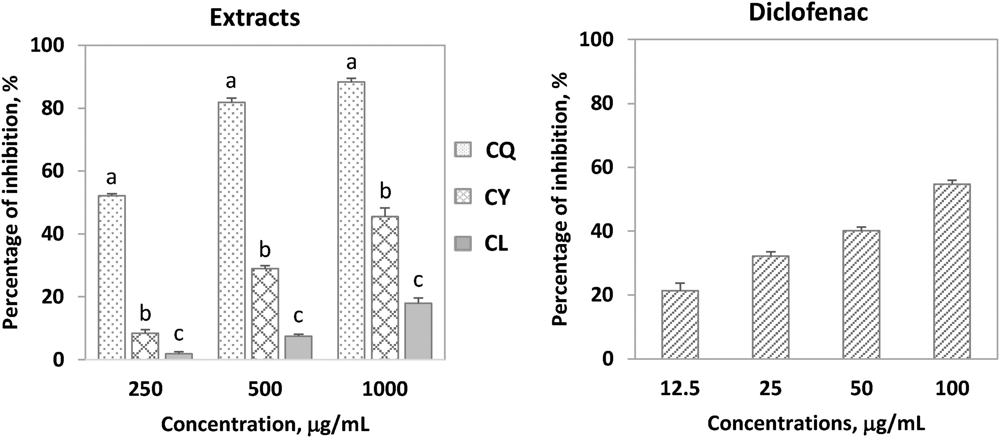
Inhibitory effects of the extracts of the Camellia species and diclofenac on albumin denaturation. CQ, CY, and CL stand for Camellia quephongensis, Camellia yokdonensis, and Camellia longii. Error bars indicate standard deviation of the means. Different letters (a, b, c) represent significant differences (P < .05) in the activity among the extracts at each concentration value.
The anti-inflammatory potential of Camellia plants has been well documented. Previous research indicated a strong inhibitory effect of aqueous extracts from C. sinensis dried leaves on albumin denaturation.38,39 C. fascicularis phenolics-rich extract down-regulated the expression of TNF-α, IL-6, and IL-1β while up-regulating the expression of IL-10 in lipopolysaccharide-induced human monocytes (THP-1) macrophages. 40 It was suggested that phenolics in Camellia extracts, such as catechins, ellagitannin, and flavonoid glycosides, contributed to the anti-inflammatory properties.
Inhibition of α-amylase
The capacity of the extracts to inhibit α-amylase was used to evaluate their potential antidiabetic properties. 41 Figure 5 illustrated the relationships between the percentage of inhibition (%) and the extract concentrations (μg/mL). It also graphically showed IC50 values of the extracts and acarbose. The extract from C. yokdonensis exerted the most potent enzymatic inhibitory activity (IC50 = 407.43 ± 1.97 μg/mL), followed by the one from C. quephongensis (IC50 = 671.26 ± 17.44 μg/mL). The C. longii extract may have the lowest activity as it had the greatest IC50 (721.47 ± 18.85 μg/mL). All the IC50 values were higher than that of acarbose (IC50 = 91.03 ± 6.47 μg/mL), implying weaker anti-α-amylase activity of the extracts. Camellia species have gained attention in recent investigations for their potential in inhibiting carbohydrate ingestion. Research has highlighted the bioactive compounds present in Camellia plants, such as flavonoid glycosides and catechins, which exhibit inhibitory efficacies on carbohydrate-digesting enzymes, including α-amylase and α-glucosidase.42,43 The findings of the present study hold promise for diabetes prevention and treatment using these Camellia species.

Inhibitory effects of the leaf extracts of the Camellia species and acarbose on α-amylase. CQ, CY, and CL represent Camellia quephongensis, Camellia yokdonensis, and Camellia longii. Error bars show standard deviation of the means. Different letters (a, b, c, d) indicate significant differences in the activity among the extracts and acarbose at P < .05.
Conclusions
This present work is the first investigation into chlorophyll and carotenoid contents, phenolic compounds, free radical scavenging properties, anti-α-amylase and albumin denaturation inhibitory activities of acetonic extracts from leaves of three Camellia species. Most of the phenolics in C. quephongensis were found at greater concentrations compared to the other species. The C. quephongensis extract showed the most potent capacity to scavenge ABTS and DPPH radicals. All the Camellia extracts could have weak α-amylase and albumin denaturation inhibitory activities. The findings of this work will provide the first evidence concerning the bioactive constituents present in the species and shed light on the potential health benefits associated with their leaves. Further research is warranted to isolate and characterize phytochemicals of the Camellia leaves as well as assess their in vivo bioactivities of importance to human health.
Experimental
Chemicals used in the study, sample collection, and determination of pigments and phenolics were described in Supplemental Material (Sections S1-S5). The antioxidant activity of the acetonic extracts was evaluated using ABTS and DPPH radical scavenging assays and reducing power assay. Inhibitory effects of the acetonic extracts on bovine albumin denaturation and α-amylase were also determined. All the bioassays and statistical analyses were detailed in Supplemental Material (Sections S6-S10).
Supplemental Material
sj-docx-1-npx-10.1177_1934578X241249090 - Supplemental material for Comparative Analysis of Pigments, Phenolics, and Bioactivities of Three Camellia Species Growing in Vietnam
Supplemental material, sj-docx-1-npx-10.1177_1934578X241249090 for Comparative Analysis of Pigments, Phenolics, and Bioactivities of Three Camellia Species Growing in Vietnam by Danh C. Vu, Trang H. D. Nguyen, Hieu Tran-Trung, Nguyen Hoang Tuan and Nguyen T. M. Nguyet in Natural Product Communications
Footnotes
Declaration of Conflicting Interests
The authors declared no potential conflicts of interest with respect to the research, authorship, and/or publication of this article.
Funding
The authors received no financial support for the research, authorship, and/or publication of this article.
Ethical Approval
Ethical approval is not applicable to the article.
Statement of Human and Animal Rights
This article does not contain any studies with human or animal subjects.
Statement of Informed Consent
There are no human subjects in this article and informed consent is not applicable.
Supplemental Material
Supplemental material for this article is available online.
References
Supplementary Material
Please find the following supplemental material available below.
For Open Access articles published under a Creative Commons License, all supplemental material carries the same license as the article it is associated with.
For non-Open Access articles published, all supplemental material carries a non-exclusive license, and permission requests for re-use of supplemental material or any part of supplemental material shall be sent directly to the copyright owner as specified in the copyright notice associated with the article.