
Abstract
A 13-year-old boy developed right-sided Horner’s syndrome following resection of a benign mediastinal schwannoma extending from T1 to T3. Postoperatively, he exhibited ptosis, miosis, and anhidrosis, confirmed by starch iodine testing. The tumor likely involved the upper thoracic sympathetic ganglia—a rare site for schwannomas. This image highlights a rare iatrogenic cause of preganglionic Horner’s syndrome. While Horner’s is classically associated with apical lung or cervical lesions, this case emphasizes the importance of recognizing postoperative Horner’s syndrome as a clinical clue to cervicothoracic sympathetic injury. It highlights the value of anatomical-clinical correlation in localizing lesions along the sympathetic pathway.
Keywords
13-year-old boy underwent right posterolateral thoracotomy for mediastinal mass located just posterior to the superior vena cava in the right paravertebral region of the superior mediastinum extending from T1 to T3. The lesion, measuring 5 × 3 × 2 cm, was incidentally detected on routine chest imaging and confirmed as a schwannoma on histopathology. On postoperative day one, the parents noted subtle drooping of the right eyelid, prompting neurology referral. Clinical examination revealed a smaller right palpebral fissure, mild right-sided ptosis, miosis, and anhidrosis affecting the right hemiface. A starch iodine test was done, which confirmed anhidrosis on the right half of the face and neck, respecting the midline and the medial aspect of the right arm and forearm and right hand (Figure 1). Figure showing starch on thinly applied and subsequently dried 2% iodine tincture turns blue in the presence of sweating on the left side of face and chest (A); ptosis and miosis in the right eye (B); and axial section of Contrast-enhanced computed tomography (CECT) of the chest showing superior mediastinal right paravertebral and paratracheal apical mass extending from T1 to T3 level- arrow (C).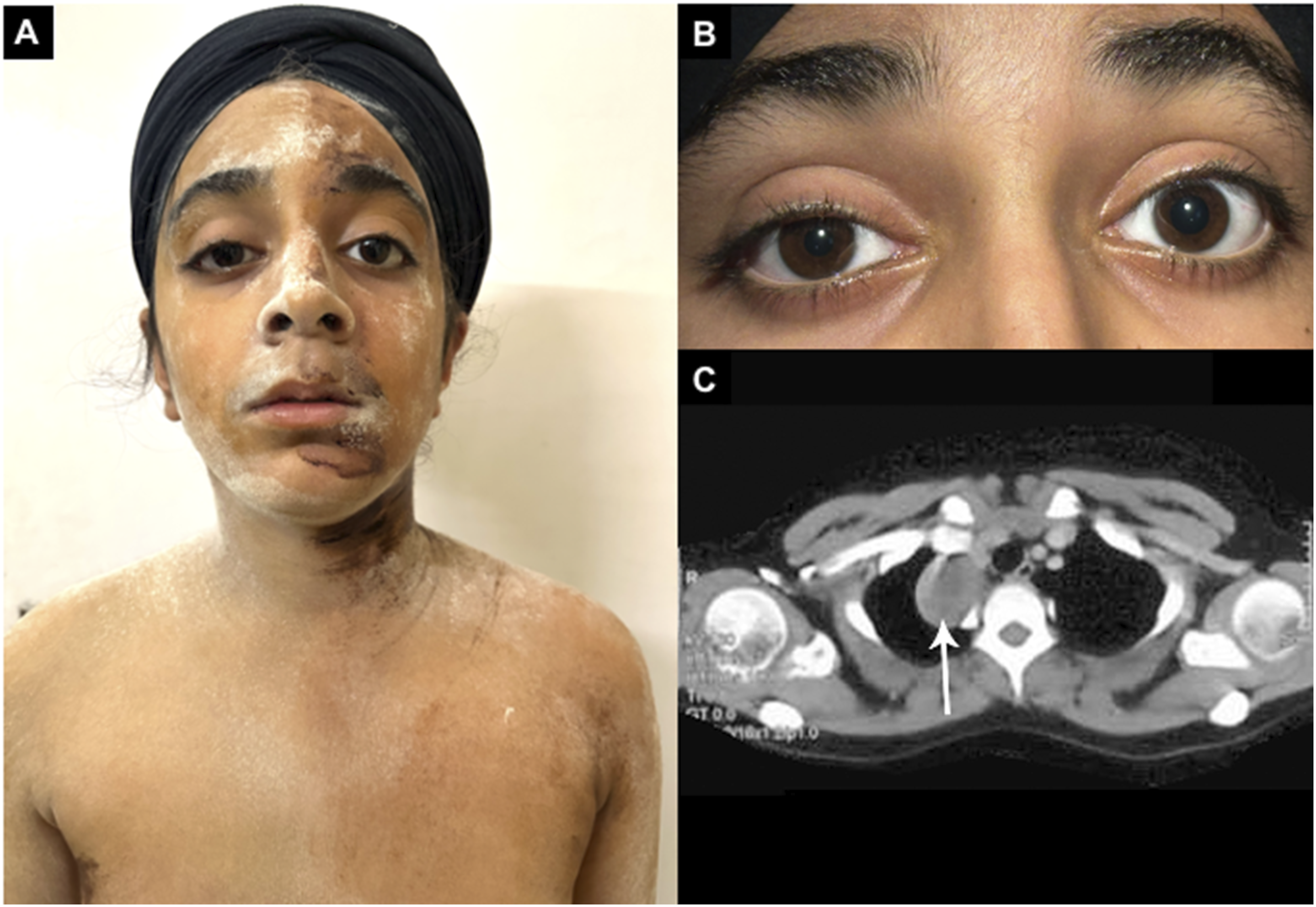
Schwannomas, though accounting for only 10% of mediastinal tumors, mostly arise in the posterior mediastinum from intercostal nerves, with rare cases involving the vagus or phrenic nerves. 1 These tumors are often discovered incidentally on chest imaging, particularly in asymptomatic young adults. 2 This case is unique for the development of Horner’s syndrome following resection of a benign mediastinal schwannoma, a rare association. Horner’s syndrome is typically seen in cervical sympathetic chain tumors or malignant apical lung masses, however can occur as a postoperative complication. The schwannoma likely originated from the first to third thoracic ganglion rather than a peripheral thoracic nerve. Sympathetic ganglia tumors more commonly include ganglioneuromas or neuroblastomas, making schwannoma at this location particularly rare. 3 Isolated preganglionic Horner’s syndrome warrants imaging of the cervicothoracic region.
Footnotes
Author Contributions
Riya Sharma MD – idea and draft with data colletion and manuscript writing. Siddharth Chand MD - data collection. Ritu Shree MD DM, Manoj Kumar Goyal MD, DM – editing and review.
Declaration of Conflicting Interests
The author(s) declared no potential conflicts of interest with respect to the research, authorship, and/or publication of this article.
Funding
The author(s) received no financial support for the research, authorship, and/or publication of this article.