






Abstract
Objective:
The aim of this study was to find the significantly altered genes in cumulus cells of women with ovarian endometriosis by using microarray and quantitative polymerase chain reaction analysis.
Methods:
Thirty women with ovarian endometriosis and 30 age–body mass index matched controls (women with infertility as a result of pure male factor) were enrolled in this study. Cumulus cells from study participants who underwent controlled ovarian hyperstimulation were isolated mechanically. Microarray comparative genomic hybridization was used to compare the transcriptome of cumulus cells from women with ovarian endometriosis and controls. According to the different expression levels in the microarrays and their putative functions, KRAS, ZNF322, and SDHA were selected and analyzed by real-time quantitative polymerase chain reaction.
Results:
There was no significant difference in the basal conditions between patients with endometriosis and controls, such as age, body mass index, basal follicle stimulating hormone and estradiol levels, and total gonadotrophin dosage. The gene expression profile of cumulus cells from patients with endometriosis was significantly different from that of controls. A total of 295 genes were significantly up- or down-regulated (p-value < 0.05 and absolute fold change > 1.5). For all of the genes adjusted p-value was found to be 0.999. Polymerase chain reaction analysis showed that KRAS and ZNF322 mRNA levels in the cumulus cells of patients with ovarian endometriosis were significantly up-regulated compared to controls (fold changes: 3.05 and 3.22, respectively).
Conclusion:
KRAS and ZNF322 mRNA levels in the cumulus cells of patients with ovarian endometriosis were significantly up-regulated.
Introduction
Endometriosis is a benign, invasive gynecological disease characterized by endometrial glands and stroma outside the uterus. 1 Endometriosis affects nearly 10%–15% of women of reproductive age and it is estimated that 30%–50% of women with endometriosis are subfertile. 2 The cause of endometriosis and endometriosis-related fertility problems are uncertain. While the fecundity rate of fertile couples is nearly 15%–20%, this rate is 2%–10% for women with endometriosis. 3 In vitro fertilization (IVF) studies have shown that endometriosis may be associated with low ovarian reserve, and low oocyte and embryo quality.4,5 Many clinical studies comparing women with endometriosis to controls have shown low implantation, pregnancy, and live birth rates. 6 This negative effect is more pronounced in ovarian endometriosis especially compared to other localizations of endometriosis. 7 In oocyte donation cycles, there are similar implantation rates between women with endometriosis and controls, with other scientific data showing disrupted oocyte and/or embryo quality causing low IVF outcomes in women with endometriosis. 8
Cumulus cells (CCs) immediately surrounding the oocyte provides nutrition and support of the oocyte. 9 There is a two-way relationship between oocyte and CCs via gap junctions which ensures protein and metabolite transportation between two cell type. This balanced relationship between the CCs and oocyte has critical importance for maturation and function of oocytes. 10 Disruption or imbalance of this relationship affects oocyte quality causing reductions in embryo quality and pregnancy rates. CCs are easily obtained during oocyte pick-up and their use as an indirect marker to evaluate oocyte quality has increased the need to understand the characteristics of CCs in recent times.11,12
Gene expression has vital importance in regulation of nearly every moment of cellular life. Revealing gene transcription profiles provides information about cellular processes and gives important information to determine the cause of biological problems. 13 CCs play important roles in oocyte development and genes expressed in CCs can be used as markers for oocyte and embryo quality. 14 Expression profiling studies showed that some genes in CCs (such as hyaluronic acid synthase 2, cyclooxygenase 2, and steroidogenic acute regulatory protein) may also be associated with the embryo quality.14–16 There are just a few studies published relating CC to infertile women with ovarian endometriosis; however, they are not very rare. 17
In this study, the CCs of women with ovarian endometriosis were compared with controls using the microarray and real-time quantitative polymerase chain reaction (RT-qPCR) to find the significantly altered genes between two groups.
Materials and methods
Study group
This case-control included a total of 60 patients, 30 with ovarian endometriosis and 30 controls attending to Division of Reproductive Endocrinology and Infertility, School of Medicine, Inonu University, between January 2016 and February 2018. This study received local ethics committee permission and patients were included in the study after giving written informed consent (Approval No: 2014/131).
The case group was women with ovarian endometriosis and diagnosis of ovarian endometriosis was placed in line with the criteria for typical ultrasonographic appearance of endometriotic cysts at least twice at two cycle intervals. 18
The control group was chosen from among couples with male factor infertility. Controls had serum follicle stimulating hormone (FSH) levels < 10 IU/mL in the early follicular stage, normal pelvic anatomy on ultrasonography and hysterosalpingography. All controls who underwent laparoscopy for the dysmenorrhea or chronic pelvic pain had normal findings on diagnostic laparoscopy. The women with poor ovarian reserve, tubal factor infertility, hyperprolactinemi, and polycystic ovarian syndrome were excluded from the study.
The inclusion criteria for both groups were age in the interval 25–39 years and body mass index (BMI) 21–29.9 kg/m2. The exclusion criteria for the present study were age ⩾ 40 years, BMI > 30 or <20 kg/m2, FSH ⩾ 10.0 IU/mL in the early follicular phase, chronic anovulation, hydrosalpinx, or chronic systemic diseases (chronic hypertension, diabetes mellitus, and thyroid diseases).
Controlled ovarian stimulation
Patients in both groups underwent controlled ovarian stimulation treatment. Standard GnRH antagonist regime began on the fifth to sixth day of a spontaneous menstrual cycle. Recombinant FSH stimulation began on the second to third day of menstruation. Gonadotropic dose was set according to serum hormone values and follicular development on ultrasonography. To induce ovulation, 10.000 hCG was administered after measurement of at least three follicles above 18 mm. Thirty-six hours after hCG administration, oocytes were collected with a needle under transvaginal ultrasound guidance. Embryo transfer was completed 2–3 days after oocyte collection.
Collection of CC samples
The cumulus oocyte complex (COC) belonging to one of the follicles above 18 mm was collected 36 h after hCG administration. The aspirate in all cases was clear without any suggestion of endometrioma cyst content fluid. After aspirating the follicle, with identification of the COC, the CCs were mechanically separated from the oocyte under a microscope. After washing with culture medium, they were transferred into a tube containing ribonucleic acid (RNA) later and immediately frozen with liquid hydrogen and stored at −80°C until RNA extraction. CCs were collected from the diseased ovaries with ovarian endometriosis and only one CC complex from mature oocytes from each patient were included in the study.
CCs obtained from 10 patients with endometriosis and 10 controls was used for microarray analysis, and CCs of 20 patients with endometriosis and 20 controls was used for RT-qPCR analysis. One control was excluded from microarray analysis due to low RNA quality (Figure 1).
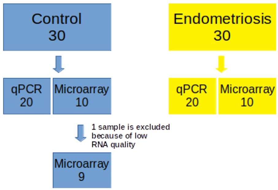
Flow chart of study cohort.
Microarray analysis
RNA purity were assessed with an ND-1000 spectrometer. The Affymetrix whole transcript expression array method was completed in accordance with the manufacturer’s instructions (Gene Chip WT Pico reagent Kit). cDNA was synthesized in line with the manufacturer’s instructions using a GeneChip WT Pico amplification kit. The GE Buffer (Genomic DNA Elimination Buffer) included in the cDNA synthesis kit destroys genomic DNA. This step is performed according to the kit protocol and performed at 42°C for 5 min before the reverse transcriptase step.
The synthesized cDNA was fragmented and labeled with biotin using a GeneChip WT Terminal labeling kit. Nearly 5.5 Mb marked DNA was hybridized with Affymetrix GeneChip Human 2.0 ST array. The hybridized array was washed, stained with GeneChip Fluidics Station 450, and scanned with a GCS300 scanner. Signal values were calculated using Affymetrix GeneChip® Command Console® software (AGCC).
Data processing and microarray data analysis
Raw data were automatically transferred to the Affymetrix data extraction protocol using software in the AGCC. Data were collected after transport in CEL files and normalized with robust multiaverage method. This process was applied within the Affymetrix® Expression Console™ Software (EC). These results were transferred for gene-level RNA analysis and differentially expressed gene analysis (DEG analysis) was performed. Analysis fold change (FC) was used to compare samples between case and controls. For the DEG set, hierarchical cluster analysis complete linkage and Euclidean distance was used. Gene-enrichment and functional annotation analyses were completed with gene ontology (http://geneontology.org/) and KEGG (http://kegg.jp).
PCR analysis
Differentially expressed mRNAs on microarray were confirmed by RT-qPCR. In short, the first chain complementary synthesis reaction was completed using an M-MLV reverse transcriptase kit. The Power SYBR Green PCR Master Mix (Applied Biosystems) and Applied Biosystem 7900HT system was used to perform amplification reactions. In this study, Kirsten rat sarcoma viral oncogene homolog (KRAS), zinc-finger protein (ZNF)322A, and succinate dehydrogenase complex subunit A(SDHA) mRNA-specific PCR primers (Qiagen, Hilden, Germany) were used. GAPDH was used as internal control. During PCR thermal cycling, conditions were 95°C for 10 min to activate the polymerase and denature the template, followed by 40 cycles of denaturation at 95°C for 15 s, annealing at 60°C for 60 s, and extension at 72°C for 30 s. Each RT-qPCR reaction was repeated three times and FC analyses were completed using the DDCt method.
Statistical analysis
Statistical analysis was completed using the SPSS program. The distribution of data was determined with the Kolmogorov–Smirnov test. Data following a normal distribution were showed as mean ± standard deviation, whereas data not following a normal distribution were showed as median and interquartile range (IQR). Categorical data were analyzed with the Pearson chi-square and Fisher exact test. For comparison of case and control groups, the student t test for independent samples or the Mann–Whitney U test was used, as appropriate p < 0.05 was accepted as statistically significant.
Results
Basal characteristics
Basic demographics and treatment characteristics of study participants are shown in Tables 1 and 2. There was no difference between women with ovarian endometriosis and control groups in terms of age, BMI, serum FSH, and estradiol levels and total gonadotropic dose used during the IVF treatment process. Similarly, there was no difference between the patients with ovarian endometriosis and controls in terms of number of oocytes, number of metaphase II oocytes, number of total embryos, and clinical pregnancy rates.
Clinical characteristics of study participants included in the microarray analysis.

BMI: body mass index; FSH: follicle stimulating hormone; E2: estradiol. Data are reported as mean ± standard deviation or median (IQR). Mature oocytes: Oocytes with the first polar body visible. Clinical pregnancy rate: presence of embryonic heartbeat visualized by ultrasound at 6 weeks after embryo transfer.
Clinical characteristics of study participants included in the PCR analysis.

BMI: body mass index; FSH: follicle stimulating hormone; E2: estradiol. Data are reported as mean ± standard deviation or median (IQR). Mature oocytes: Oocytes with the first polar body visible. Clinical pregnancy rate: presence of embryonic heartbeat visualized by ultrasound at 6 weeks after embryo transfer.
Microarray analysis
In this study, 53.618 transcripts were researched in ovarian endometriosis and control patients. Of these, 295 genes were identified to have different expression in both groups. Cluster analysis of top nine differently expressed genes in patients with ovarian endometriosis and controls is presented in Figure 2.

A heat map of top nine differentially expressed genes shows that KRAS, ZNF322, SNAP23, FAM8A1, SUZ12P1, and CFLP1 are up-regulated, while MIR4718, SPRR2G, and SDHA are down-regulated in endometriosis compared to the controls.
A total of 295 genes were significantly up- or down-regulated (p-value < 0.05 and absolute FC > 1.5). For all of the genes adjusted p-value was found to be 0.999. Among the 295 different genes, the genes with highest differential expression between the two groups were identified as kirsten rat sarcoma viral oncogene homolog (KRAS), zinc-finger protein (ZNF) 322, succinate dehydrogenase complex subunit A (SDHA), family with sequence similarity 8 member A1 (FAM8A1), SUZ12 pseudogene 1 (SUZ12P1), small proline-rich protein 2G (SPRR2G), cofilin 1 pseudogene 1(CFL1P1), microRNA 4718 (MIR4718), and synaptosomal-associated protein 23 (SNAP23). Principal components analysis (PCA) of DEGs and entire gene set are provided in Figure 3.

(a) Principal components analysis (PCA) of differentially expressed genes and (b) PCA of entire gene set.
The gene ontology results for the differently expressed genes are given in Figure 4(a)–(c) as a bar graph. The gene ontology and KEGG analysis results identified that these different genes play roles in cellular and metabolic processes, especially, and in biological processes like protein and ion binding.
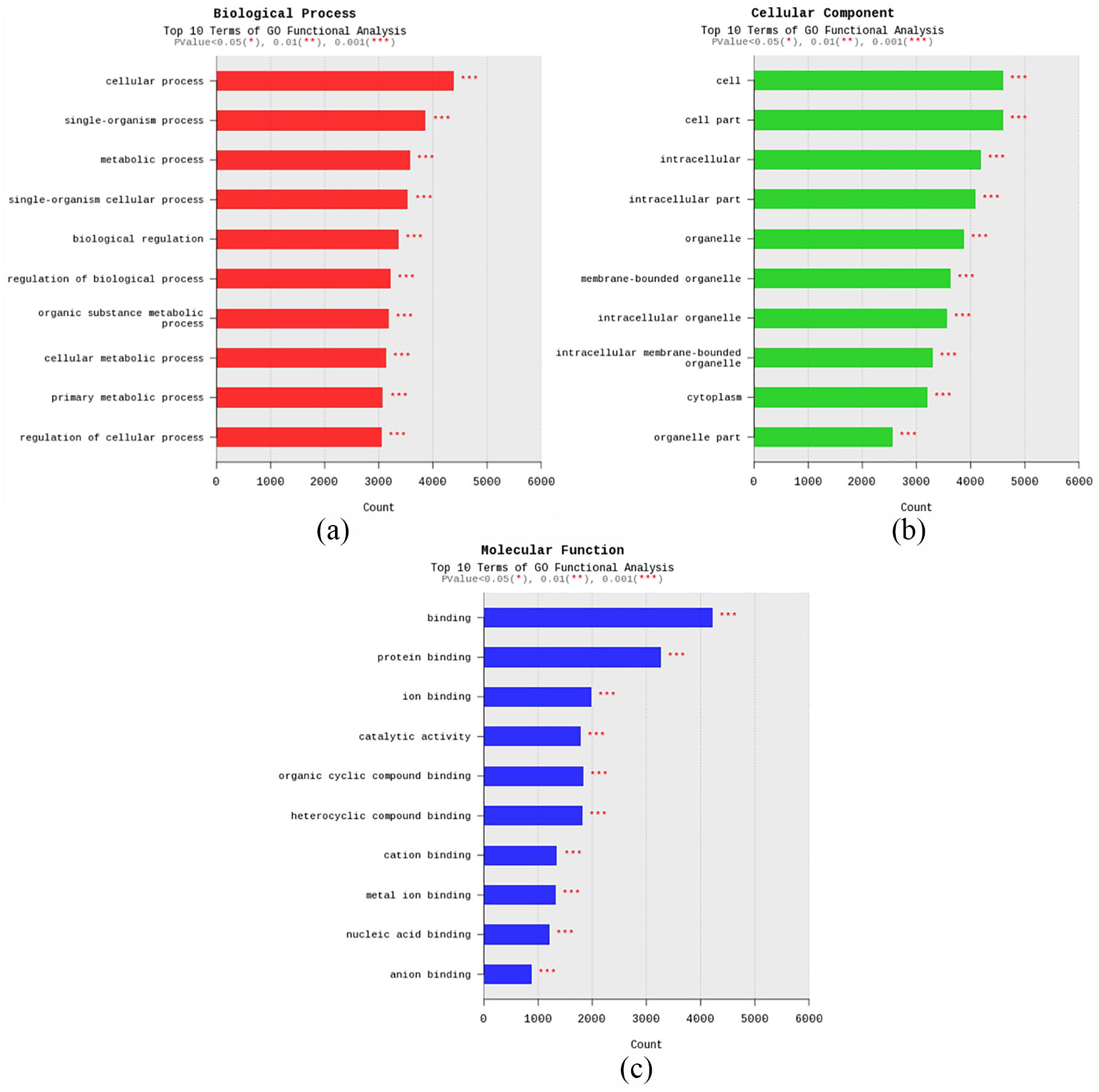
(a) GO functional analysis (biological processes), (b) GO functional analysis (cellular components), and (c) GO functional analysis (molecular functions).
RT-qPCR validation
The results of PCR analysis identified that KRAS and ZNF322 mRNA gene expression level in CCs of patients with ovarian endometriosis is up-regulated compared to controls (FCs 3.05 and 3.32, respectively, both p values < 0.01). On the contrary, there was no statistically significant difference regarding SDH mRNA expression levels in the CCs of patients with ovarian endometriosis and controls.
Discussion
The present study found that KRAS and ZNF322 gene expression was higher in the CCs of endometriosis patients compared to controls.
Based on the previous studies, it is clear that RAS activation (most likely KRAS) in the ovary is cell and context specific. 19 KRAS, a well characterized oncogene, has been implicated in the pathogenesis of endometriosis. 20 Activation of an oncogenic KRAS gene in the murine ovarian surface epithelium results in the de novo formation of lesions with endometriotic morphology. 19 The authors of that study speculate that RAS pathway activation by different mechanisms may play an important role in the development of endometriosis in humans. While mutational changes to KRAS appears to be a pivotal change in endometriosis-related ovarian cancers, 21 KRAS up-regulation was found in CC from patients with ovarian endometriosis suggesting that KRAS expression in CC is a potential key biomarker for ovarian endometriosis-related infertility.
In addition to the role of KRAS on the pathogenesis of endometriosis, it may have a main role on endometriosis-related infertility. Activation of the endogenous RAS pathway plays an important physiological role in both ovulation and luteinization of granulosa cells. 22 RAS is a physiological mediator of gonadotropin signals in ovarian follicles and has critical importance for follicular rupture and oocyte meiotic maturation and COC expansion. 23 However, animal studies have shown that activation of the KRAS gene in ovarian surface epithelium in mice causes lesions similar to endometriosis. 20 In addition, inappropriate expression of a constitutively active KRAS mutant in CCs gives rise to arrest of the cell cycle in granulosa cells, abnormal follicular growth, and premature ovarian insufficiency. 23 Premature exposure of granulosa cells to constitutively active KRAS leads to the formation of abnormal follicles, in which granulosa cells cease to divide, are non-apoptotic, and fail to granulosa cell differentiation.22,24
ZNF322 is a member of zinc-finger transcription factor family and may regulate transcriptional activation in mitogen activated protein kinases (MAPKs) (extracellular signal-regulated kinases 1 and 2 (ERK 1/2) signaling pathway). 25 ERK1/2 may act as the intrafollicular mediators to stimulate the COC expansion and oocyte maturation. 26 MAPK signaling pathway plays an important role in ovulation, oocyte maturation, COC expansion, and luteinization.26,27 The effect of ERK1/2 activation in granulosa cells of preovulatory follicle at earlier stages of growth indicate that activation of these kinases is controlled tightly by specific mechanisms. Inappropriate activation of ERK1/2 in granulosa cells of small growing follicles might disrupt normal follicular development. 27 Overexpression of ZNF322A promoted self-renewal ability and increased stemness-related gene expressions in vitro and in vivo. 28 ZNF322 up-regulation was found in CC from patients with ovarian endometriosis suggesting that ZNF322 expression in CC is a potential key biomarker for ovarian endometriosis-related infertility. Although there was a difference regarding SADH gene expression in microarray analysis, SADH gene expression was not different in RT-qPCR. PCR is a commonly used validation tool for confirming gene expression results obtained from microarray analysis; however, microarray and PCR data often result in disagreement. 29
Women with ovarian endometriosis ovulate fewer oocytes than healthy controls 30 and studies have shown that compromised follicular development was reported in women with endometriosis. 31 While fecundity in normal couples is 15%–20%, this rate reduces to 2%–10% in women with endometriosis. With endometriosis, 3-year cumulative pregnancy rates 32 are lower compared to unexplained infertility. Especially low pregnancy rates and live birth are associated with ovarian endometriosis compared to other forms of endometriosis. 7 The cause of this negative effect on oocyte and embryo quality in ovarian endometriosis may be due to differences in the expression of genes like KRAS in COC. Increases in knowledge accumulation in this area will contribute to enlightening the correlation of ovarian endometriosis and infertility and development of new strategies to improve the low clinical outcomes of IVF reported in these patients.
One of the limitations of this study is that endometriosis diagnosis was not made histopathologically. Contrary to this, characteristic ultrasonographic appearance of ovarian endometriosis with transvaginal ultrasonography was reported to have high sensitivity (84%–100%) and specificity (90%–100%) in previous studies. 18 Another limitation of the study is that CCs of women with endometriosis but without endometrioma were not evaluated. The basic features of the two groups such as age and BMI were matched which is a strong aspect of our study. This study is the first in the literature to use microarray analysis of CCs from ovarian endometriosis patients.
In conclusion, microarray analysis of CCs belonging to metaphase II oocytes in endometriosis patients show different gene expression profiles compared to controls. Validation with PCR shows that these patients had increased KRAS and ZNF322 gene expression. There is a need for larger series studies to explain the etiopathogenesis of infertility related to ovarian endometriosis. It is assumed that a better understanding of the underlying molecular mechanism of infertility related to ovarian endometriosis may provide novel diagnostic and therapeutic approaches for endometriosis and infertility.
Footnotes
Acknowledgements
The authors would like to thank the TUBITAK (The Scientific and Technical Research Council of Turkey) and Scientific Research Projects Unit of Inonu University. The authors would also like to thank Ismail Tas, MSc, for helping them with PCR analysis.
Authors’ note
The summary of the article was presented as an oral presentation at the 20th World Congress on in Vitro Fertilization (ISIVF 2019).
Author contributions
A.K. and G.T. researched literature and conceived the study. A.K, G.T., and N.T. were involved in protocol development and patient recruitment. Y.C. was involved in genetic analysis and B.D. was involved in bioinformatics analysis. All authors reviewed and edited the manuscript and approved the final version of the manuscript.
Declaration of conflicting interests
The author(s) declared no potential conflicts of interest with respect to the research, authorship, and/or publication of this article.
Ethical approval
This study received Malatya Clinical Ethics Committee permission and patients were included in the study after giving written informed consent (Approval No: 2014/131).
Funding
The author(s) disclosed receipt of the following financial support for the research, authorship, and/or publication of this article: This study was supported by TUBITAK (The Scientific and Technical Research Council of Turkey) (Project number: 214S667) and Scientific Research Projects Unit of Inonu University (Project number: TSA-2018-1361).
Informed consent to participate
Written informed consent was obtained from all subjects before the study.
Informed consent to publish
Written informed consent was obtained from the patient(s) for their anonymized information to be published in this article.
