






Abstract
Purpose:
To describe cytomegalovirus (CMV) retinitis outcomes in HIV-infected and non–HIV patients at a tertiary care center.
Participants:
Twenty-six eyes from 20 patients with active CMV retinitis were included in this study. Patients were immunocompromised due to HIV or secondary to immunosuppressive therapy for malignancy and organ transplantation.
Methods:
This is a retrospective, observational study of patients with newly diagnosed active CMV retinitis. The main outcomes included the incidence of visual acuity loss, the loss of visual acuity to 20/200 or worse, and the loss of 3 lines of vision or more. Secondary outcomes included the identification of risk factors for these visual results and the development of various complications such as retinal detachment (RD) and cataract formation.
Results:
In all, 51.9% (n = 14) of eyes lost vision from baseline at most recent follow-up and 34.6% (n = 9) lost 3 lines or more of vision, 34.6% (n = 9) of the eyes lost significant vision at most recent follow-up and had a final vision of 20/200 or worse, and 22.73% of phakic eyes underwent cataract surgery, whereas 23.1% (n = 6) of eyes developed RD. Patients presenting with a CD4+ T-cell count <100 cells/µL were more likely to lose vision when compared to those presenting with a count >100 cells/µL (P = .0440). Although not statistically significant, patients who were immunocompromised due to HIV were less likely to lose 3 or more lines of vision (P = .1881) and less likely to have a final visual acuity of 20/200 or worse (P = .1881), when compared to patients who were immunocompromised due to other reasons. There was also a nonsignificant trend for eyes affected by a larger area of CMV retinitis at baseline (>25%) to have a final visual acuity of 20/200 or worse when compared to eyes with CMV retinitis involving <25% of the total retina (P = .089). We did not detect trends or associations between any other risk factors tested and visual outcomes. We did not identify an association between HIV status and baseline area or zone affected by CMV retinitis.
Conclusions:
Our cohort demonstrated that CMV retinitis remains a vision-threatening problem among patients who are immunocompromised due to HIV or other conditions. Immunocompromised patients are still at a significant risk of vision loss and complications from CMV retinitis and should be managed by a multidisciplinary team of physicians. In the immediate future, improved therapies are necessary to achieve immune recovery in patients, particularly for those remaining chronically immunosuppressed.
Introduction
Cytomegalovirus (CMV) retinitis is a potentially blinding infection affecting immunocompromised patients unable to generate a sufficient T-cell response against the organism. 1 The CMV retinitis is the most frequent ocular opportunistic infection in patients living with HIV/AIDS and is a significant cause of vision loss in this population. 2 In the pre–highly active antiretroviral therapy (pre-HAART) era, 20% to 40% of patients with HIV were affected, with a median time of 13 months from HIV diagnosis to 20/200 or worse visual acuity. 3,4 The use of HAART resulted in an 80% to 90% reduction in the incidence of CMV retinitis, and the Longitudinal Study of Ocular Complications of AIDS (LSOCA) found that immune recovery was associated with a 61% reduction in vision loss to 20/200 or worse. 4
Despite the availability of HAART and declining incidence, CMV retinitis still remains a vision-threatening problem among immunocompromised patients. In the era of HAART, the rate of vision loss to 20/200 or worse among the HIV-infected population is 3.4/100 eye-years, 2 and a significant percentage still develop complications such as retinal detachment (RD). 5 In addition, patients who are immunocompromised due to reasons other than HIV, such as organ transplant recipients or patients with cancer, may be chronically immunosuppressed and at risk of CMV retinitis and vision loss.
The goal of this study is to describe the outcomes of eyes with CMV retinitis in the era of HAART at Weill Cornell Medical College (WCMC), New York Presbyterian Hospital (NYP; New York, New York). This is the first study, to our knowledge, looking at a cohort of patients who are immunocompromised due to both HIV and non-HIV etiologies at a tertiary care center.
Methods
This was a retrospective, observational study of patients with newly diagnosed CMV retinitis who presented to WCMC and NYP for management between the years 2007 and 2015. The study protocol was approved by the institutional review board of WCMC. Patients were verified to have newly diagnosed active CMV retinitis based on examination by an experienced retina or uveitis specialist along with a positive serum or intraocular CMV titer. All patients were 18 years or older and required at least 6 weeks of follow-up to be included in this study. All etiologies of immunosuppression were included such as HIV or immunosuppressive therapy for organ transplantation and cancer. Eyes were excluded if the CMV retinitis was inactive at presentation or had a follow-up of less than 1 month.
Data collection at enrollment and at the follow-up time points included demographic information, medical and ophthalmic history, and ophthalmologic examination. The examination included best-corrected visual acuity, intraocular pressure (IOP), slit lamp examination, indirect ophthalmoscopy, and ocular imaging using optical coherence tomography (OCT), fluorescein angiography, and ultra-widefield fundus photography (OPTOS plc, Scotland, United Kingdom). Laboratory review included plasma, aqueous, or vitreous CMV viral load and CD4+ T-cell count. Immune recovery was defined as CD4 T-cell count ≥ 100 cells/µL. 2
Eyes were characterized based on location and size of retina affected. Zone 1 was defined as an area within 1500 μm from the nerve or 3000 μm from the center of the fovea, whereas all other eyes were grouped together into zones (2 + 3). 5 Eyes were grouped into having less than or greater than 25% of the total retina being affected by retinitis. 5
Four treating physicians participated in this study, determining the best management for each patient given the clinical presentation. The therapies that were utilized included ganciclovir implant, systemic medication (oral valganciclovir, intravenous ganciclovir sodium, intravenous foscarnet sodium, or intravenous cidofovir), intravitreal ganciclovir, intravitreal foscarnet, and/or CMV-specific T-lymphocyte infusion therapy. 1 The main outcome measures included visual acuity loss, visual acuity loss to 20/200 or worse, 2 and the loss of 3 or more lines of vision. Secondary outcomes included the identification of risk factors for these visual results and the development of various complications such as RD and cataract formation.
Statistical Analysis
The Wilcoxon signed rank test was used to test whether the median difference in the continuous variables at baseline and most recent follow-up was zero. Wilcoxon rank sum test was used to test the association between vision outcomes and age, whereas Fisher exact test was used for all other variables. McNemar test was used to test for associations between the outcomes of interest at baseline and at the last follow-up. Fisher exact test was also used to determine whether RD was associated with HIV statuses, baseline area, or zone of CMV retinitis. All P values were 2-sided and a P value ≤ .05 was considered statistically significant.
Results
The charts of 75 patients with a diagnosis of CMV retinitis were reviewed, and of these, 26 consecutive eyes from 20 patients with active CMV retinitis were included in this study. Six patients had bilateral disease that was active during data collection. Eyes were excluded if the CMV retinitis was inactive at presentation. Baseline characteristics of participants are detailed in Table 1. The median age in our cohort was 49.5 years with a median follow-up of 68.29 weeks. Seventy percent of the patients were men. The majority of patients were immunocompromised due to HIV (14 patients, 19 eyes). Five patients (6 eyes) had a history of hematologic malignancy and were immunocompromise post stem cell transplant, whereas 1 patient was on immunosuppressive therapy for organ transplantation. At initial ophthalmic visit, 3 patients presented with an initial T-cell CD4 count >100 cells/µm, 12 presented with a CD4 count <100 cells/µm, and 4 had an unknown CD4 count. During follow-up, the median CD4+ T-cell count increased (P = .004).
Baseline Characteristics.a
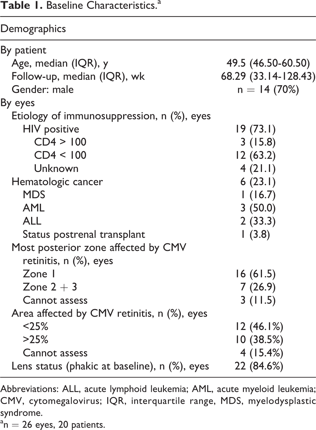
Abbreviations: ALL, acute lymphoid leukemia; AML, acute myeloid leukemia; CMV, cytomegalovirus; IQR, interquartile range, MDS, myelodysplastic syndrome.
an = 26 eyes, 20 patients.
At initial presentation (Table 1), 22 (84.6%) eyes were phakic. The majority of eyes presented with zone 1 disease (n = 16, 61.5%), whereas the rest were grouped as zones 2 and 3 (Figures 1 and 2). Of the eyes with zone 1 disease, 11 had foveal involvement based on OCT imaging (Figure 3). At baseline, 12 (54.5%) eyes presented with CMV retinitis affecting less than 25% of the total retina, and 10 (45.5%) eyes presented with retinitis affecting greater than 25% of the total retina (Figures 4 and 5).
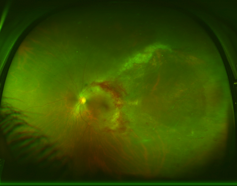
Color photograph (OPTOS plc, Scotland, United Kingdom) of zone 1 cytomegalovirus retinitis.
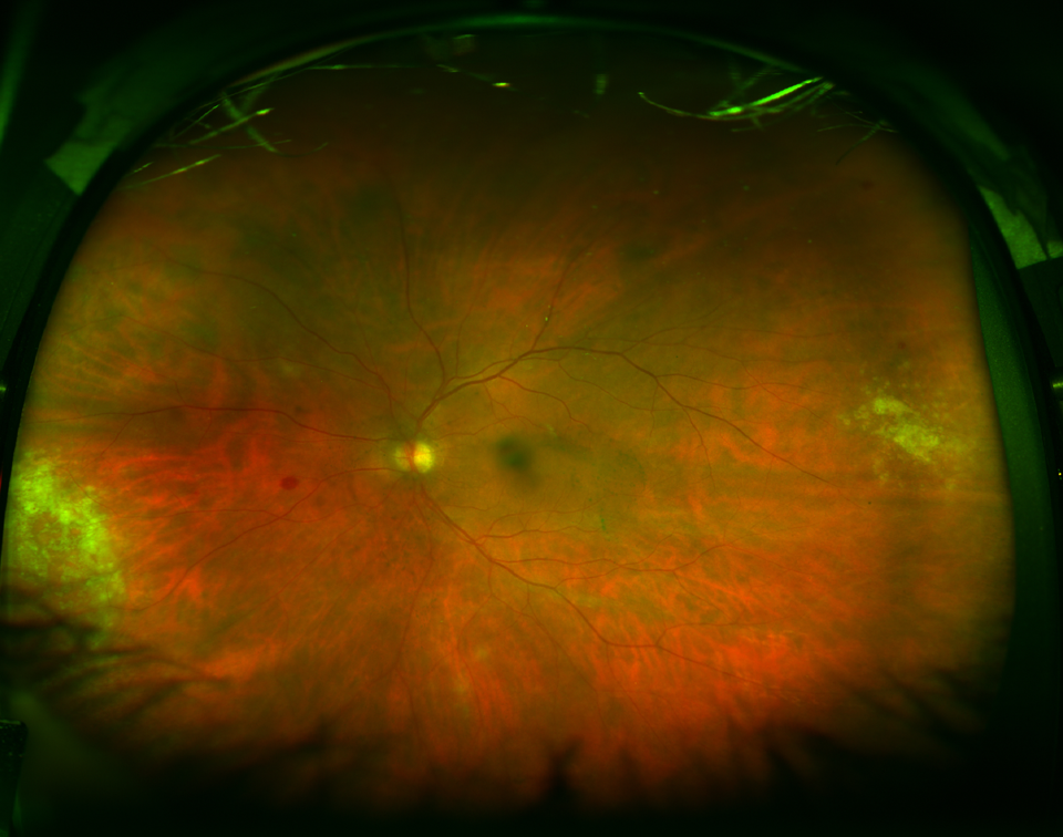
Color photograph (OPTOS plc, Scotland, United Kingdom) of zone 2/3 cytomegalovirus retinitis.
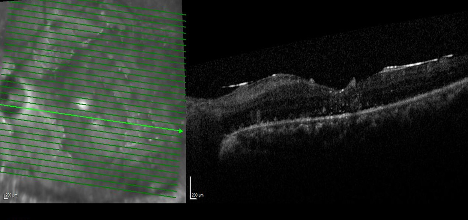
Optical coherence tomography (Heidelberg Engineering, Heidelberg, Germany) of foveal involving cytomegalovirus retinitis infection.
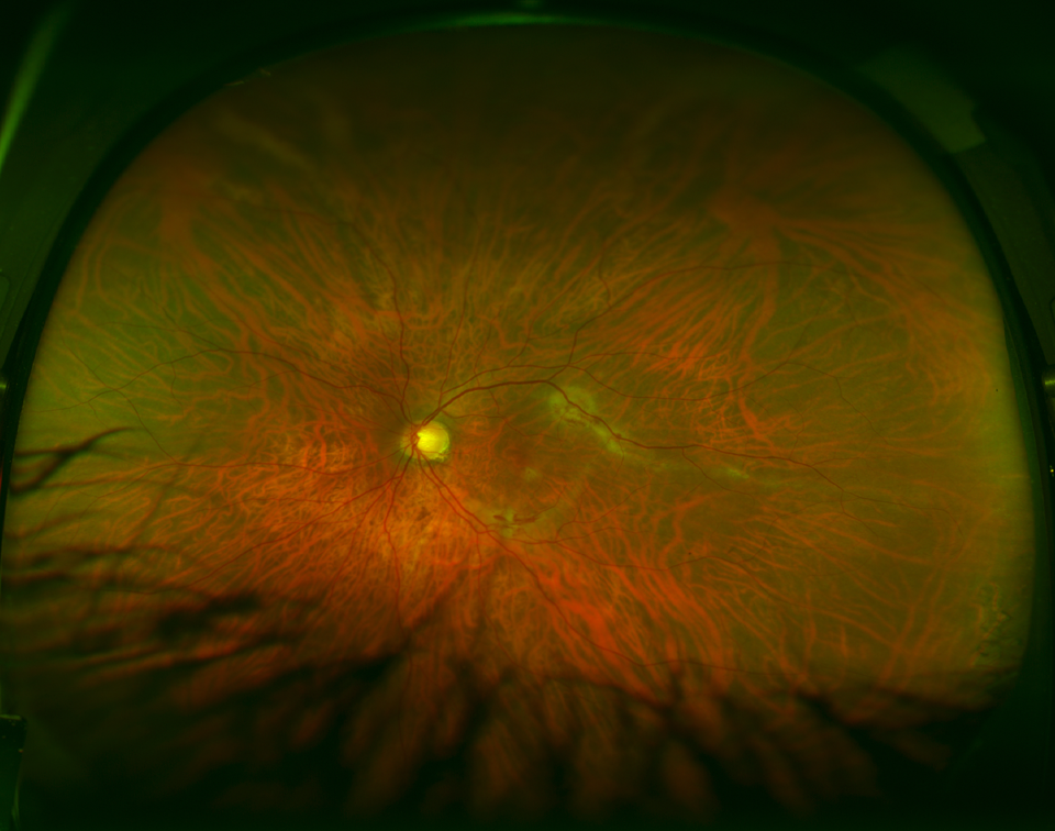
Color photograph (OPTOS plc, Scotland, United Kingdom) demonstrating cytomegalovirus retinitis affecting less than 25% of the retina.
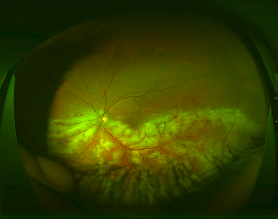
Color photograph (OPTOS plc, Scotland, United Kingdom) demonstrating cytomegalovirus retinitis affecting more than 25% of the retina.
During follow-up (Table 2), the median Snellen visual acuity decreased from 20/44 (logarithm of the minimum angle of resolution [logMAR] equivalent, 0.349) at initial visit to 20/54 (logMAR, 0.438) at most recent follow-up (P = .84). A total of 42.3% of eyes (n = 11) improved vision during follow-up and 15.4% (n = 4) gained 3 or more lines of vision; 51.9% (n = 14) of eyes lost vision from baseline at most recent follow-up with 34.6% (n = 9) having lost 3 or more lines of vision; and 34.6% (n = 9) of the eyes had severe loss of vision, with a visual acuity of 20/200 or worse at most recent follow-up. Patients were more likely to be phakic at baseline when compared to most recent follow-up (P = .03), and 22.73% of the phakic eyes underwent cataract surgery. One eye developed neovascular glaucoma and vitreous hemorrhage with an elevated IOP that ultimately required multiple bevacizumab injections followed by an Ahmed glaucoma implant surgery combined with pars plana vitrectomy and endolaser. A total of 23.1% (n = 6) of eyes developed RD, which were repaired surgically with pars plana vitrectomy and oil placement. One eye did not undergo surgery because the visual acuity was no light perception, and the RD was felt to be inoperable. Of the 5 eyes that underwent RD repair, 2 eyes were successfully repaired with 1 surgery, whereas 2 required a second RD surgery for recurrent RD and 1 eye required 2 additional vitrectomy surgeries.
Outcomes.a
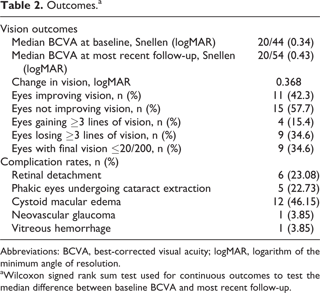
Abbreviations: BCVA, best-corrected visual acuity; logMAR, logarithm of the minimum angle of resolution.
aWilcoxon signed rank sum test used for continuous outcomes to test the median difference between baseline BCVA and most recent follow-up.
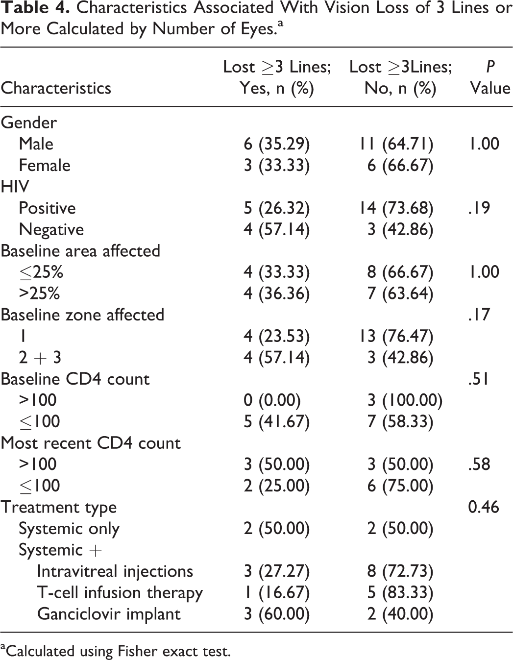
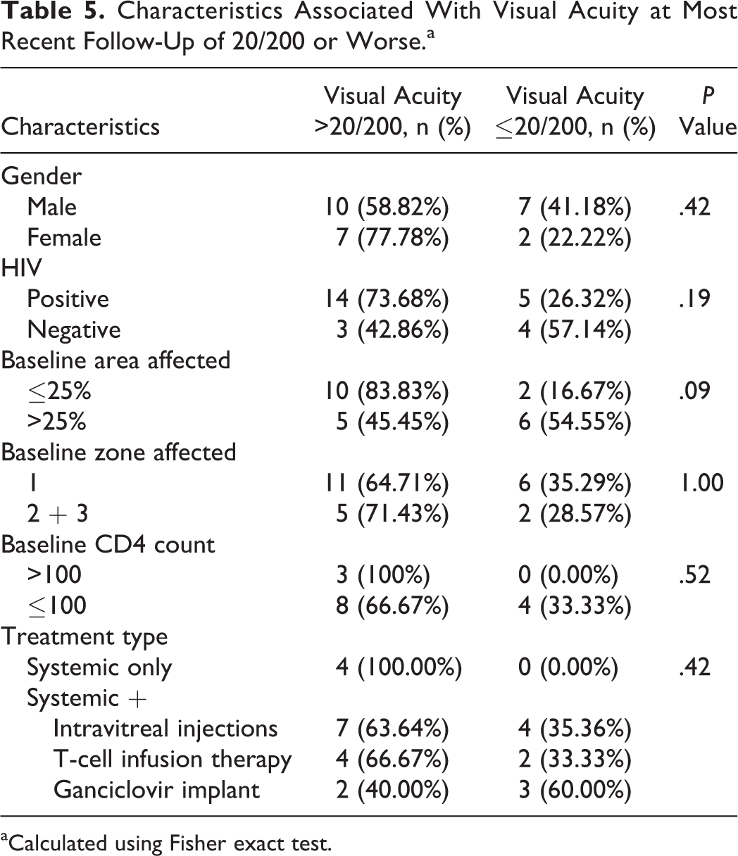
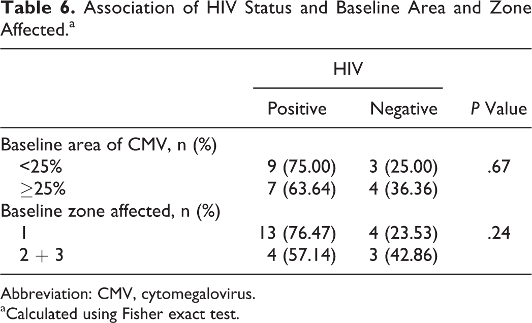
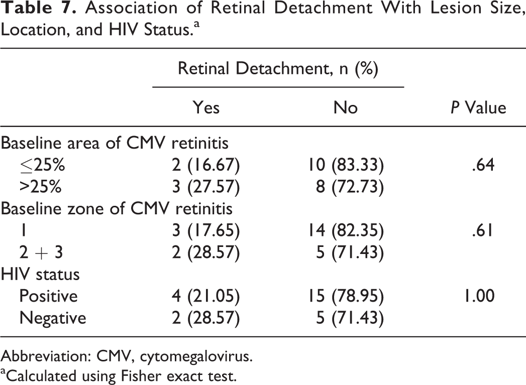
Four eyes underwent treatment with antiviral systemic therapy alone. In addition to systemic therapy, 11 eyes were treated with intravitreal injections, 6 eyes with T-cell infusion therapy, and 5 eyes had ganciclovir implant placement (Table 3). When testing risk factors against vision outcomes (Table 3), we found no association between vision loss and gender, cause of immunosuppression (HIV vs other), baseline area or zone of retina affected, or treatment type. Eyes presenting with zone 1 disease were more likely to have intravitreal injections when compared to eyes with zone 2 or 3 disease (P = .0035). Those with a CD4 count <100 were more likely to lose vision when compared to patients presenting with a CD4 count >100 (P = .04) (Table 3). Although not statistically significant, patients who were immunocompromised from therapies related to cancer and organ transplantation treatment were more likely to lose 3 or more lines of vision (P = .19) (Table 4) or have a decline in vision to 20/200 or worse (Table 5) when compared to patients with HIV. There was also a nonsignificant trend demonstrating that eyes affected by a larger area of CMV retinitis (>25%) were more likely to have a final visual acuity of 20/200 or worse when compared to eyes with CMV retinitis involving less than 25% of the total retina (P = .089) (Table 5). We did not find trends or associations between any of the other risk factors tested and visual outcomes. We also found no association between HIV status and baseline area or zone affected by CMV retinitis (Table 6). There were no associations between HIV status or baseline area or zone of retina affected and risk of RD (Table 7).
Characteristics Associated With Vision Changes Calculated by Number of Eyes.a
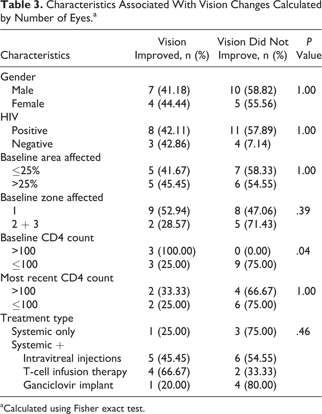
aCalculated using Fisher exact test.
Characteristics Associated With Vision Loss of 3 Lines or More Calculated by Number of Eyes.a
aCalculated using Fisher exact test.
Characteristics Associated With Visual Acuity at Most Recent Follow-Up of 20/200 or Worse.a
aCalculated using Fisher exact test.
Association of HIV Status and Baseline Area and Zone Affected.a
Abbreviation: CMV, cytomegalovirus.
aCalculated using Fisher exact test.
Association of Retinal Detachment With Lesion Size, Location, and HIV Status.a
Abbreviation: CMV, cytomegalovirus.
aCalculated using Fisher exact test.
Discussion
The CMV retinitis is a potentially blinding infection affecting immunocompromised patients such as those with AIDS, hematologic malignancy following stem cell transplant, or who are on immunosuppressive therapy for organ transplantation. 6 Prior to the development of HAART, CMV retinitis was most frequently encountered in patients with AIDS. The advent of HAART, and, in turn, immune reconstitution in patients with HIV, has dramatically altered the incidence and course of CMV retinitis in patients with HIV. As compared to an incidence of 25% among HIV-positive patients with CD4 count <100 cells/mL in the pre-HAART era, the rates in the post-HAART era were reported to be 7%. 7,8 Rates of adverse visual outcomes such as vision loss to 20/200 or worse have likewise decreased. 4 Despite this, CMV retinitis continues to remain a vision-threatening problem among patients with HIV, particularly among patients without immune recovery. 4 In addition, patients who are immunocompromised due to reasons other than HIV, such as those on immunosuppressant therapy after stem cell transplant or organ transplantation, still remain chronically immunosuppressed and at risk of CMV retinitis. This is the first report, to our knowledge, of CMV retinitis in patients both with HIV and without HIV. In our cohort of newly diagnosed active CMV retinitis at WCMC, a significant number of eyes lost vision. More than half (51.6%) lost vision during follow-up, whereas 34.6% of eyes lost 3 lines or more of vision and 34.6% of eyes ended up with a final vision of 20/200 or worse.
We found that patients who presented with a CD4+ T-cell count >100 cells/µL had better visual outcomes than those with a count <100 cells/µL. These data confirm the previously reported findings from the LSOCA cohort who showed that a lower CD4+ T-cell count was also associated with increased rates of visual acuity loss. 4 Although it was not statistically significant, we found eyes that presented with larger areas of CMV retinitis (>25%) were more likely to have a visual acuity of 20/200 or worse. These findings illustrate the importance of treating CMV retinitis in patients with a multidisciplinary team, to optimize immune recover and to monitor and treat patients early when the lesions may be smaller.
Patients in our cohort were treated with systemic anti-CMV therapy. In addition, those with HIV were also taking HAART. Many eyes in our cohort were also treated with intravitreal therapy, related to the significant number of cases presenting with zone 1 disease. We found no differences in outcomes based on treatment type (Tables 3 and 4), although the study was not specifically designed to investigate this. Six eyes in 4 patients received CMV-specific T-lymphocyte infusion therapy, as described by Gupta et al. 1 This therapy has been shown to be effective in treating systemic infection 9,10 and may be particularly useful in patients who remain chronically immunosuppressed, are intolerant to current CMV therapy, or whose infection is resistant to standard therapies. The T-cell infusion therapy has thus far been safe and efficacious in our cohort, and 1 patient showed a sustained and significant improvement in retinitis without concomitant systemic or intravitreal anti-CMV therapy. 1 A study to further investigate T-cell infusion therapy in a larger cohort with a longer follow-up is underway.
Eyes with CMV retinitis are at risk of various ocular complications. Retinal detachment is one of the most common causes of vision loss, and various studies have reported an incidence rate of approximately 33% per eye per year. 5,11,12,13 In past studies, patients treated with HAART were less likely to develop RD, 5 whereas larger lesion sizes 5,11,13 and a more anterior location of retinitis 5,13 were associated with increased risk of RD. Our findings are in agreement with these prior studies. In all, 23.1% of eyes in our cohort had RD during follow-up, and although it was not statistically significant, eyes with larger areas and more peripheral retinitis were at higher risk of RD (Table 7). It is possible that with a larger cohort, these associations may have been significant. In addition to RD, many eyes in our cohort developed cataract formation during follow-up. A significant number of eyes in our cohort underwent major ophthalmic surgery during follow-up including RD repair and cataract extraction (26.3% of phakic eyes).
All patients who were 13 years or older, with a diagnosis of AIDS, were eligible to enroll in the LSOCA study. Thorne et al included all patients diagnosed with CMV retinitis by clinical examination. 4 They showed that 40.2% of eyes presented with zone 1 CMV retinitis. In our cohort, the majority of eyes presented with zone 1 disease (61.5%) when compared to zone 2 and zone 3 disease. This difference may be secondary to patient selection and referral bias. Our cohort consisted mainly of newly diagnosed CMV retinitis and included HIV-negative patients. In addition, WCMC is a tertiary care center and it is possible that we see more patients who present with visually significant central defects or were referred to us for intravitreal treatment. We did not find any significant difference in visual acuity outcomes based on zone location, whereas Thorne et al demonstrated that eyes with baseline zone 1 disease had higher rates of vision loss during follow-up. The sample size in our cohort is small compared to the LSOCA one, and it is possible that a larger sample size may have illustrated a difference. It is also possible that zone 2 or 3 eyes are at higher risk of RD and subsequent significant vision loss, negating the effect that posterior involvement has on vision. We demonstrate that all eyes with CMV retinitis, regardless of location, are at high risk of significant vision loss and should be monitored carefully and treated accordingly.
We have a unique cohort of immunocompromised patients with a wide range of underlying conditions including, but not limited to, HIV. Although not statistically significant, patients who were immunocompromised other than reasons secondary to malignancy and organ transplantation were more likely to lose 3 lines of vision or more (P = .1881) (Table 4) and have a final vision of 20/200 or worse when compared to patients with HIV. There was no difference in RD risk (P > .999) or baseline area of retina affected by CMV (P = .67) between HIV-positive and -negative patients. Patients without HIV remain at high risk of complications related to CMV retinitis. They are at risk of chronic immunosuppression and poor tolerability to systemic anti-CMV medications. In patients with hematologic malignancies and/or history of stem cell transplant, systemic ganciclovir can cause severe myelotoxicity. 6 Nephrotoxicity from foscarnet remains a concern in these patients as well and is particularly of concern in patients with a history of renal transplantation. Agarwal et al found that intravitreal ganciclovir alone can be used to treat non-HIV patients successfully. 6 Consistent with our and others’ findings that improved immune function correlates to better visual outcomes in CMV retinitis, immune reconstitution is the center stone of long-term control of CMV retinitis. Thus, it is important for patients with CMV retinitis to be managed concurrently with a multidisciplinary team of treating physicians to improve immune recovery. It may not be possible, however, to titrate immunosuppressive agents based on the underlying etiology, which makes CMV-specific T-lymphocyte infusion therapy a very attractive potential future therapy, particularly in this patient population. 1
There are several limitations to the current study including those biases intrinsic to a retrospective one. This study was conducted at WCMC, a tertiary care center, and thus subject to a referral bias. Our sample size was relatively small, which may be due to the dramatic decrease in the incidence of CMV retinitis in the era of HAART, although we feel the number of subjects included in this study is comparable to other studies. 6 It is possible that with a larger cohort, other statistically significant associations with visual outcomes could have been detected. The goal of this study is to describe the outcomes of eyes with CMV retinitis in the era of HAART at WCMC. Four treating physicians participated in this study, each determining the best management for each patient given the presenting clinical situation. This was not a comparative treatment trial designed to test for treatment differences in a randomized manner. Although this is a potential limitation, we do feel that having multiple treating physicians makes our results clinically applicable to other practices that also utilize multiple specialists. We are in the process of conducting a separate study looking at outcome differences based on treatment, including T-cell infusion therapy.
In conclusion, although the incidence of CMV retinitis has decreased, our cohort demonstrates that this condition remains a vision-threatening problem among those who are immunocompromised due to HIV or other reasons. Patients are still at significant risk of vision loss and complications such as RD and cataract formation. 14 Improved future therapies are necessary, specifically related to immune recovery in patients who remain chronically immunosuppressed.
Footnotes
Authors’ Note
This study was conducted at Weill Cornell Medical College, New York, NY, USA.
Declaration of Conflicting Interests
The author(s) declared no potential conflicts of interest with respect to the research, authorship, and/or publication of this article.
Funding
The author(s) disclosed receipt of the following financial support for the research, authorship, and/or publication of this article: This study was funded in part by an unrestricted departmental grant from Research to Prevent Blindness. Memorial Sloan-Kettering Cancer Center and Atara Biotherapeutics have entered a licensing agreement for the CMV cytotoxic T-Lymphocyte program. Amanda Artis was partially supported by the following grant: Clinical and Translational Science Center at Weill Cornell Medical College (UL1-TR000457-06).