
Abstract
The aim of the present study was to determine the influence of doping rare earth ion on strontium calcium aluminate (CaO–SrO–SiO2–Al2O3). Therefore, the authors have manufactured luminescent material consisting of 40CaO–5SrO–5SiO2–50Al2O3 doped with Dy3+. The compositions have been selected on the basis of chemical stability. Five pellets were prepared with different calcination temperatures and times, namely 400 and 600°C for 1 and 2 h, in order to shed light on their luminescence behaviour. X-ray diffraction, energy dispersive X-ray analysis and scanning electron microscopy elaborate and characterise the formation of small particle of photoluminescent material in the phosphor matrix host material.
Keywords
Introduction
The luminescence of rare earth (RE) ions in the silicate host has been studied for a long time. 1 Lin et al. 2 prepared M2MgSi2O7:Eu, Dy (M = Ca, Sr, Ba) phosphors with melilite structure, which exhibits a good long afterglow property. Kodama et al. 3 reported that Ca2Al2SiO7:Ce single crystal had long afterglow phenomenon and could be used for tunable solid state laser materials. In this work, a new long afterglow phosphor was first synthesised by solid state reaction. The increased concentration of emission ions is usually effective to obtain high brightness. However, the emission is often quenched when the concentration is greater than a critical concentration. This phenomenon results from migration of excitation energy among the emission ions and is closely dependent on the crystal structure. 4 Therefore, high concentration doping of emission ions for matrix is possible for such a case. 5 The Dy ions substituted for Ca/Sr sites in 40CaO–5SrO–5SiO2–50Al2O3 are located in the two-dimensional site with a fairly low concentration.
In recent years, studies on RE ion doped glasses have gained much interest of researchers for the reason that the particular 4f electronic configuration of RE in varied glass matrices leads to emissions from ultraviolet to infrared 6 with many potential uses, including fluorescent lamps, two-dimensional X-ray sensors, solar control devices, solid state laser, optical amplifiers, etc. 7 Nowadays, white light emitting diodes have emerged as an important class of lighting devices and show high potential for replacement of conventional lighting sources like incandescent and fluorescent lamps, the advantage being their long lifetime, lower energy consumption, higher reliability and environmental friendly characteristics. 8
Recently, luminescence materials doped with Dy3+ have drawn much interest for its white emission. Dy3+ with 4f (Ref. 9) configuration has complicated f-block energy level; therefore, various possible transitions between f levels are expected. However, the transitions between these levels are highly selective and show sharp line spectra. 9 The Dy3+ is a good activator because of two dominated bands in the emission spectra, and its position depends strongly on the crystal field of the host lattice used. 10 One is the blue band (480 nm) corresponding to the 4 F9/2– 6 H15/2 transition, and the other is the yellow band (580 nm) ascribed to the 4 F9/2– 6 H13/2 transition. 11 Luminescent materials doped with Dy3+ will present white emission by adjusting the yellow/blue intensity ratio value, which can be used as potential white phosphors. 12 So many researchers have been engaged in the luminescent property of Dy3+ in different composition. 13 As for hosts, phosphates have been shown to be useful hosts for RE ions to fabricate phosphors emitting a variety of colors. 14 In the present work, the authors report on the photoluminescence properties of Dy3+ doped 40CaO–5SrO–5SiO2–0Al2O3 phosphors.
Materials and methods
Long afterglow phosphors of 40CaO–5SrO–5SiO2–50Al2O3 doped with Dy3+ were prepared using a solid state reaction. Al2O3, CaO, SrCO3, SiO2 and Dy2O3 were employed as the raw materials. Small quantities of B2O3 (5 mol.-) were added as a flux. The raw materials were mixed homogeneously with ball mill for 2 h and prefired at 900°C for 2 h. Then, the powder was reground and pressed into pellets and finally sintered at 1300°C for 2 h with a weak reductive atmosphere of 5H2–95N2 gas flow. The final product phosphor were reground, pressed into pellets and calcined at 400 and 600°C for 1 and 2 h for each temperature in air.
Phase identification was performed using X-ray diffraction (XRD) carried out with Cu Kα radiation operating at 40 kV and 30 mA with Bragg–Brentano geometry at room temperature using Siemens diffractometer D5000, equipped with diffraction software analysis. Diffraction patterns were collected in the range from 10 to 80°, in steps of 0·04° and 4 s counting time per step. The scanning electron microscopy (SEM) was observed with JEOL (JSM-840 SEM, Tokyo, Japan) equipped with an energy dispersive X-ray analyser (EDAX). High vacuum evaporation (SCD 004 from Balzers Union, Balzers, Leichthenstein) was used to coat a thin gold film to make the surface of the specimens electrically conductive. Their luminescence properties were studied using excitation and emission spectra obtained from photoluminescence spectroscopy.
Results and discussion
After the phosphor was irradiated with a UV lamp (254 nm) for 10 min, a visible blue light could be clearly seen with the naked eye in the dark 1 h after removal of the activating light. Figure 1 showed the photos of the as prepared phosphor sample of CaOAl2O3 doped with Dy3+ before and after emission under UV irradiation. Dy3+ has emissions due to the atomic energy levels of itself and emissions due to the acceptor levels of defect sites formed by Dy3+. The emission and excitation spectra of the Dy3+ codoped phosphor are shown in Fig. 2. The results indicate that they are both broadband spectra. The emission spectra at 376 and 420 nm of unheated powder sample (curve a) shifted to 386 and 419 nm at 400°C for 1 h (curve b) and back to 376 and 420 nm at 400°C for 2 h (curve c) with the excitation spectrum peaks at 390, 412 and 475 nm (curve d). It is believed that Dy3+ ions are considered to act as the trap levels located at a suitable depth. The trap can capture the free holes and release the trapped holes. Therefore, the depth of Dy3+ traps will influence the lifetime of the trap holes and, thus, the after glow lifetime. The detail mechanism is still being studied. 15
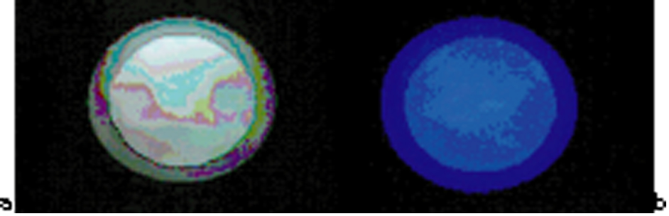
Photos of as prepared phosphor sample of CaO–Al2O3 doped with Dy a before and b after emission under UV irradiation
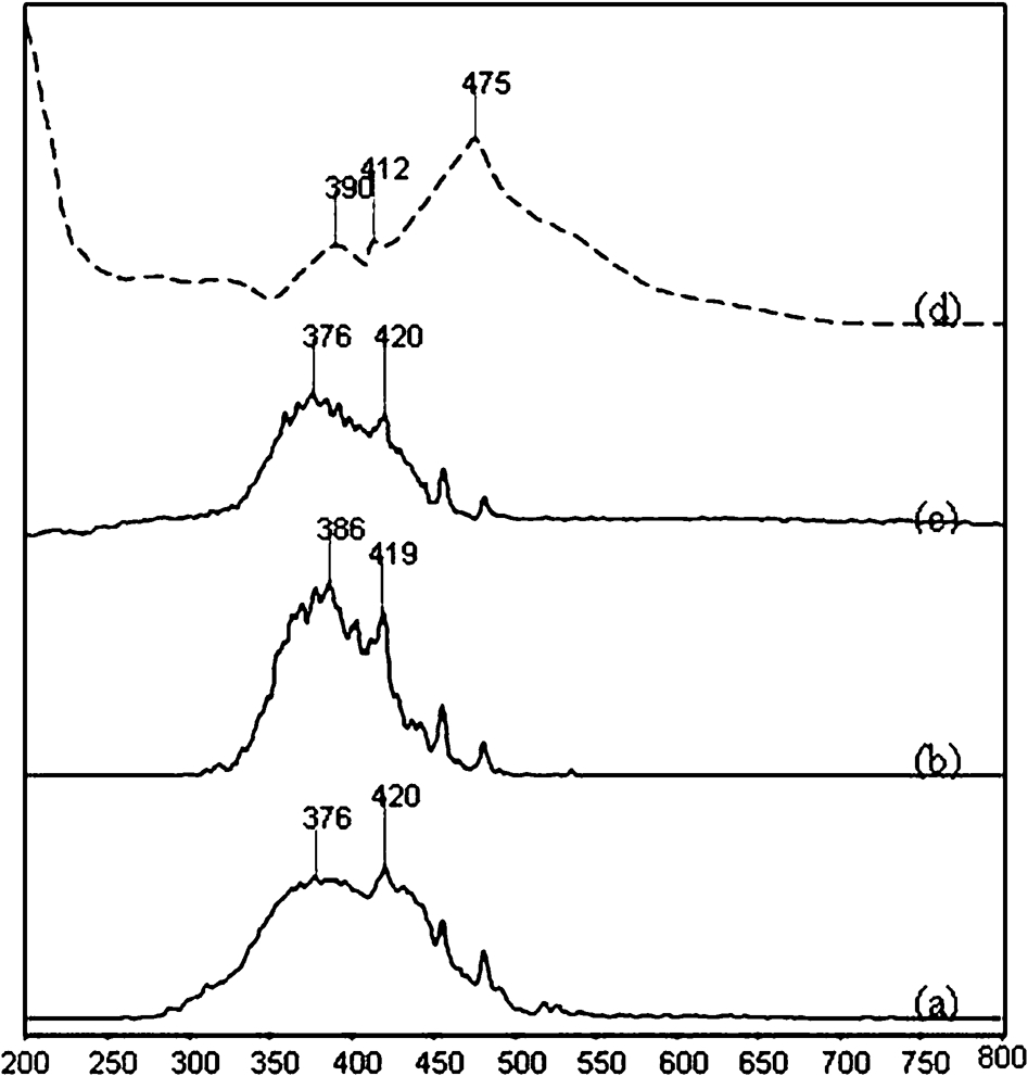
a emission spectra of unheated powder sample, b emission spectra of powder sample heated at 400°C for 1 h, c emission spectra of powder sample heated at 400°C for 2 h and d excitation spectra of CaAl2O4:Dy
The diffraction patterns of doped and undoped 40CaO–5SrO–5SiO2–50Al2O3 are shown in Fig. 3. The patterns show well defined peaks, which indicate the crystalline and phase formation of the synthesised compounds. The peaks of doped can be indexed based on the space group P21/n of monoclinic phase of calcium aluminium oxide CaAl2O4 (JCPDS no. 70-0134) and space group P421m of gehlenite Ca2Al2SiO7 (JCPDS no. 35-0755). It also indicates that the small amount of doped Dy2O3 cannot form any new phase. In addition to CaAl2O4, some impurity phases like Ca3Al2O6 (JCPDS no. 38-1429) and Ca12Al14O33 (JCPDS no. 09-0413) are also seen as secondary phases. This behaviour is attributed to the fact that heat needed to form the single phase CaAl2O4 is very low at the end of heating process. Even after sintering at 1300°C, some secondary phases are seen due to the slow conversion of these phases to form single phase CaAl2O4. 16
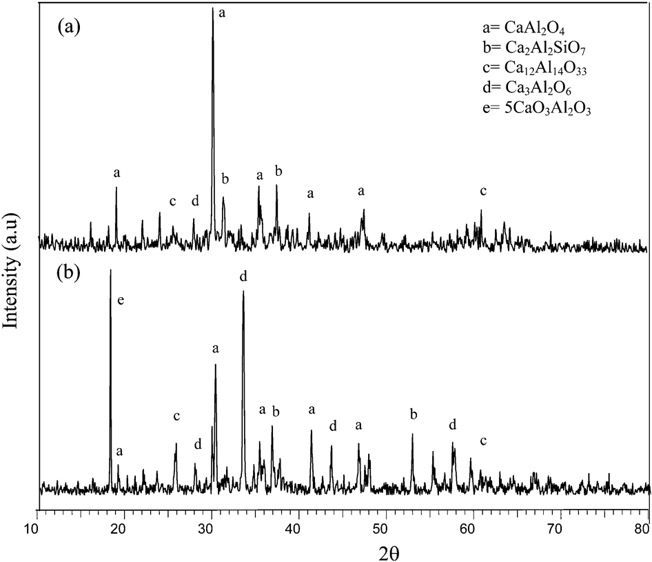
X-ray diffraction pattern of 40CaO–5SrO–5SiO2–50Al2O3 a doped in reducing atmosphere and b undoped sample heated in air
The diffraction patterns of doped 40CaO–5SrO–5SiO2–50Al2O3, heated under reducing conditions revealed significant differences compared to those of undoped samples heated in air. Multiphases of powder sample can be easily formed, even if the compositional ratio is slightly deviated from the equilibrium, which may be due to the firing conditions and the mole ratio of CaO to Al2O3. In this experiment, since 1 mol.- Dy2O3 was added, the actual composition of 40CaO–5SrO–5SiO2–50Al2O3:Dy2O3 was 99∶1, leading to the deviation from the equilibrium composition for single phase. Moreover, by adding a flux, B2O3 facilitated the reactions and lowered phase transformation temperature, resulting in the coexistence of solid and liquid phases. Besides, 40CaO–5SrO–5SiO2–50Al2O3 systems easily generate the multiphase compounds, such as CaAl4O7, Ca3Al2O6 and Ca12Al14O33, according to Lea and Desch 17 and Jerebtsov and Mikhailov. 18
The sintered powder samples were investigated by SEM for surface morphology and crystallite sizes. Particle size of phosphor plays an important role in determining the luminescence quality of the material. Uniform particle size distribution and fine particles in the range of micrometre to nanometre are some of the requirements of good luminescent materials. The SEM image of the prepared powder sample sintered at 1300 and 1350°C are shown in Fig. 4. Polycrystalline morphology is clearly observed in these micrographs for that composition. From the micrographs, it can be seen that the sample mainly consists of polygonal shaped grains with small incrustations on the surface and solid micrometer polycrystalline structures, which exists with some conglomeration and foamy structure among the crystalline grain for the high temperature thermal decomposition. The presence of these incrustations and foamy structure is probably due to the influence of secondary phases and reflects the inherent nature of the reaction. The typical crystalline irregular grain shape with size of several tenths of a micrometre to several micrometres is observed, and the agglomerate sizes are <2 μm. This morphology is similar with the findings observed by Singh et al. 19
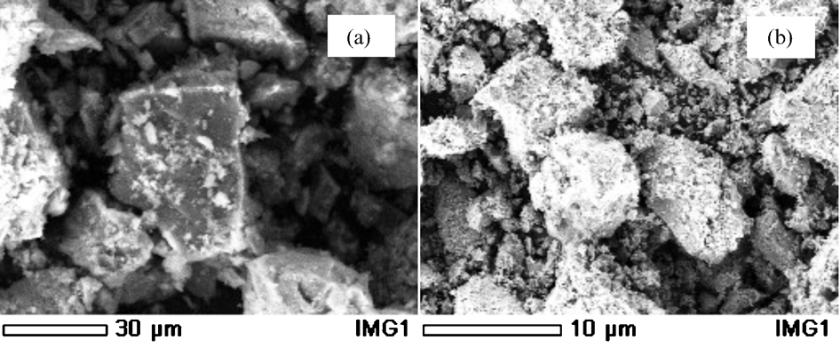
Images (SEM) of undoped 40CaO–5SrO–5SiO2–50Al2O3 sample sintering at a 1300°C and b 1350°C
This result showed that the process of high temperature solid state reaction produces sample with the grain size in the range of several tens of micrometres, the distribution of components is asymmetrical, and it is difficult to crush the hard phosphor blocks into small particles. To further confirm the concentration of 40CaO–5SrO–5SiO2–50Al2O3 content in the powder sample, EDAX was performed on the sample. Ca, Al and O were detected on the powder sample. The Dy3+ element was not detected clearly due to its low concentration, although it can be detected by the emission spectra (not shown here).
Conclusions
The XRD, EDAX and SEM elaborate and characterise the formation of photoluminescent material in the phosphor matrix host material. Topology and morphological characteristics of the material and the results of XRD show that, as the sintering temperature increases, the morphology has been modified and the grains are finer in size.
Footnotes
Acknowledgements
This work is supported by the Ministry of Science Technology and Innovation under research grant project no. 03-01-06-SF0363. The authors gratefully acknowledge the support provided by Universiti Malaysia Sarawak and Universiti Teknologi Malaysia.