
Abstract
This work performs metal–metal bonding using CuO nanoparticles prepared with salt base reaction in aqueous solution. A colloid solution of CuO nanoparticles was prepared by mixing Cu(NO3)2 aqueous solution and NaOH aqueous solution. Submicrometre sized leaf-like aggregates composed of CuO nanoparticles were produced at a Na/Cu ratio of 1·7 and at 20°C, though Cu2(OH)3NO3 was also obtained. An aging process, which is a process composed of preparation of the particles at 20°C and then aging them at 80°C, provided transformation from Cu2(OH)3NO3 to CuO with no damage of the leaf structure. The shear strength, which was required for separating discs bonded using the particles as a filler at 400°C in H2 gas, was 32·5 MPa at the maximum for the particles prepared at the Na/Cu ratio of 1·7 with the aging process. These results indicated that the formation of leaf-like aggregates of CuO particles with high purity led to efficient metal–metal bonding.
Introduction
Metal–metal bonding with the use of solder is very common among various metal–metal bonding techniques. Since metals or metallic alloys with low melting points are usually used as solders, the bonding can be made at low temperatures. A conventional solder based bonding technique has used metallic alloys composed mainly of lead and tin as solders or fillers between the materials to be bonded because of their low melting points. Since the toxicity of lead for living bodies has been pointed out in recent years,1– 3 the use of lead is showing a tendency to be limited. Accordingly, lead free alloys based on tin, which are available for the bonding, are quite required, and various tin based alloys have been developed as new solders.4– 6 The tin based alloys still have a problem on their low melting point. Metallic materials can be bonded at low temperatures using the alloy. If the bonded materials are kept at temperatures higher than their melting point, they may be released due to remelting of the alloy.
High quality metal–metal bonding will be achieved using metallic materials such as Au, Ag and Cu as fillers because of their high electric conductivity and thermal conductivity. Since their melting points are quite higher than those of the conventional lead and tin based solders, a high temperature is required for metal–metal bonding. Materials to be bonded are exposed to a high bonding temperature during the bonding, so that they may be damaged.
The melting point of metallic particles decreases with a decrease in their size down to several nanometres.7– 9 Paying attention to this property of the decrease in melting point, bonding can be successfully performed at a low temperature using metallic nanoparticles. After the bonding is completed at the low temperature, the nanoparticles become metallic bulk. Since the metallic bulk should have a melting point higher than that for the nanoparticles, the bonded materials are unreleased if they are used below their melting points. Low temperature bonding with the use of metallic nanoparticles has been examined by various researchers.10– 12 These works have focused on metallic Ag nanoparticles for realising a new filler in metal–metal bonding because of not only high electric and thermal conductivities, but also high chemical stability. Apart from metallic Ag nanoparticles, Ag oxide nanoparticles have been also examined for bonding, in which the bonding was performed in reducing gas such as H2.13, 14 Though those Ag based nanoparticles helped the bonding, the bonding faces problems of high cost and migration property of Ag.
Metallic Cu nanoparticles are also promising materials for metallic bonding, because metallic Cu has high electric conductivity and high thermal conductivity and is available at low cost. In addition, metallic Cu is superior in migration property to Ag. However, the metallic Cu nanoparticles are oxidised easily in air,15, 16 which spoils bonding properties. Methods using surfactants or stabilisers have been proposed for preparing chemically stable metallic Cu nanoparticles.17– 19 Our group has also studied the preparation of aqueous colloid solution of metallic Cu nanoparticles by reducing Cu2+ with hydrazine in the presence of citric acid as a stabiliser and CTAB as a surfactant.20 In the methods, surfactants or emulsifiers may be left in the obtained samples, which also will spoil the bonding properties.
Apart from the metallic Cu nanoparticles, the nanoparticles of compounds of Cu in oxidative state may be available as a filler for metal–metal bonding, since it can be reduced to metallic copper with reducing agent or reducing atmosphere. From this view point, our group has performed metallic bonding with the use of Cu salt (CuI) nanoparticles as the filler.21 However, metal–metal bonding could not be made, since the existence of iodine, which was derived from CuI, probably prevented formation of metallic Cu. Copper oxide nanoparticles are another candidate as the filler. Various methods for producing copper oxide nanoparticles have been proposed by many researchers, which were based on reactions of Cu ions derived from Cu salt and base.22– 24 In our previous work, copper oxide nanoparticles prepared with the salt base reaction in aqueous solution have been examined as the filler for metal–metal bonding, and a shear strength as high as 25·4 MPa has been recorded.25 The previous work implied that the formation of leaf-like aggregates composed of CuO nanoparticles resulted in large shear strength. The aim of the present work is to prove the importance of formation of leaf-like CuO aggregates by investigating metal–metal bonding properties of leaf-like CuO aggregates prepared in aqueous solution with various base/Cu molar ratios and ways to heat CuO particle colloid solution. Since a preliminary experiment implied that the impurity contained in the aggregates spoiled their bonding property, their purity was also discussed.
Experimental
Chemicals
Cu(II) nitrate trihydrate [Cu(NO3)2.3H2O] [Kanto Chemical Co. Inc., 77·0–80·0%, as Cu(NO3)2] and sodium hydroxide solution (NaOH) (Kanto Chemical Co. Inc., 1M) were used as CuO precursors. All chemicals were used as received. Water that was ion exchanged and distilled with Yamato WG-250 was used in all the preparations.
Preparation
Colloid solutions of CuO nanoparticles were synthesised using a reaction between copper ions and base. An aqueous solution of NaOH was added to Cu(NO3)2 aqueous solution under vigorous stirring at 20 and 80°C. Initial concentrations of Cu(NO3)2 and NaOH were 0·01 and 0·017–0·021 mol L−1 in the final solution respectively, which resulted in Na/Cu molar ratios of 1·7–2·1. The reaction times were 24 and 12 h for the reaction temperatures of 20 and 80°C respectively. For detailed investigation into the effect of the reaction temperature, the particles prepared at 20°C were aged at 80°C for 3 h (aging process).
Characterisation
The CuO particles were characterised by TEM, X-ray diffractometry (XRD) and thermal analysis [thermogravimetry differential thermal analysis (TG-DTA)]. Photographs (TEM) were taken with a JEOL JEM-2000FX II microscope operating at 200 kV. Samples for TEM were prepared by dropping and evaporating the particle colloid on a collodion coated copper grid. Dozens of particle diameters in TEM images were measured to determine the number averaged particle size and standard deviation of particle size distribution. Measurements using XRD were carried out with a Rigaku RAD-B X-ray diffractometer at 50 kV and 150 mA with Cu Kα1 radiation. For preparing a powder sample for XRD measurement, the supernatant of the particle colloid was removed with decantation, and then the residue of the colloid was dried at room temperature for 24 h in vacuum. Thermal analysis was performed in 3% (v/v) H2/N2 gas at a heating rate of 10°C min−1 with a Mettler-Toledo TGA/SDTA851 thermal analyser. Samples for thermal analysis were obtained in the same manner as that for the XRD samples.
Metallic bonding property was investigated by the same set-up used in our previous works.10, 13, 14, 26 Powder samples were sandwiched between copper discs [a stage (diameter, 10 mm; thickness, 5 mm) and a plate (diameter, 5 mm; thickness, 2·5 mm)] and pressed at 1·2 MPa under annealing in H2 at 400°C for 5 min with a Shinko Seiki vacuum reflow system. For investigation of the microstructures of the plate to stage joints, they were sectioned, metallographically polished and observed with a Hitachi S-4800 SEM. For examination of bonding strength, shear strengths, which were required to separate the bonded plate and stage, were measured with a Seishin SS-100KP bond tester. The copper plate was observed with a JEOL JSM-5600LV microscope after the measurements of shear strengths.
Results and discussion
Morphology of particles
Effect of Na/Cu ratio
Figure 1a–c shows photographs of particle colloid solutions prepared at 20°C with various Na/Cu ratios. The colours of the colloid solutions were greyish brown at a ratio of 1·7, brown at 1·9 and greyish black at 2·1, i.e. a tone of the colloid solution became dark as the Na/Cu ratio increased. This observation indicated that a large amount of CuO, which is black, was produced at the large Na/Cu ratio. At 2·1, the grey blackish colloid solution was not highly dispersed, and precipitation took place immediately after preparation. The large Na/Cu ratio corresponded to a large NaOH concentration, which resulted in high ionic strength of the solution. Since an increase in the ionic strength compresses double layer on solid materials such as colloidal particles,22,27– 29 the double layer repulsion between CuO nanoparticles was probably reduced at the large Na/Cu ratio. Thus, at the large Na/Cu ratio, CuO nanoparticles probably aggregated and grew because of the increased ionic strength that would favour the precipitation of CuO nanoparticles.
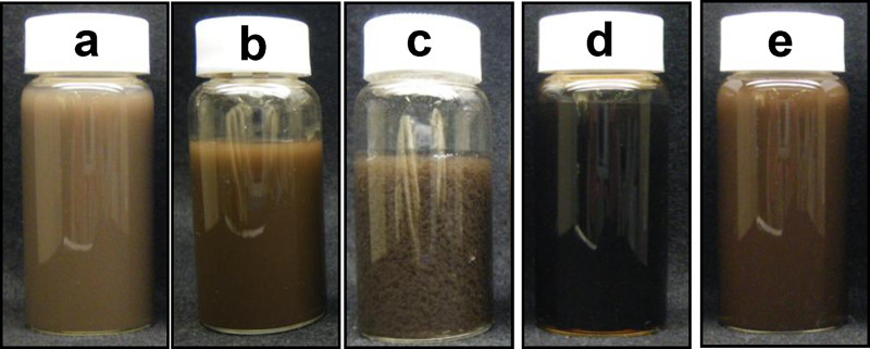
Photographs of various particle colloid solutions. Samples a–c were prepared at 20°C with Na/Cu ratios of 1·7, 1·9 and 2·1 respectively. Sample d was prepared at 80°C with Na/Cu ratio of 1·7. Sample e was obtained employing the aging process for sample a, i.e. aging the as prepared sample a at 80°C
Figure 2a–c shows TEM images of the particles prepared at 20°C with various Na/Cu ratios. At the ratios of 1·7 and 1·9, leaf-like aggregates composed of nanoparticles with a size of ∼10 nm were produced. The longitudinal and lateral sizes of the aggregates at 1·7 were 1000 and 400 nm, respectively. For the ratio of 1·9, those sizes were 600 and 400 nm respectively. Our previous work showed that the pH of a Cu(NO3)2 aqueous solution increased and approached an iso-electric point (i.e.p.) of CuO nanoparticles with the addition of an NaOH aqueous solution at an Na/Cu ratio of 1·9.25 Since the ratio of 1·9 was below the ratio of 2·0 that is the stoichiometric ratio of NaOH for Cu(NO3)2, the pH was ranged below the i.e.p. of CuO nanoparticles. A similar tendency of pH change should be observed in a reaction of metal ions and base in aqueous solution if the base/metal ion ratio is below its stoichiometric ratio. At the ratio of 1·7 in the present work, the pH value of the Cu(NO3)2 aqueous solution should have approached the i.e.p. of CuO nanoparticles with the NaOH addition, similar to the case of the ratio of 1·9, because the ratio of 1·7 was also below the stoichiometric ratio of 2·0. The approach to the i.e.p. resulted in a decrease in surface potential of the CuO nanoparticles, which provided a decrease in repulsion strength among the CuO nanoparticles. Consequently, the aggregation of the CuO nanoparticles took place. At 2·1, micrometre sized precipitates were produced. Besides them, nanoparticles with a size of 1200 nm and their aggregates with a size of 20 nm were also produced. Since the ratio of 2·1 was close to the stoichiometric ratio of 2·0, surface potential of the as prepared CuO nanoparticles appeared to be quite small just after the NaOH addition, which resulted in the formation of aggregates of the CuO nanoparticles and succeeding precipitation of the aggregates. With the increase in reaction time, it was possible that the pH of the solution gradually moved away from the i.e.p. since the ratio of 2·1 was above the stoichiometric ratio of 2·0. Consequently, the aggregation of the CuO nanoparticles was partially controlled. However, the precipitates were not separated after they were once produced.
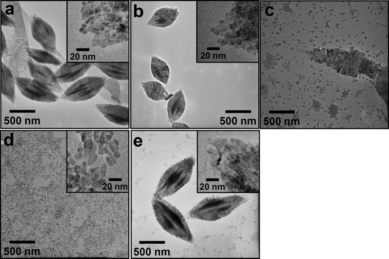
Images (TEM) of various particles: samples a–e were the same as in Fig. 1
Figure 3a–c shows the XRD patterns of particles prepared at 20°C with various Na/Cu ratios. At the ratio of 1·7, two sharp peaks were recorded mainly at 12·8 and 25·8°. They were attributed to Cu2(OH)3NO3. Besides the Cu2(OH)3NO3 peaks, peaks assigned to monoclinic CuO were also detected at 35·6, 38·7 and 48·8°. At 1·9, the CuO peaks were intensified, and the Cu2(OH)3NO3 peaks were still detected. For 2·1, the Cu2(OH)3NO3 peaks disappeared, and the peaks due to CuO were mainly detected with faint peaks at 16·7 and 23·8° attributed to Cu(OH)2. Possibly, the reaction for formation from Cu(NO3)2 to CuO was not completed at the ratios smaller than the stoichiometric ratio of 2·0.
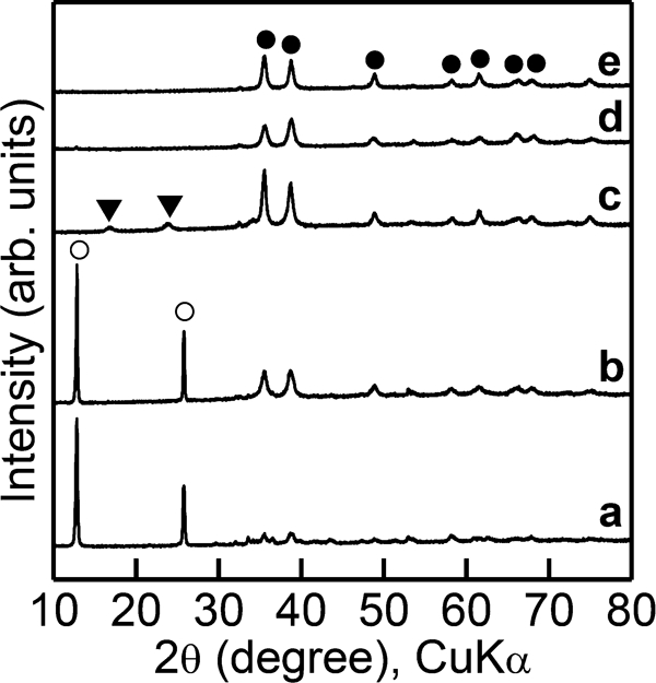
X-ray diffraction patterns of various particle powders: samples a–e were the same as in Fig. 1; • CuO; ○ Cu2(OH)3NO3; ▾ Cu(OH)2
Curves of TG-DTA for the particles prepared at 20°C with various Na/Cu ratios are shown in Fig. 4a–c. Around 200°C, endothermic peaks and weight losses were detected for all the ratios examined. According to the XRD measurement shown in Fig. 3, the as prepared CuO nanoparticles contained Cu2(OH)3NO3 for the ratios of 1·7 and 1·9. According to Lee et al.'s work,22 Cu2(OH)3NO3 begins to decompose into CuO at ∼225°C under an increase in temperature, which results in a weight loss due to the elimination of water and NO3 group. Thus, the weight losses accompanying the endothermic peaks detected in the present work were assigned to the decomposition of Cu2(OH)3NO3 into CuO. Though Cu2(OH)3NO3 peaks were not observed in the XRD pattern for 2·1, the weight loss with the endothermic peak was also detected. This result indicated that the amount of Cu2(OH)3NO3, which was contained in the as prepared CuO nanoparticles, was too small to detect the Cu2(OH)3NO3 with the XRD measurement. The temperature for the endothermic peak was lowered from 240 to 210°C as the ratio increased from 1·7 to 1·9. Because the amount of Cu2(OH)3NO3 should have been small for the small Na/Cu ratio, the decomposition of Cu2(OH)3NO3 was probably completed with low energy, i.e. at low temperatures. In the range of 250–320°C, exothermic peaks and weight losses were detected, and the weights did not change above 320°C, for all the particles examined. It was implied that the CuO was reduced in the reducing gas to form metallic Cu in the temperature range. The reduction probably resulted in removal of oxygen from the Cu oxide, which corresponded to the weight loss. In our previous work,25 it was confirmed that Cu oxide was reduced to metallic Cu under annealing at 400°C in H2 gas, which supported the implication given by the thermal analysis.
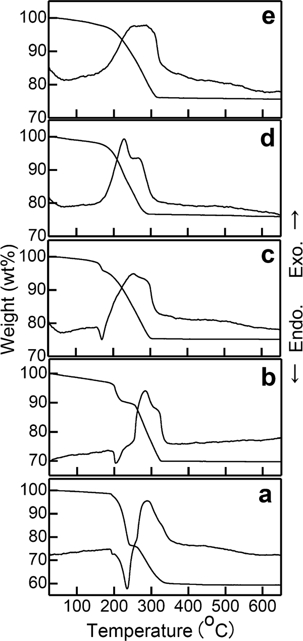
Curves of TG-DTA in 3% (v/v) H2/N2 gas for various particle powders: samples a–e were the same as in Fig. 1
Effect of reaction temperature
To investigate the effect of reaction temperature on morphology of particles, the reaction temperature was varied under a constant Na/Cu ratio of 1·7 in all the preparations of particles examined in this section. Figure 1d shows a photograph of the particle colloid solution prepared at a reaction temperature of 80°C. In contrast to the sample prepared at 20°C (Fig. 1a), the sample prepared at 80°C was blackish, which indicated that a large amount of CuO was produced with the increase in the reaction temperature to 80°C. Figure 1e shows a photograph of the particle colloid solution obtained with the aging process. A tone of the colloid solution was darker than the sample before the heating (Fig. 1a). This observation implied that the aging accelerated the production of CuO even for the colloid solution prepared at the low temperature.
Figure 2d shows a TEM image of the particles prepared at 80°C. With increasing the reaction temperature up to 80°C, such leaf-like aggregates as were produced at 20°C disappeared, and particles with a size of 10 nm were obtained with no aggregation. In a preliminary experiment, in the case of 80°C, a value of pH decreased after reaching a maximum rapidly compared with low temperatures. This meant that the pH moved away from the i.e.p., and electrostatic repulsion between the particles became strong. Consequently, aggregation of particles was controlled at the high temperature. Figure 2e shows a TEM image of the sample prepared with the aging process. Leaf-like aggregates with the longitudinal size of ∼1100 nm and the lateral size of ∼500 nm were produced. There was no large difference in morphology of aggregates between the particles with and without aging at 80°C. This result indicated that the aging did not damage the morphology of leaf-like aggregates.
Figure 3d shows an XRD pattern of the particles prepared at 80°C. Such peaks due to Cu2(OH)3NO3 as were detected for the particles prepared at 20°C disappeared with the rise of reaction temperature up to 80°C. Possibly, the transformation from Cu2(OH)3NO3 to CuO was promoted efficiently at the high temperature. Figure 3e shows an XRD pattern of the sample prepared with the aging process. Peaks attributed to CuO were also detected, and no peaks due to Cu2(OH)3NO3 were detected. Even for the particles that were once prepared at the low temperature, the rise of reaction temperature promoted the transformation from Cu2(OH)3NO3 to CuO.
Figure 4d shows the TG–DTA curves for the particles prepared at 80°C. The dominant weight loss accompanying the endothermic peak, which was assigned to the decomposition of Cu2(OH)3NO3 into CuO, was detected for the particles prepared at 20°C, as shown in Fig. 4a. In contrast, such weight loss and endothermic peak were not detected for 80°C. This result indicated that the Cu2(OH)3NO3 containing such species as NO3 group and the H2O to be eliminated at ∼225°C were not present in the particles prepared at 80°C, which corresponded to the speculation obtained from the observation in Fig. 1e and the conclusion resulting from the XRD measurement in Fig. 3e. In the range of 200–300°C, an exothermic peak and a weight loss assigned to the reduction of CuO to metallic Cu were detected similarly for 20°C (Fig. 4a). The weight did not change above 300°C. Figure 4e shows the TG-DTA curves of the sample prepared with the aging process. A weight loss accompanying an endothermic peak assigned to the decomposition of Cu2(OH)3NO3 into CuO was not detected either. This result also supported the promotion effect of aging at 80°C on transformation from Cu2(OH)3NO3 to CuO. The CuO was reduced to metallic Cu in the range of 200–300°C similarly for 20°C (Fig. 4a) because of the detection of weight loss accompanying an exothermic peak.
Bonding properties
Figure 5 shows photographs of the copper stages after the measurement of shear strength. Reddish brown products that were obviously metallic Cu were observed in widespread area on the stages for all the samples examined. This observation indicated that the as prepared particles were reduced to metallic Cu annealing at 400°C in H2 gas, which was supported with the XRD measurement (Fig. 3) and the thermal analysis (Fig. 4). Consequently, the metallic Cu bonded the copper discs.
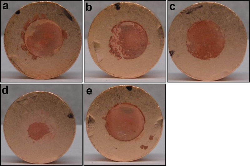
Photographs of copper stages after measurement of shear strength: particles used for measurements were the same as in Fig. 1
Effect of Na/Cu ratio
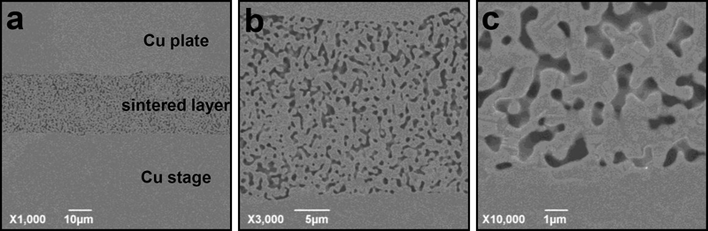
Table 1 shows the shear strengths for the Na/Cu ratios. The shear strengths were 21·8, 27·9 and 15·8 MPa for Na/Cu ratios of 1·7, 1·9 and 2·1 respectively. The shear strength for the Na/Cu ratio of 1·9 was larger than that for 1·7. In the TEM observation (Fig. 1a and b), the aggregate size for the Na/Cu ratio of 1·7 was larger than that for 1·9. Our previous work showed that large leaf-like aggregates gave large shear strength.25 The result in the present work did not correspond to that in the previous work. As given in the XRD measurement (Fig. 3a and b), the main component of the leaf-like aggregates prepared at 1·7 was Cu2(OH)3NO3, and the formation of CuO was promoted with the increase in the Na/Cu ratio to 1·9. Since the groups of NO3 and H2O in Cu2(OH)3NO3 eliminated heating at 250–320°C according to the thermal analysis (Fig. 4), many pores were probably produced during bonding at 400°C. The production of pores probably prevented particles from sintering during bonding, which decreased the shear strength. The increase in Na/Cu ratio to 2·1 provided the decrease in shear strength to 15·8 MPa. As given in the XRD measurement (Fig. 3c), the increase in Na/Cu ratio to 2·1 diminished Cu2(OH)3NO3, and consequently, CuO was made the main component of the leaf-like aggregates. Nevertheless, the shear strength for the Na/Cu ratio of 2·1 was smaller than that for 1·9. The increase in the Na/Cu ratio to 2·1 not only promoted the formation of CuO, but also decreased the aggregate size, as shown in Fig. 2. Large shear strength can be obtained for large leaf-like aggregates. Accordingly, in the present work, the large shear strength was recorded at 1·9, since the large leaf-like aggregates were produced, compared with those for 2·1.
Shear strengths for Na/Cu ratios: reaction temperatures were 20°C
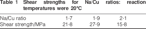
Figure 6a–c shows the SEM images of surfaces of the copper plates separated with shear stress. Sintering appeared to take place among particles with a size of ∼1000 nm for the Na/Cu ratio of 1·7. In contrast, for 1·9, many dimples were observed accompanying with sharp tips on the surface. Though dimples were also formed at 2·1, the dimples were somewhat unclear compared with the Na/Cu ratio to 1·9. Dimples are formed in the bonded region when metals that are strongly bonded are separated with shear stress. Accordingly, this observation supported that the coppers could be strongly bonded using the CuO particles prepared at 1·9.
Images (SEM) of copper stages after measurement of shear strength: particles used for measurements were the same as in Fig. 1
Effect of reaction temperature
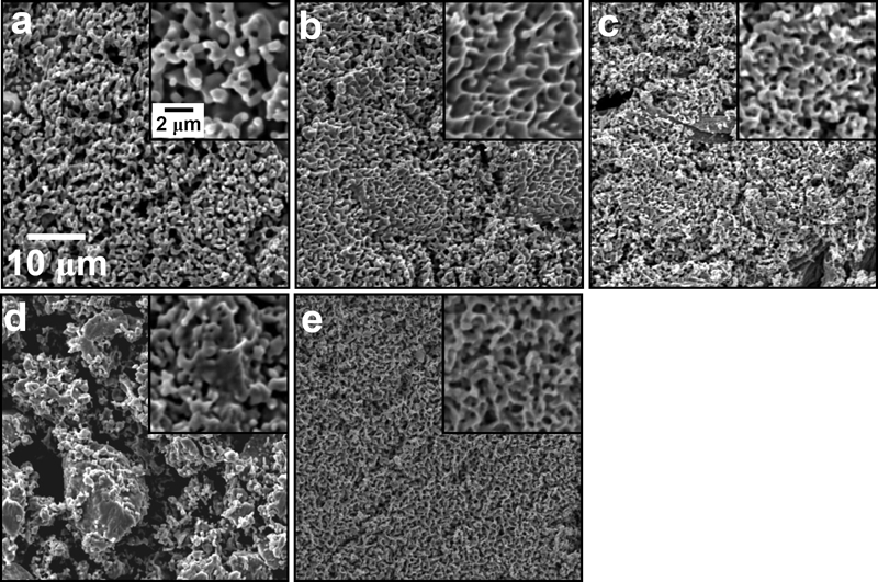
Table 2 shows shear strengths for the reaction temperatures. The shear strength for 20°C (21·8 MPa) was larger than that for 80°C (15·9 MPa). The leaf-like aggregates were produced for 20°C, in contrast to no production of such aggregates for 80°C in the TEM images (Fig. 1a and d). Since a large shear strength is obtained in large leaf-like aggregates, shear strength as large as 21·8 MPa was recorded for 20°C in the present work. According to the XRD measurements, the particles contained Cu2(OH)3NO3. As discussed above, the presence of Cu2(OH)3NO3 deteriorated the bonding property. Accordingly, removal of Cu2(OH)3NO3 with no damage of leaf-like aggregate structure may improve the bonding property. As concluded in the section on ‘Effect of Na/Cu ratio’, the aging process resulted in the transformation from Cu2(OH)3NO3 to CuO (Fig. 1e). With the aging process, the shear strength achieved a value as high as 32·5 MPa. According to the discussion in the section on ‘Effect of Na/Cu ratio’, the production of pores by annealing at the high temperature resulted in low shear strength. The aging process also might have produced pores in the particles through elimination of groups of NO3 and H2O in Cu2(OH)3NO3 during the transformation from Cu2(OH)3NO3 to CuO. Though the pore production may deteriorate the bonding ability of particles, the effect of formation of leaf-like structure on bonding probably overcame the effect of pore production in the present work. Consequently, large shear strength was obtained for the aging process. Figure 6a, d and e shows SEM images of the surfaces of copper plates separated with shear stress. In both cases of 20 and 80°C, sintering appeared to take place among particles with a size of ∼1000 nm. In contrast, many dimples were observed accompanying with sharp tips on the surface of the aging process, which supported the strong bonding with the use of particles prepared with the aging process. Figure 7 shows microstructures of the plate to stage joint made using the CuO particles. The particles were sintered, and consequently, micrometre sized domains were formed. The domains were so fused with the Cu stage that a border between the domains and the Cu stage could not be observed clearly, which confirmed the strong bonding. Some voids, whose formation may spoil bonding, were also observed. A bonding method without production of such voids is waited to be developed for better bonding.
Images (SEM) of the plate to stage joint made using Cu nanoparticles. Images a–c were taken with various magnifications shown in the images. The particles used for the observation were the same as in Fig. 1e
Shear strengths for reaction temperatures: Na/Cu ratios were 1·7
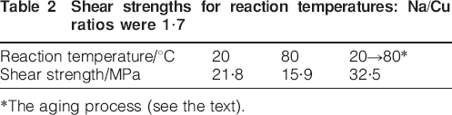
*The aging process (see the text).
Conclusions
The CuO nanoparticles were used as fillers for metallic bonding. The CuO nanoparticle colloid solutions were prepared by reacting 0·01M Cu(NO3)2 with NaOH in aqueous solution at Na/Cu ratios of 1·7–2·1 and reaction temperatures of 20–80°C. Conditions of an Na/Cu ratio of 1·7 and a reaction temperature of 20°C resulted in the formation of submicrometre sized leaf-like aggregates composed of CuO nanoparticles containing Cu2(OH)3NO3. Neither Cu2(OH)3NO3 nor clear leaf-like aggregates were produced at high Na/Cu ratios and a reaction temperature as high as 80°C. In contrast, Cu2(OH)3NO3 contained in the particles prepared at the Na/Cu ratio of 1·7 and 20°C was transformed to CuO with no damage of the leaf structure of aggregates with the process composed of aging the particles at 80°C after preparation at 20°C (the aging process). The bonding examinations in H2 gas at 400°C revealed that the largest leaf-like aggregates without Cu2(OH)3NO3, which were prepared at the Na/Cu ratio of 1·7 with the aging process, had the largest shear strength of 32·5 MPa in all the CuO nanoparticles examined. Accordingly, it was found that the formation of leaf-like aggregates of CuO nanoparticles with high purity was required for efficient metal–metal bonding.
Footnotes
Acknowledgements
This work was partially supported by Hitachi Ltd. We thank Professor T. Noguchi (College of Science, Ibaraki University, Japan) for his help in TEM observation.