
Abstract
The effect of rare earth metals (REMs) on the microstructure and segregation during solidification in a highly alloyed Ni based superalloy was investigated. Rare earth metals considerably decrease the amounts of coarse columnar grains and increase the amounts of equiaxed grains. They are segregated to the interdendritic regions mainly as the precipitate of Ni5Ce. Both the dimension and distribution of γ′ particles and MC carbide are modified by REM. Eutectic (γ+γ′), Laves phase, δ phase and σ phase are precipitated in the interdendritic regions because of the serious segregation of niobium and titanium aggravated by REM. The differential thermal analysis indicates that the precipitation temperature of phases and the solidification sequence of the superalloy can be changed due to the additions of REM.
Introduction
Rare earth metals (REMs) are significantly utilised as refining and microalloying elements to improve the high temperature properties of steels and superalloys, such as oxidation resistance, hot workability, ductility and creep rupture.1–4 Meanwhile, numerous investigations have revealed that minor elements, such as phosphorus, sulphur, boron and zirconium, can greatly affect the microstructure and solidification behaviour in different Fe and Ni based superalloys.5–8 However, the effect of REM on the solidification behaviour in Ni based superalloys remains to be clearly known.
To distinguish the effect of REM on solidification and to reveal the mechanism by which they influence the Ni based superalloys, the present work was designed using a highly alloyed Ni–Cr–Co superalloy with trace additions of REM.
Experimental
Two ingots (90 mm in diameter and 200 mm in height) without REM (named alloy 1) and with REM (named alloy 2) were prepared via vacuum induction melting. Both alloys were melted at 1540°C, held for 10 min and then poured into a cast iron mould followed by air cooling. The compositions of both alloys are listed in Table 1.
Compositions of superalloys with and without REM/wt-
Samples for optical microscopy (Nikon Optiphot-100S optical microscope) and scanning electron microscopy (SEM; JEOL JSM6301F microscope operated at 15 kV) coupled with energy dispersive X-ray analysis system were ground to 2000 grit, mechanically polished and finally electroetched in a solution of 10 mL H3PO4 and 90 mL H2O. Samples for transmission electron microscopy (TEM; Tecnai 20 electron microscope) were prepared using a twin jet electropolishing unit Struers TenuPol-5 with an electrolyte of 10 perchloric acid in ethanol solution at −20°C and 20 V. The TEM observations were operated at an accelerating voltage of 200 kV. The degrees of elemental segregation and distribution of REM were studied by a Cameca SX100 electron probe microanalyser (EPMA) operated at a beam intensity of 15 nA and an accelerating voltage of 20 kV. The compositions of at least three dendrite cores and three interdendritic regions were determined by EPMA using the discrete point measurement technique.
Differential thermal analysis (DTA) was carried out with cylindrical samples (3 mm in diameter and 3 mm in height) using a SETSYS Evolution 18 TG-DTA analyser in a dynamic Ar atmosphere. The samples were heated up to 1450°C at a rate of 10°C min−1 and held for 3 min, followed by cooling at 10°C min−1 to room temperature.
Results and discussion
As cast microstructure
The macrostructures of alloys 1 and 2 are comprised of fine equiaxed grains near the edge of ingots, followed by columnar grains and then equiaxed grains at the centre. The length of each zone in the radial direction is shown in Table 2. Rare earth metals greatly decrease the amounts of coarse columnar grains and increase the amounts of central equiaxed grains, which is the same with REM on steels.9
Range of zones in as cast structure of alloys 1 and 2/mm
The microstructures of alloys 1 and 2, including dendrite cores and interdendritic regions, are shown in Fig. 1. The main phases in both alloys are γ, γ′ and MC carbide,10 and γ′ is the main strengthening phase, as shown in Fig. 2. The morphology and size of γ′ particles in the dendrite cores are quite different from those in the interdendritic regions in both alloys, which are spherical and small in the dendrite cores while cubic and much larger in the interdendritic regions. In addition, the particle size in alloy 1 is smaller than that in alloy 2 in both the dendrite cores and the interdendritic regions. MC carbide in both alloys is blocky, as observed by TEM analysis (Fig. 3a). The energy dispersive X-ray system analysis listed in Table 3 shows that it consists of niobium, titanium as well as molybdenum. The carbides in the REM containing alloy are smaller and more dispersed than those without REM.
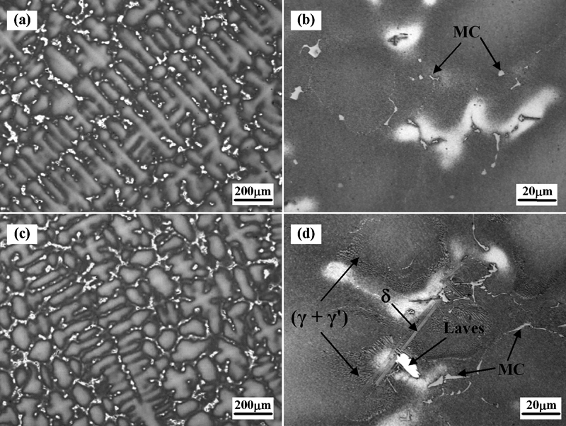
Dendritic microstructures of a, b alloy 1 and c, d alloy 2
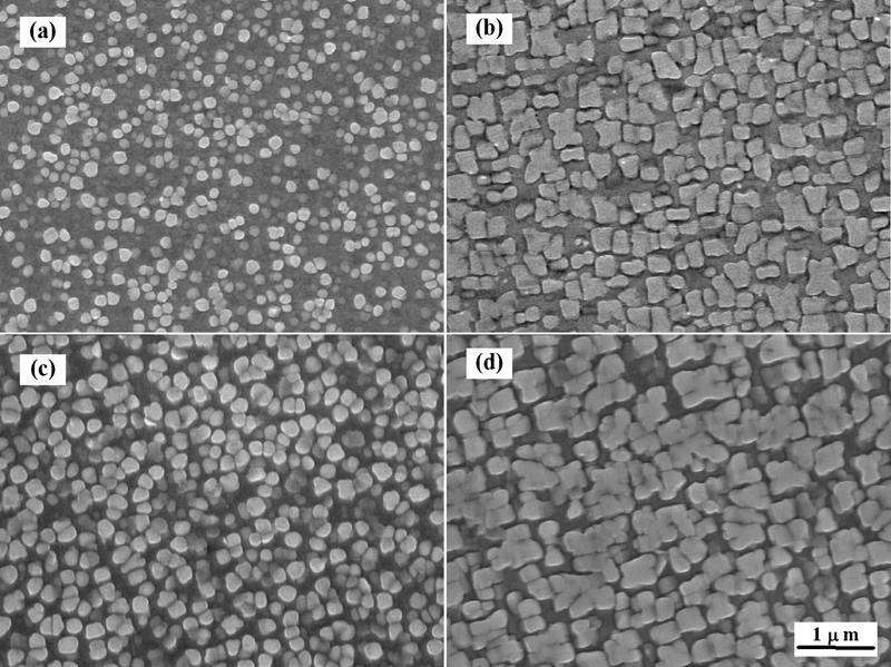
Morphologies of γ′ in dendrite cores and interdendritic regions in a, b alloy 1 and c, d alloy 2
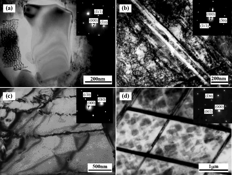
Images (TEM) of precipitates in alloy 2 with corresponding selected area diffraction patterns
Compositions of precipitates in interdendritic regions in alloy 2/wt-
In alloy 1, the phases include γ, γ′ and MC carbide and are relatively simple, and no more phases are identified. However, the precipitates in alloy 2 are more complex due to the additions of REM, as indicated in Fig. 1d.
Eutectic (γ+γ′) in alloy 2 is precipitated in the transition area between the dendrite cores and the interdendritic regions, as shown in Fig. 1d. As seen in Fig. 4a, the dimension of γ′ particles in eutectic (γ+γ′) is much larger than those in dendrite cores and interdendritic regions. Meanwhile, TEM analysis shows that the fine lamellar δ (Ni3Nb) phase is formed parallel to each other in alloy 2 (Fig. 3b). It is precipitated near eutectic (γ+γ′) in the interdendritic regions, as shown in Fig. 4a, and its EDX spectrum is listed in Table 3.
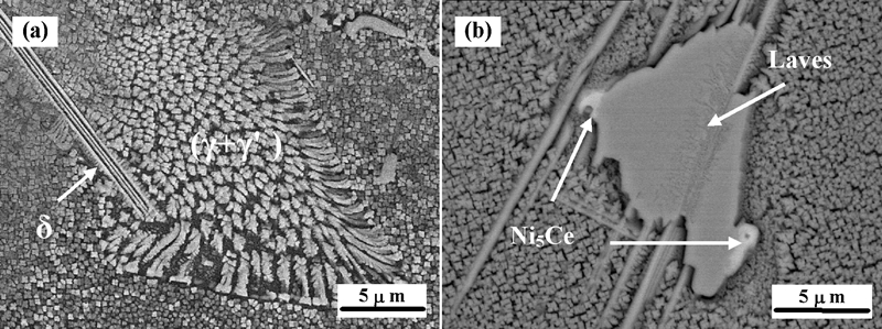
Images (SEM) of precipitates in alloy 2
The bulk Laves phase is formed in alloy 2, as determined by TEM analysis (see Fig. 3c). It is precipitated inside the interdendritic regions, as shown in Figs. 1d and 4b, and its EDX spectrum is shown in Table 3. The compositions of niobium and titanium of Laves as well as the δ phase are much higher than their average compositions in the superalloy. The plane scan analysis by EPMA indicates that an REM enriched phase is formed in the interdendritic regions. The REM enriched phase that is brightly white in the backscattered electron image of SEM (Fig. 4b) is always located on the edge of the Laves phase, indicating that it is precipitated after the formation of Laves phase. It is mainly comprised of nickel and cerium as well as some lanthanum, as listed in Table 3, which corresponds to the intermetallic phase of Ni5Ce.
A topologically closed packed phase, known as σ phase, is created in alloy 2, as determined by TEM analysis. Figure 3d shows its morphology and [3 1 0] zone diffraction. Because the precipitation of MC carbide, eutectic (γ+γ′), δ phase and Laves phase in the interdendritic regions depletes lots of aluminium, titanium and niobium, resulting in the enrichment of chromium, cobalt and molybdenum in the matrix, then σ phase is precipitated.
Elemental segregation
The solute distribution ratio K, calculated by dividing the composition in the interdendritic regions by that in the dendrite cores, was introduced to evaluate the degree of elemental segregation. The results are listed in Table 4. Niobium and titanium are segregated more in the interdendritic regions as positive segregation elements. In contrast, chromium and cobalt are segregated more in the dendrite cores as negative segregation elements. Elements such as aluminium and molybdenum are not segregated obviously.
Contents of main elements in dendritic microstructure of alloys 1 and 2/wt-
As shown in Table 4, the segregation tendency of each element in both alloys does not vary with REM, but the degree of elemental segregation is affected. The REMs aggravate the elemental segregation, which results in K values much larger for positive segregation elements or smaller for negative segregation elements. Among all the elements, the segregation of niobium and titanium is the most influential, which is aggravated by 13·74 and 7·83 respectively due to the additions of REM.
Solidification sequence
The DTA cooling curves of both alloys are shown in Fig. 5. There are three obviously exothermic peaks marked with A, A′, B, B′, C and C′, but one more small exothermic peak marked with D′ between 1210 and 1190°C exists in alloy 2. As observed from the microstructure of DTA samples, alloy 1 consists of γ, γ′ and MC carbide, while alloy 2 consists of γ, γ′, MC carbide, Laves phase and a small account of eutectic (γ+γ′). On cooling from above the liquidus temperature, two large exothermic peaks A and A′ initiate at 1346°C, which is associated with the formation of primary austenite dendrites. The secondary peaks B and B′ may represent the eutectic reaction L→γ+MC, and the first deviation from the local baseline is taken as the onset temperature. Peaks C and C′ are rather broad, indicating that the corresponding reaction is the precipitation of γ′ particles.
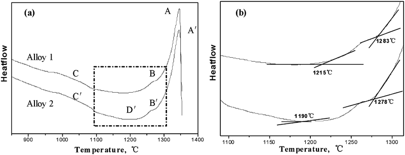
a DTA cooling curves of alloys 1 and 2 and b magnification of dotted rectangular area in a
However, another two reactions take place in alloy 2: L→γ+(γ+γ′) and L→γ+Laves. The latter one (D′) indicates the termination of solidification, which is in agreement with many Ni–Nb–C system superalloys.11–13 For the low quantities of eutectic (γ+γ′), no sufficient energy can be detected by DTA scan, so no exothermic peak is discerned. However, it can be conjectured that the reaction appears to occur below B′ and above D′ because eutectic (γ+γ′) is distributed in the transition regions between dendrite cores and interdendritic regions. Thus, the solidification sequence of alloy 2 can be described possibly by a four-step process: first is the L→γ at 1346°C in which the interdendritic liquid becomes enriched in niobium, titanium and carbon, followed by a eutectic type reaction L→γ+MC at 1278°C depleting the interdendritic liquid of carbon, then L→γ+(γ+γ′) as a second eutectic reaction and, in the end, termination of solidification with the last eutectic type reaction L→γ+Laves at 1210°C. Accordingly, the solidification path of alloy 1 may be as follows: solidification initiates with freezing of γ dendrite and terminates with L→γ+MC beginning at 1283°C.
The effect of REM on the solidification can be illustrated as follows. First of all, REMs appear to not alter the liquidus, but they may lower the solidus temperature, which is 1215°C for alloy 1 and 1190°C for alloy 2, consequently broadening the solidification interval by 25°C. Meanwhile, REM can depress the onset temperature of MC carbide formation by 5°C and induce the precipitation of eutectic (γ+γ′) and Laves phase. Furthermore, as γ′ particles are precipitated between 1085 and 950°C in alloy 1 and between 1100 and 955°C in alloy 2, REM can elevate the precipitation temperature of γ′ by 15°C and enlarge its interval by 10°C, which interprets the fact that the dimension of γ′ particles in alloy 2 is much larger than those in alloy 1, in both dendrite cores and interdendritic regions.
Conclusions
Rare earth metal additions considerably promote columnar to equiaxed transition in the as cast ingots. Phases such as eutectic (γ+γ′), Laves, δ, σ and Ni5Ce are precipitated in the interdendritic regions due to the additions of REM. Rare earth metals are segregated to the interdendritic regions, aggravating the segregation of niobium and titanium. Rare earth metals appear to broaden the solidification interval and depress the onset temperature of MC carbide formation, consequently modifying the distribution and dimension of γ′ phase and MC carbide.