
Abstract
Surface textured Si doped TiCN coatings were synthesised on Ti–6Al–4V alloy by laser cladding technique. Phase constituent examination by X-ray diffraction revealed the formation of similar phases of TiC0.2N0.8 and Ti5Si4 within all coated samples. Laser coated samples presented much higher surface free energy compared to Ti–6Al–4V control due to the textured structure, which in turn demonstrated a better wettability and improved biomineralisation. Variation of silica content presented no significant influence on surface free energy, indicating that the silica content can be varied in a large range. The mineralised samples obtained after immersion in simulated body fluid were characterised to understand the mechanism and kinetics of Ca–P precipitation. The results confirmed that the precipitation kinetics of Ca–P was influenced by the substitution of silica.
Introduction
As commonly used bioinert materials, titanium and its alloys are widely used as load bearing implant materials for both dental and orthopaedic applications1,2 owing to their excellent properties such as mechanical properties, biocompatibility, corrosion resistance, and tissue compatibility. However, the poor interfacial bonding between the metallic surface and the surrounding bone,3,4 along with their inferior tribological properties and bioactivity constitute a major obstacle limiting the longevity and usage of the implants. Meanwhile, the poor wear resistance of Ti and its alloys results in generation of wear debris, and further in osteolysis, 5 which is one of the main limiting factors affecting the longevity of load bearing implants. 6 An alternative and more effective solution to improve the corrosion and wear resistance and enhance the formation of bone-like layer on the metal implant surface is to deposit a multifunctional bioactive coating.
In recent years, different types of hard ceramic coatings owing to their excellent wear and corrosion resistance and high hardness continue to make inroads into many biological applications by overcoming the drawbacks mentioned above. Titanium nitride (TiN) is one of the most frequently used ceramic coatings due to its excellent wear and corrosion resistance and high hardness. Nowadays, TiN coatings have been widely used in a wide range such as heart valve replacements, dental prosthesis, and materials for hip joint. 7 This is also due to an excellent haemocompatibility of TiN films. 8 Compared to TiN coating, TiCN possesses even superior wear and corrosion resistance, thermal conductivity, and hardness. These properties make it a very interesting candidate employed as a protective layer in tool steels and semiconductors. 9 Besides, its non-cytotoxic character and biocompatibility combined with mechanical and corrosion properties make it a very interesting material for biomedical applications. Recently, Serro and his coworkers 10 reported an investigation of TiCN coatings used for improving the wear resistance of orthopaedic joint implants. In the past work, one of the few studies on the corrosion of TiCN as a biomaterial was reported by Hollstein and Louda. 11 The results revealed that TiCN film presented a slow kinetics of the corrosion processes and a good biocompatibility. Recently, Antunes et al. 12 reported an investigation on the corrosion resistance and in vitro biocompatibility of TiCN coatings on AISI 316L austenitic stainless steel for orthopaedic applications. In their work, both cytotoxity and genotoxicity tests revealed the biocompatible nature of the TiCN film which is a primary indication of its potential use as a coating for biomedical devices. However, in order to obtain artificial implants with enhanced physical, chemical, mechanical, tribological, and biological properties for accelerated self-adaption in human body and long term performance, it is necessary to prepare a multifunctional biocoating with various properties, such as excellent corrosion resistance, high wear resistance, high fatigue and tensile strength, and especially excellent biocompatibility and bioactivity. 13
Over recent years, the effect of silicon substitution on material bioactivity has attracted significant interest. This is due to the fact that the presence of aqueous Si has been shown to involve directly in the mineralisation process, where aqueous Si, in the form of functional groups such as Si–OH, is able to induce the precipitation of HA from electrolyte solutions in the presence of proteins that normally inhibit its precipitation.14,15 In addition, aqueous Si has been shown to enhance osteoblast proliferation, differentiation and collagen production, and to have dose dependent effects on osteoblast cells under in vitro conditions.16–18 In light of this, silicon containing materials are increasingly used in biocoating. Si doped TiN films were found to demonstrate superior performance over binary nitrides in tribological tests. 19 In the work by Veprek and Reiprich, 20 TiN/Si3N4 film was used to form the ternary Ti–Si–N system due to the fact that Si3N4 owns the superior combination of fracture toughness and hardness, which in turn makes it a very attractive candidate for high load medical applications, such as metal prostheses replacement. 21 Recently, Shtansky et al. 22 designed a Si doped multifunctional bioactive film using the composite TiC0.5+CaO+Si and TiC0.5+CaO+Si3N4 as target. The Ti–Si–Ca–C–O–N films exhibited low fiction coefficient, hydrophilic properties, improved electrochemical characteristics, and furthermore a high level of biocompatibility and biostability. The film also did not reveal any bactericidal and toxic activity.
Although, TiCN and other Si doped bioactive coating have received extensive studies, there are not many efforts towards understanding effects of Si addition in different amounts on the wetting and in vitro bioactivity of these composite coatings. In the meanwhile, most of the techniques, including DC magnetron sputtering, 19 plasma enhanced magnetron sputter deposition, 23 plasma enhanced chemical vapour deposition, 24 and plasma vapour deposition (PVD) 12 have been applied to obtain Si doped biocoating and TiCN coating for orthopaedic applications. In the present work, a highly intense laser beam was used to melt the precursor (SiO2/TiCN coating) and the portion of Ti–6Al–4V substrate to get a microtextured multiphase coating and a metallurgical bonding at the interface. The effects of SiO2 addition in different weight ratios on the wetting, Ca–P deposition process, and the mechanisms are studied. The chemical processes in simulated body fluid (SBF) immersion are also discussed.
Materials and methods
Fabrication and characterisation
Ti–6Al–4V coupons (100×50×3 mm) were used as substrate. They were prepared for coating by initially polishing using silicon carbide emery paper to remove the oxide film, followed by rinsing with acetone. A mixture of commercially titanium carbonitride (TiC0.3N0.7) and silica (SiO2) powder was used as the precursor materials. TiCN powder had a spherical morphology with particle size of about 3 μm, and that of SiO2 is between 30 and 45 μm. The precursors were mixed in an organic solvent to form slurry and then sprayed onto the substrate coupons. The sprayed coupons were then air dried to remove the moisture and get a uniform thickness of 20–30 μm for all pre-coating deposits. Finally, the samples were scanned using a JHM-1GY-700 pulsed YAG laser processing system equipped with a 700 W Nd–YAG laser and a powder feeder to obtain a metallurgical bond between the precursor and the substrate material. Pulse height, pulse width, and pulse repetition rate were controlled through the control panel of the laser. The lens assembly is equipped with a 200 mm focal length convex lens, which gives a spot diameter of approximately 300 μm at focus. The focused spot is kept at approximately 15 mm above the surface of the sample surface. The laser processing parameters employed in the current investigation are that: the laser beam scan speed of 3.0 mm s−1, pulse frequency of 15 Hz, and average laser power input of 100 W.
Nondestructive phase evaluation of the samples before and after immersion in SBF was conducted using a TD-3500 X-ray diffractometer, with Cu Kα radiation of wavelength 0.15406 nm. The system was operated at 30 kV and 20 mA in a 2θ range of 20–90 using a step size of 0.02°. Microstructure and morphological evolutions of the coated samples before and after immersion in SBF were characterised using a SSX-550 scanning electron microscope. An energy dispersive spectrometer with the scanning electron microscope was used to study the elemental analysis of the mineralised samples. The microhardness measurements along the depth of the cross-section were performed using a 401MVD microhardness tester with a normal load of 200 g for 10 s.
Wetting and in vitro studies in SBF
Wetting is the first and foremost event when a biomaterial is implanted into the biological system. Hence, it is very essential to investigate the wettability of a biomaterial before conducting further biological studies. To evaluate the wettability and in vitro bioactivity of the coated samples, SBF solution with similar ionic composition as that of human blood plasma was prepared by mixing reagent grade chemicals in the following order: NaCl (8.026 g), NaHCO3 (0.352 g), KCl (0.225 g), K2HPO4.3H2O (0.230 g), MgCl2.6H2O (0.311 g), CaCl2 (0.293 g) and Na2SO4 (0.072 g) in distilled water (700 mL). The fluid was then buffered to pH 7.4 at 37°C with trihydroxymethyl aminomethane (6.063 g) and hydrochloric acid (40 mL).
Before in vitro assays, contact angle measurements were conducted by a static sessile drop technique using a HARKE-SPC contact angle goniometer, equipped with a video camera for imaging. A 5 μL liquid droplet with the diameter of 2 mm was suspended from the tip of the hypodermic syringe on the thoroughly cleaned sample and the advancing contact angle was taken as a measure of wettability. The liquid droplet placed on the sample was allowed to stabilise for 10 s before the reading was taken. The test was conducted at room temperature and a minimum of 10 contact angle readings were taken on each sample. For the calculations of surface energy, an apolar liquid (dioiodomethane) and two polar liquids (distilled water and formamide) were used. In order to correlate the surface energy calculations to the mineralisation behaviour of the samples, and to explain the in vitro bioactivity of the coatings in SBF solution, contact angle between the samples and SBF was also measured. The surface energy components of different samples were calculated according to Van Oss et al.'s
25
approach using the following equation

In this model, it is assumed that the surface energy of a solid (or a liquid) can be divided into the Lifshitz-van der Waals surface tension (γLW) and Lewis acid base surface tension (γAB) components, hence, the surface energy γS can be expressed as following equation


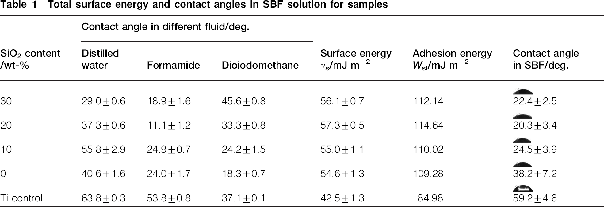
Total surface energy and contact angles in SBF solution for samples
In vitro studies were performed by immersing the samples in SBF. Samples were immersed in a plastic vial containing 15 mL of SBF solutions and were kept under static conditions inside an incubator at 37°C for 8 and 14 days. To study the kinetics of HA precipitation on the coated samples, a set of four samples from each processing condition was immersed in the SBF solution. The SBF solution was refreshed every 24 h to maintain the pH value of 7.4. After exposure, samples were dried at 100°C for 24 h to remove the moisture. Phase constituents and surface feature of samples after SBF immersion were studied using X-ray diffraction (XRD) and scanning electron microscopy (SEM) equipped with energy dispersive X-ray spectroscopy (EDS), respectively.
Results and discussion
Phase and microstructure analysis
X-ray diffraction patterns of laser processed x wt-SiO2–TiCN coatings (x = 0, 10, 20 and 30) are presented in Fig. 1. The analysis revealed no difference in the phase type with variation of SiO2 content in the coating. All samples demonstrated the same phases as marked in the diagram. The major phases identified within the detectable limits of the instrument were TiCN and Ti5Si4. The presence of identical phases in the coatings of all laser processed samples is expected to provide the similar wetting characteristics during SBF immersion. However, as will be stated in the following section, all the coatings demonstrated a significant increase in wetting compared to uncoated Ti–6Al–4V control.
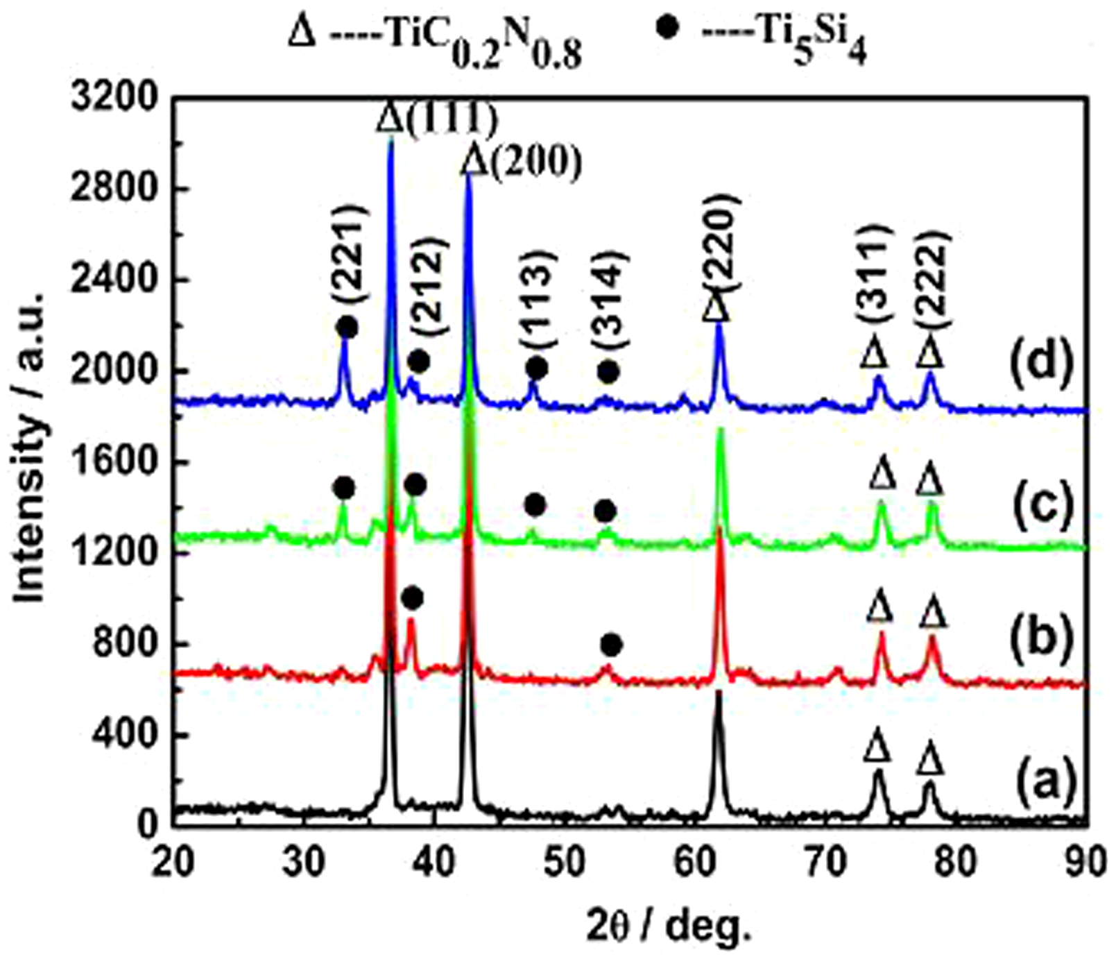
a 0 wt-; b 10 wt-; c 20 wt-; d 30 wt-
A representative cross-sectional microstructure and microhardness distribution of laser cladded 20 wt- SiO2–TiCN coating are shown in Figs. 2 and 3, respectively. The structure exhibited sound bonding between the coating and the substrate. No unmelted or partially melted powders were observed throughout the structure's cross-section (Fig. 2a). However, non-uniform microstructure and few minor cracks were observed (Fig. 2b–d). This indicates large temperature fluctuations in the liquid metal pool due to changes in the composition across the sample cross-section. It was reported that large temperature fluctuations can result in non-uniform or inadequate intermixing of molten powders in the transition region. 26 In the present work, the large temperature fluctuations could be due to large difference between melting points of TiCN (>2950°C) and SiO2 (1670°C), and their different laser absorption coefficients. It was observed that the microstructure in the top portion of the coating is more porous than that beneath it (Fig. 2b). In fact, the presence of porosity in the top portion of the coating being in contact with bone, likely to improve cell–material interactions.27,28
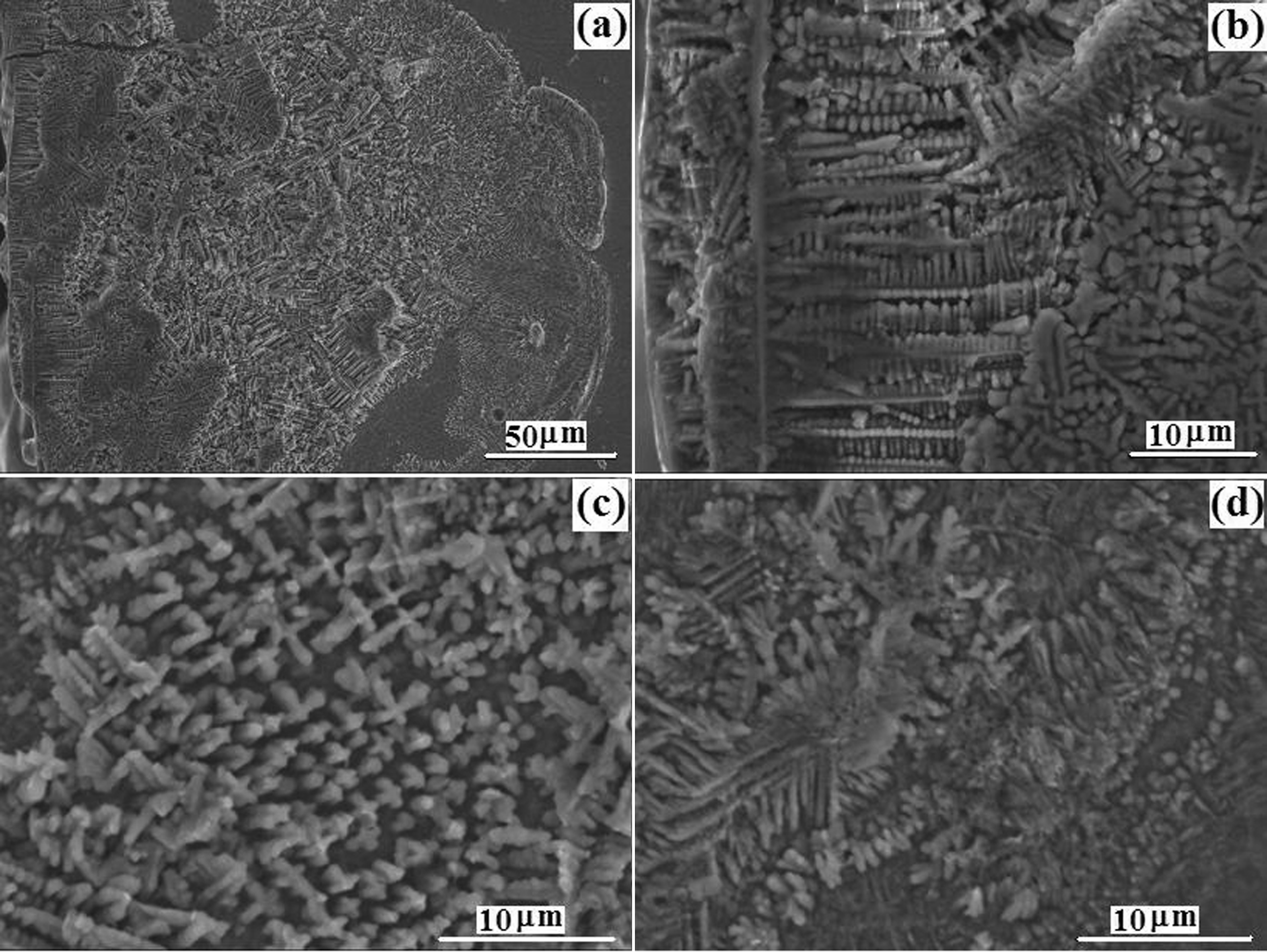
a low magnification overview; b within upper portion of coating; c within middle portion of coating; d within bottom portion of coating (near interface between coating and substrate)
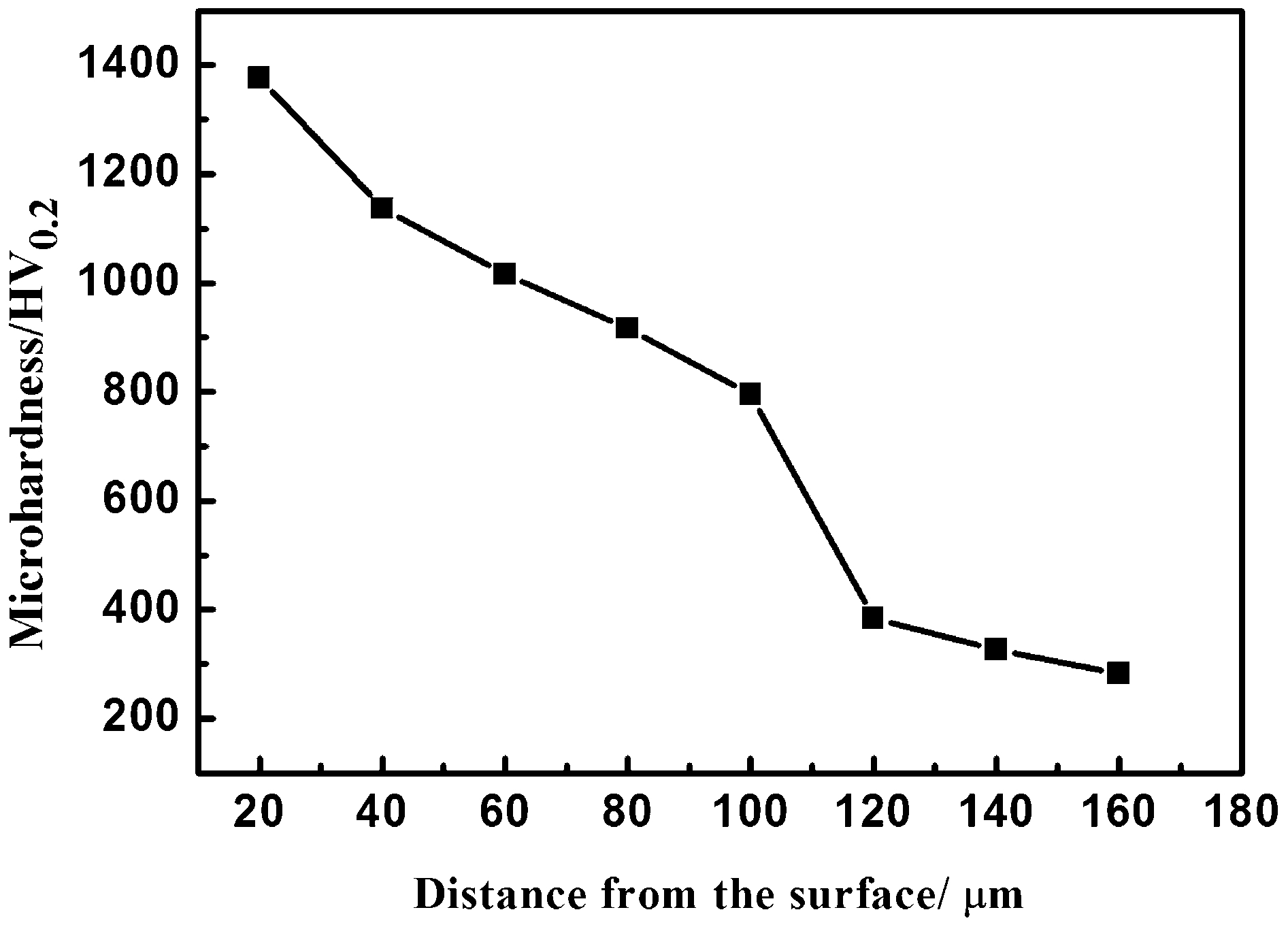
Typical cross-sectional microhardness distribution of laser processed 20 wt-SiO2–TiCN coating on Ti–6Al–4V
Figure 3 illustrates that a gradient distribution of hardness value was observed. The microhardness in the coating zone is about 1400 HV0.2. This enhancement of microhardness value was mainly resulted from the crystallite size refinement due to the addition of SiO2 and proper laser treatment as well as the formation of TiCN hard phase coating. This enhancement is expected to improve the friction and wear properties of Ti–6Al–4V substrate. The corresponding research is now in progress and will be reported later.
Wetting study
The substitution of silicon can not only improve the apatite precipitation, but also can significantly alter surface properties such as wettability and surface energy, which in turn have strong influence not only on cell–material interactions but also on the ability to form chemisorbed lubricating films. Hence, before in vitro assays, contact angle measurements were conducted to evaluate the wetting behaviour. The contact angle of distilled water, Forma-mide, and Dioio-domethane for the coatings and the bare Ti is listed in Table 1 used for calculation of surface free energy. The contact angle for the coatings and bare Ti in SBF is also measured as listed in Table 1. The contact angle measurements were carried out at least for 10 times at different locations to obtain average values.
Surface energy components as a function of silicon content calculated from equations (1) to (3) are illustrated in Fig. 4. The surface free energy γs varies in a slight range of 55.0–57.3 mJ m−2 with an increasing first and then a slight decreasing with increasing silica content in the range of 10–30 wt- in the precursor, indicating that in present work the silica content can be in a large range. It also can be seen that the surface free energy γs for all the x wt-SiO2–TiCN (x = 0, 10, 20 and 30) coatings is significantly higher than that of Ti control, indicating a higher wettability was achieved by silicon substitution and laser treatment for the coatings. The higher wettability is attributed to a high density of grain boundaries serving as high energy adsorption sites. It was reported that the addition of SiO2 can lead to more grain boundary grooves formation 29 and in turn further may have contributed to the increase in surface free energy and improve the wetting behaviour.
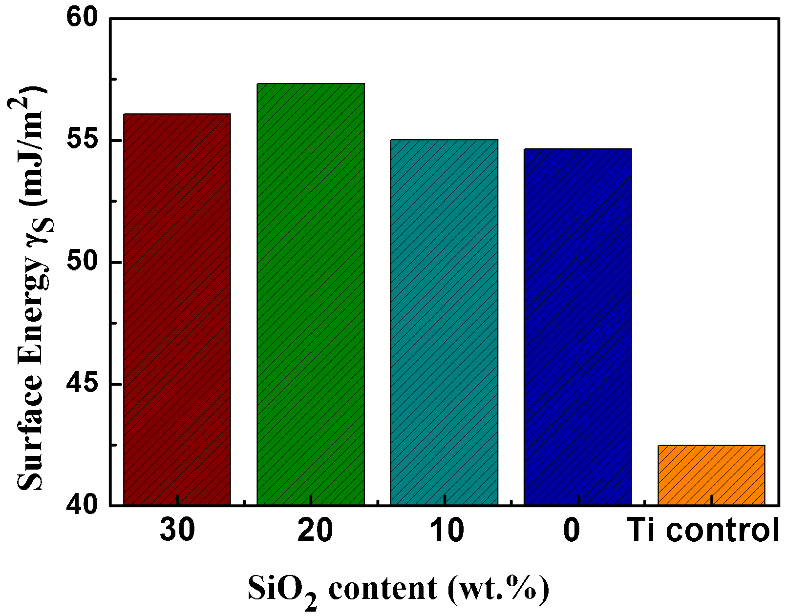
Surface energy components as function of silicon content
Wetting is the first and the foremost event when a biomaterial is implanted into the biological system. Hence, on the basis of surface free energy examination, understanding the wettability with SBF and the effect of SiO2 addition on the wetting behaviour were important in this work. Table 1 presents the contact angles and corresponding light optical images of the liquid droplet shadow on x wt-SiO2–TiCN (x = 0, 10, 20 and 30) samples, and the Ti–6Al–4V control. It is obvious that the laser treated coatings shows significant decrease in the contact angle compared to Ti control. The different wetting behaviour for the three probe liquids can be attributed to a difference in the surface free energy. For the bare Ti, the contact angle in SBF is 59.2±4.6°, which is much higher than that of the laser treated coatings ranging from 38.2±7.2 to 20.3±3.0°, indicating the laser treated coating are more hydrophilic with SBF than the bare Ti. Furthermore, the laser treated coating with silica addition presents a decrease first and then a slight increase in contact angle in SBF with increasing silica content. It indicates that the addition of silica is benefit for increasing the surface free energy, which in turn improves the wetting. The improved wetting behaviour can further be explained by adhesion energy Wsl, which is given by the following equation (4),
30
supposing that the total surface tension of the probe liquid results solely from the dispersion interactions and no hysteresis was considered

In vitro bioactivity studies and kinetics of Ca–P precipitation
Figure 5 represents the XRD studies after immersion in SBF for 8 days (Fig. 5a) and 14 days (Fig. 5b) for coated samples with different silica content, respectively. The base spectrum (before immersion in SBF) is not included in each of these, because they have been illustrated in Fig. 1 separately. It is obvious that a maximum attributed to the apatite phase at 2θ = 31.75° and 2θ = 25.68° corresponding to planes (211) and (002), respectively, were detected after 8 and 15 days immersion of each sample in SBF (Fig. 4a and b). In addition to the above peaks (2θ = 31.75 and 25.68°), one more apatite peak appeared at 2θ = 46.71° corresponding to plane (222), for the sample with SiO2 content of 20 and 30 wt- after 8 days (Fig. 5a) and 15 days (Fig. 5b) of immersion in SBF. Besides this peak, another more apatite peak was detected at 2θ = 49.46° corresponding to plane (213), for the sample with SiO2 content of 30 wt- after 8 days (Fig. 5a) and 15 days (Fig. 5b) of immersion in SBF. This clearly indicates that the samples with higher SiO2 content have a pronounced biomineralisation compared to the other samples with lower SiO2 content.

Patterns (XRD) of coatings after immersion in SBF for a 8 days and b 15 days
It can also be observed that there is an increased crystallographic texturing along the planes (002) and (211) with increasing SiO2 content both for 8 and 15 days of immersion in SBF. Such a phenomenon may be attributed to the improved wetting ability by SiO2 addition and the role of Si on the biomimetic precipitation. As stated earlier, the contact angle between SBF and the samples decreased from 59.2±4.6° for Ti control to 20.3±3.4° for laser cladded samples due to SiO2 addition, meanwhile, the contact angle between SBF and laser cladded samples presented a decreasing first and a slight increasing with increasing SiO2 content. Lower contact angle also suggests higher surface energy of coated surfaces with varying SiO2 content. The higher hydrophilicity and surface energy of these surfaces improved the apatite forming ability in SBF. On the other hand, some researchers pointed out that Si promotes biomimetic precipitation by a combination of increasing the solubility of the materials via creation of defects in the lattice,31–33 by generating a more electronegative surface 34 and by generating a smaller grain size with more triple point junctions per unit area, facilitating increased dissolution at the surface. 31 Similar biomimetic precipitation has been observed at the surface of Si substituted HA and α-TCP materials.31,35,36 Compared Fig. 5a to b, it can also be observed that there is an increased crystallographic texturing along planes (002) and (211) with increasing immersion time for any sample with certain SiO2 content. This result is in accordance with the XRD result given earlier. It revealed that the texturing may have happened due to the epitaxial grain structure ((220 and (221)) on the Ti alloy substrate as seen in Fig. 1.
Scanning electron microscopy observations for the samples with different SiO2 addition demonstrated (Fig. 6) the formation of an apatite like layer, following immersion in SBF for 8 and 15 days. After immersion in SBF for 8 days (Fig. 6(a–1) to (d–1)), fewer apatite layer with globular morphology was observed, while after immersion for 15 days (Fig. 6(a–2) to (d–2)), the apatite layer presented well distributed globular morphology. Hence, it can be concluded that the apatite layer presented well distributed globular morphology with increasing immersion time. Furthermore, it can be seen from the SEM images that, the samples with lower SiO2 content exhibited the similar porous morphologies after 15 days’ immersion in SBF. A number of micropits (<0.5 μm) on each globular particle were observed on the coatings with lower SiO2 content (Fig. 6(a–2) and (b–2)). Similar phenomenon has been reported by Zhang et al., 37 and they pointed out that such pits may facilitate incorporation and nutrient transportation. In contrast to these samples, the samples with a higher SiO2 content presented a different surface morphology, as shown in Fig. 6(c–2) and (d–2). As can be seen, with increasing SiO2 content, the micropits disappeared. The coating surfaces were homogeneously distributed with spherical apatite with diameters of 2–5 μm. It can also be observed from the EDS spectra (presented as an inset within the SEM images) that there is a strong presence of Ca and P atoms following immersion in SBF. These studies therefore further supplement the earlier results from the XRD studies (Fig. 6a and b). It is worth noting that a small amount of Na and Cl from the SBF solution were also found by EDS analysis. This may be resulted from the incomplete washing operation after exposure in SBF. This enhancement in mineralisation or bioactivity for the laser coated samples is, as explained in earlier sections, a result of their improved wettability with SBF solution due to the addition of SiO2. Besides that, Paital et al. 38 have demonstrated that such laser processing can produce a so called textured surface, which in turn further improve the wettability of the laser processed sample.

Images (SEM) with EDS results of coatings with SiO2 addition of a 0 wt-, b 10 wt-, c 20 wt- and d 30 wt-, after 8 and 15 days of immersion in SBF


The mechanism for improvement in mineralisation can also be explained by the similar theory given by Paital et al.
38
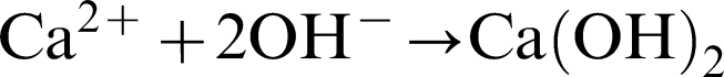
In their work, they pointed out that the laser processed samples present a textured surface features with radial grooves owing a smaller value of surface texture parameter described as a ration of depth to radius of the groove. This smaller value of the surface texture parameter, in turn, can make the liquid drop placed on the surface to easily overcome the energy barriers and completely wet the surface, and decrease the contact angle, thereby, improving the wettability in SBF. This improvement in wettability further enhances the reaction of SBF solution with Ti5Si4 phase present on the surface of the sample. The silicon ions react with water molecules and in turn lead to the generation of negatively charged Si–OH group. It has been widely reported that the generation of Si–OH group or the application of a negative potential to a Si wafer will introduce the precipitation of an apatite-like phase in physiological media.39,40 The precipitation of OH− ions attracts the calcium ions (Ca2+) from SBF towards the negatively charged (OH−) surface, followed by the formation calcium hydroxide. Then, the formed calcium hydroxide reacts with the phosphate ions ( ) present in the SBF solution, and consequently form apatite nuclei on the surface. The above procedures can be described as the following possible equations


Conclusions
Si doped TiCN microtextured coatings were deposited by laser cladding technology with variable silica content (0 to 30 wt-). The process of laser cladding resulted in the formation of TiC0.2N0.8 and Ti5Si4 within the coated samples. There was no significant variation in the phase types and amounts with variation of SiO2 content from 10 to 30 wt- in the precursor. The structure exhibited sound typical fine dendrites in the coating. Laser coated samples presented much higher surface free energy compared to Ti–6Al–4V control due to the laser textured structure. Variation of the surface free energy γs in a narrow range of 55.0–57.3 mJ m−2 with an increase first and then a slight decrease with increasing silica content in the range of 10–30 wt- in the precursor, indicated that the silica content can be varied in a large range. The coatings substituted by silica demonstrated a better wettability. Hence, the samples substituted by silica demonstrated improved biomineralisation compared to the uncoated Ti–6Al–4V and 100 wt-TiCN coated sample.
Acknowledgements
The work described in this paper is financially supported by Fundamental Research Funds for the Central University (grant no. N120405002), the 45th Scientific Research Foundation from the State Education Ministry for the Returned Overseas Chinese Scholars, and the 8th University Student's Innovation Training Project (grant no. 140012).