
Abstract
Currently, custom made heart implants suffer from critical imperfections in terms of mechanical incompatibility and uncontrolled degradation rate, which leads to premature in vivo failure. Interpenetrating polymer networks (IPNs) are a success story in drug delivery and regenerative medicine where they offer fascinating morphologies and tailored properties like controlled degradation rate. This review will critically discuss the current state of the art with respect to the two most important biopolymers found in heart valve, i.e. collagen and hyaluronic acid formulated IPNs as tissue engineered heart valve (TEHV). Comparison of the existing experimental approaches and recent technical challenges will be demonstrated. Finally, multifaceted proposals for improvements in design and performance of TEHVs will be presented as a route map for future research.
This review is the winning entry in the 2015 Materials Literature Review Prize of the Institute of Materials, Minerals and Mining, run by the Editorial Board of MST. Sponsorship of the prize by TWI Ltd is gratefully acknowledged.
Introduction
Heart is an organic pump that transports oxygenated blood from the respiratory system to the whole body, and then pumping the blood stream saturated with metabolic byproducts back to the respiratory system. Like any mechanical pump, the working cycle of the heart critically depends on the proper functioning of heart valves. Heart valves can repeat their working cycle (opening and closing) ∼3 × 10⁹ times during one's lifetime. 1 Therefore, heart valves play a key role in sustaining the smooth working of the heart. When a heart valve does not work the way it should do, the condition is usually associated with valvular heart disease. The overall death rate in the USA corresponds to one death every 40 s due to valvular heart disease. 2 According to the American Heart Association, the number of heart surgeries has increased by 28% in 2010 as compared to 2000 with a total direct cost reaching up to 315.4 billion dollars in the USA alone. Around 290 000 patients worldwide receive heart valve replacements annually, and the number is expected to increase up to 850 000 by 2050 due to an increase in the average life expectancy.3,4 In addition, a recent survey by the British Heart Foundation reports ∼4 million deaths/year due to cardiovascular disease in Europe. 5 Cardiovascular disease could be either congenital or acquired. Minors born with congenital defects usually have irregularities associated with valve shape, size and number of leaflets. For example, a patient born with two cusps instead of three suffers from bicuspid aortic valve disease. 6 According to the British Heart Foundation's report, 1 of 180 babies is born with congenital heart defect. 7 Acquired heart valve diseases, on the other hand, are caused by age related degeneration. The disease is usually manifested as improper valve opening or closing. When there is a restriction with full opening of the valve, the disease is called valvar stenosis. Valvular insufficiency is associated with lack of proper seal of cusps during valve closing. In both cases, heart is forced to work more than usual to meet the standard stroke volume. This condition gradually leads to heart valve dysfunction and may become a life threatening condition leading to heart failure.1,8,9 For instance, aortic valve disease is one of the major causes of heart failure affecting ∼5 million people in USA. 10
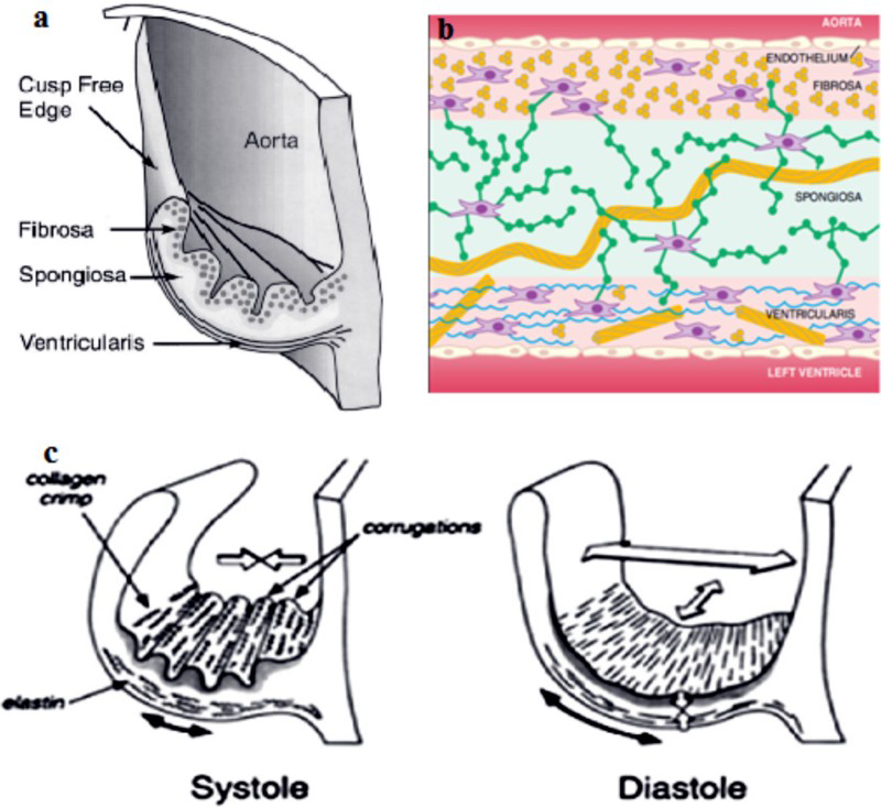
The trilayer microstructure of the heart valve leaflet comprises three different natural polymers: collagen, elastin and glycoaminoglycans (GAGs), each of which plays an important role as shown in Fig. 1a and b. For example, circumferentially aligned collagen fibres bear the load during systole–diastole, while elastin makes the large strains possible during opening and closure of heart valve (Fig. 1c). The presence of GAG ensures tissue hydration and smooth load transition between the layers, and aids in cellular activities. 11 At a molecular level, the distribution and conformations of these entities closely resemble the intertwined mesh of individual polymeric chains. Such an interlinked microstructure gives heart valve enough strength (2.6 MPa) to undergo steady systole–diastole cycle 3 × 10 9 times during one's lifetime.1,12
There lies a niche where tissue engineers are currently facing a challenge to develop tissue engineered heart valves (TEHVs) resembling the natural one in terms of microstructure and biomechanical performance. Interpenetrating polymer networks (IPNs) are smart materials that offer a pathway to build natural polymer based implants with interlaced microstructure and hence improved biomechanical properties. This review will discuss the prospects of exploiting IPNs based on natural heart tissue components such as collagen and GAGs towards the development of an ideal TEHV.
Heart implants/prosthesis
The first ever implantable heart device was a left ventricular bypass pump that was implanted at Baylor College of Medicine in 1963. 15 All the heart implants used in cardiac surgery are either mechanical or biological, each of which has their own limitations. 16
Biological prosthesis
A discussion of biological substitutes used for the heart valve repair now follows.
Autografts and allografts
Diseased aortic valves are usually replaced by allografts/homografts isolated from human cadavers. However, the potential risk lies in the ultimate heart failure due to the degenerative changes in the valve. The Ross procedure is an alternative procedure that involves the replacement of aortic valve by pulmonary valve (autograft) and takes the advantage of structural similarity between the two valves. Pulmonary valve then in turn is replaced either by an allograft or a xenograft. 17 This procedure is still in practice, and patients usually have a better quality of life. 18 However, it is technically challenging and is feasible for a small group of patients only. 19
Xenografts
Xenografts, one of the bioprosthesis still in practice, are derived from animal, e.g. porcine aortic tissue or bovine pericardial tissue, and mounted in a supporting frame. These are usually treated with glutraldehyde (GA), which enhances the degradation resistance of the xenograft by forming the crosslinks between complex tissue proteins.17,20 Glutraldehyde treated xenografts have more stable mechanical properties than mechanical prosthesis, offer unlimited supply from donors and do not involve the risk of thrombosis, elimination of anticoagulation treatment and hemolysis.8,9 However, xenografts are prone to immunogenic response and transmission of certain type of disease such as Creutzfeldt-Jakob. 21 In addition, animal heart valve is inferior to the human heart valve in terms of mechanical properties, which can facilitate only short term durability. 11 For example, the elastic modulus of human aortic valve (15.55 MPa) is almost twice to that of porcine aortic valve (7.78 MPa). 22
Decellularised grafts
Decellularised grafts have recently emerged as a template for TEHVs. 23 Decellularised valves when applied as natural scaffolds exhibit the natural anatomy of heart valve and preserve important physical features like protein chemistry and mechanical strength.24,25 Interestingly, decellularised heart valves can also have longer shelf life up to 18 months without deterioration of properties. 26 However, transfer of decellularised xenografts to human is challenging. This is the reason for the limited success achieved so far with decellularised TEHVs in clinical applications, especially with children. 27
Mechanical prosthesis
A mechanical prosthesis is made of metal or synthetic polymer, which makes it superior in mechanical properties. Although these implants are currently being utilised by surgeons in an appreciable number, complications associated with their use is more than the benefits they offer. 28
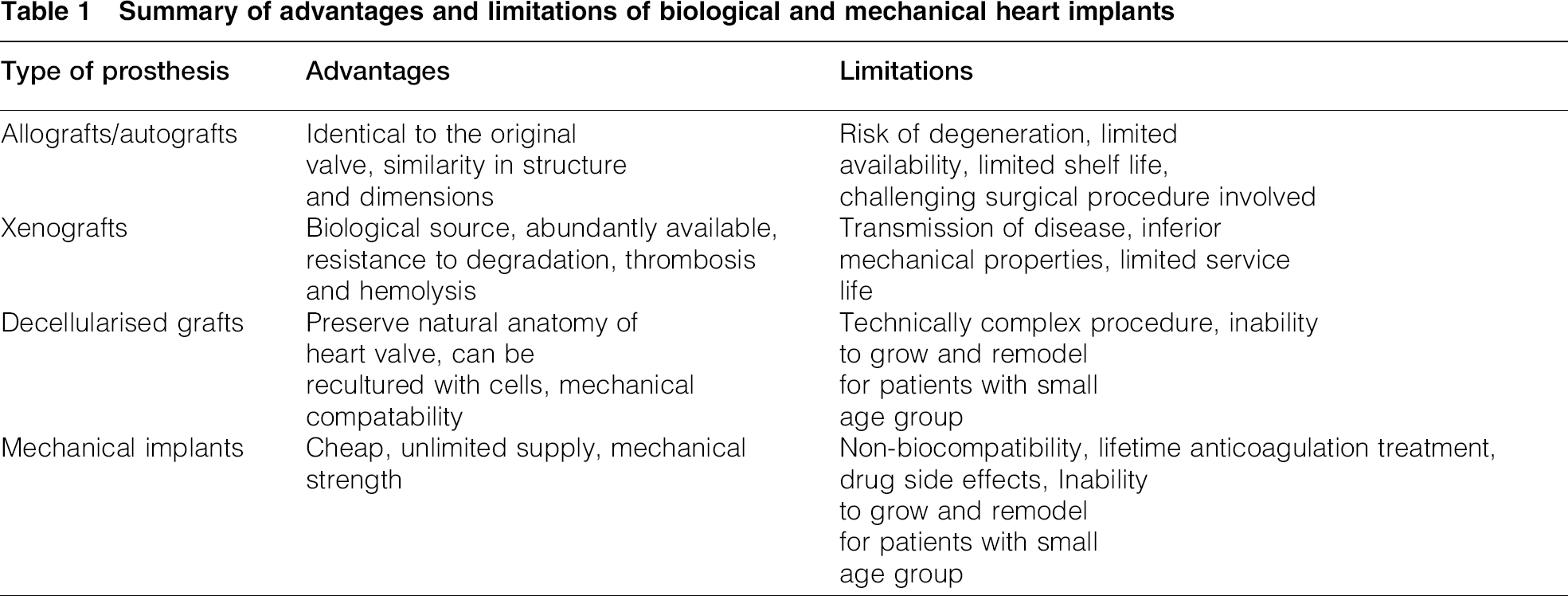
Biocompatibility is the most important limitation of synthetic prosthesis. In addition, mechanical heart valves are thrombogenic in nature due to the lack of endothelial cell lining. Therefore, oral anticoagulation treatment is necessary in patients receiving mechanical valve replacements. Such a treatment may cause severe or fatal bleeding.8,29 Another limitation for such implants is the limited use for children, as these implants are not able to grow or remodel themselves over the course of time. 30 Advantages and limitations of all types of heart implants have been summarised in Table 1.
Summary of advantages and limitations of biological and mechanical heart implants
Tissue engineered heart valves
The term ‘tissue engineering’ for the first time was used by Fung at a National Science Foundation Workshop in Washington, DC in 1987. 31 Tissue engineering has been defined in a number of ways, e.g. a comprehensive definition coined by World Technology Panel Report states
‘The application of principles and methods of engineering and life sciences to obtain a fundamental understanding of structure–function relationships in novel and pathological mammalian tissues and the development of biological substitutes to restore, maintain or improve tissue function.’ 32
A tissue engineering system is based on three vital components: cell, signal and scaffolds. Cells are seeded inside the scaffold, which provides the support in three dimensions during the tissue growth. Signals provide a pathway for gene expression and extracellular matrix (ECM) development. An ideal scaffold material for heart valve must be biocompatible, biodegradable and should have adequate strength to provide necessary support to growing tissue and favourable surface chemistry for cell attachment and proliferation. 33 Recent tissue engineering technology is investigating both synthetic and natural polymer scaffolds. 9
Synthetic polymeric scaffolds
Early synthetic polymers used in the human body such as celluloid, bakelite and paraffin were abandoned soon after their first trial because of the immune response of the body. Later on, Teflon and Dacron were used for heart valve applications again with little success. Silicon rubber has been a success story in the past for the construction of balls or discs in the heart valve, but it is not in use now due to the development of new materials with superior tear strength. Propylene has been used in a limited number in similar heart valve applications, whereas polycarbonate has been attempted as material of construction in a substandard mitral valve. 34
The polyglycolic acid (PGA)/polylactic acid (PLA) system along with its copolymers offers a tailored biodegradability. It has found many applications in tissue engineering because it is a Food and Drug Administration approved material for medical devices.8,31 Polyglycolic acid has also emerged as a resorbable suture, but later on, it was modified by copolymerisation with PLA to increase its degradation resistance. 35 However, these polymers are not mechanically compatible (elastic modulus of PGA is 7 GPa, whereas elastic modulus of PLA is 3.5 GPa), as heart valve substitute has poor surface chemistry for cell attachment.8,9,36 The design of the suture was further modified to have an extended degradability period for adequate tissue growth by introducing a layer of polyhydroxyoctanoate between two PGA sandwich layers. However, the design was only feasible for low pressure pulmonary valve. 37
Polyurethane has been another choice of material for the construction of heart leaflet. The native polyurethane has good biocompatibility, but the degradation rate is too high to maintain the structural integrity of the implant. Currently, efforts are in progress to modify the back bone chain with various monomers like polycarbonate and poly(dimethylsiloxane) and poly(hexamethylene oxide) to tailor the degradation rate to suit the regeneration of tissue. 25
Natural polymeric scaffolds
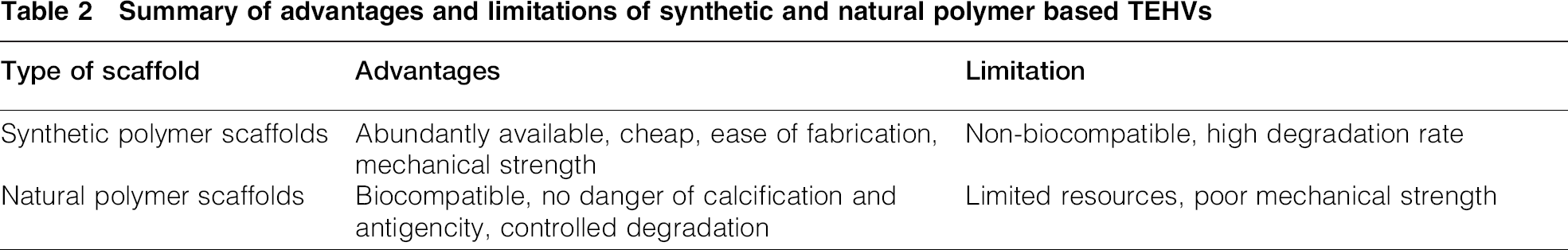
The supporters of natural materials for tissue regeneration claim that the human body can withstand only those materials that are chemically and biologically identical to human tissues. 34 In this regard, decellularised scaffolds such as allografts or xenografts possess great potential to substitute for diseased heart valves due to their similar chemical and biological character to host tissue. These have been reported with minimal danger of calcification and antigencity. 24 Small intestinal submucosal matrix and fibrin, derived from patients’ own blood, are other competing natural polymers being investigated for similar heart valve tissue engineering applications.9,24,28 Advantages and limitations of synthetic and natural polymer based TEHVs have been summarised in Table 2.
Summary of advantages and limitations of synthetic and natural polymer based TEHVs
Collagen is a major component of the heart valve. Total collagen present in the human heart valve has been naturally classified as 74% type I, 24% type III and 2% type V collagen. 38 Glycoaminoglycans are the second most abundant ingredient (60% collagen, 20% GAGs and 10% elastin) on the bases of dry weight of heart valve. 39 The human heart valve contains a variety of GAGs, of which hyaluronic acid (HA) is the most abundant. Other GAGs present in minor quantities in the heart valve are dermatansulphate, chondroitin-4-sulphate and chondroitin-6-sulphate. 40
Interpenetrating polymer network as novel biomaterial for heart tissue engineering
Human organs such as tissues, muscles and bones are composed of polymers. The most abundant polymers found in human body are nucleic acid and proteins, e.g. proteins alone make 12–15% of human body by mass. 41 The heart valve uses collagen, elastin and GAGs as the building blocks for its unique anisotropic structure. Hence, natural polymers can be thought of as the most suitable materials of choice for heart tissue regeneration.
The first IPN was devised by Aylsworth in 1914 for the manufacturing of phonograph records. 42 The term IPN was later proposed in the 1960s by Miller on the basis of interconnected structure of this class of polymers. 43 Recently, these novel materials have gained attention for smart biological applications. Structural conformations of IPNs, their current applications in medicine and potential utilisation in heart tissue engineering will have been outlined and discussed further.
Classification of IPNs
Interpenetrating polymer networks can be considered as analogous to ‘alloys’ of polymers where two polymer networks are not bonded to each other. The International Union of Pure and Applied Chemistry definition of IPNs is as follows:
‘A polymer comprising two or more networks which are at least partially interlaced on a molecular scale but not covalently bonded to each other and cannot be separated unless chemical bonds are broken. A mixture of two or more preformed polymer networks is not an IPN.’ 44
Interpenetrating polymer networks can be classified on the basis of either how the two networks are arranged with respect to each other or how the bonds are formed:
(i) when only one of the polymers forms a network and the other is simply penetrating through it, the system is called semi IPN (ii) when both the polymer systems are independently crosslinked, the system is called full IPN.
If the polymerisation of both polymer systems takes place at the same time by two non-interfering chemical reactions, the product is called simultaneous IPN. On the other hand, if the polymerisation of second polymer takes place after the completion of polymerisation of the first polymer, the product is called sequential IPN. In the later case, the polymer to be polymerised first is called the primary network, and the polymer to be polymerised later is referred as the secondary network.
The component polymers of an IPN may be synthetic or natural or a combination of both types of polymer. Similarly, the bonds present in an IPN may be either chemical/physical or covalent/non-covalent.45,46
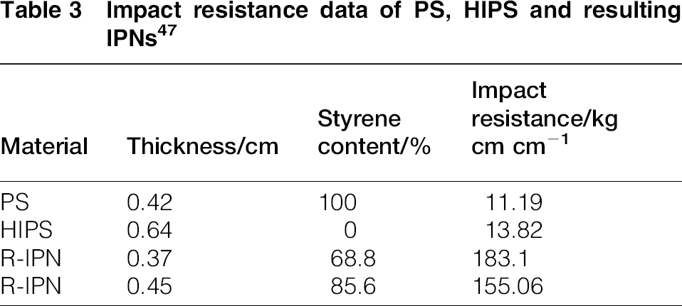
Being a combination between two different polymers, IPNs may exhibit the beneficial properties of its constituent elements. It has been observed by that certain properties of component polymers contribute in the resulting IPN structure in an extraordinary manner. For example, when homopolymer polystyrene (PS) was combined with high impact polystyrene (HIPS), the impact strength of resulting IPN increased to 15 times that of PS and HIPS as shown in Table 3. 47 Recently, similar findings have been reported for polyacrylamide–alignate IPNs system where the values of elastic modulus and tensile strength exceeded the summation of those for individual polymers alone.48,49
Impact resistance data of PS, HIPS and resulting IPNs 47
Applications in medicine
The inherent entangled structure of IPNs offers excellent ‘tailorability’ with respect to the choice of components, nature and extent of crosslinking. Therefore, this class of materials has a great potential for the customised design and properties for a particular biomedical application. For example, IPNs have shown promising features for potential use as a drug delivery system. Such a system is designed carefully to liberate the drug in a fashion required by the target site. The release of drug is usually stimulated by changes in the surrounding environment (pH, body temperature, etc.). 46 Chen et al. have reported the development of alignate/chitosan based pH sensitive IPN structured hydrogels. These pH driven hydrogels have been applied for drug administration to the intestine (alkaline pH). 50 Various studies have shown the successful application of such complex composite hydrogels systems based on biodegradable polymers as drug carriers in the form of microspheres, sponges, sheet, film, tablet, capsules and nanoparticles or drug delivery and wound management.51–53
Hyaluronic acid–collagen based IPNs as scaffolds for heart tissue engineering
Many experimental techniques have been attempted to manufacture tailored biomaterials to encourage three-dimensional tissue growths. Natural polymer based IPNs can serve this need to an appreciable extent due to the flexibility in control of microstructure, customised physical and specific biological properties. A number of IPNs based on natural polymers such as proteins (collagen, fibrin, silk and gelatin), polysaccharides (HA, chondroitin sulphate, dextran, agarose and chitosan) and DNA have been developed in the last two decades. Initial in vitro trials on IPNs have indicated their long term potentiality as three-dimensional scaffolds for skin, bone, cartilage and nerve tissue regeneration.54–58
Collagen and HA are the two major components of ECM, and hence are the essential components of heart valves. 59 Collagen has been utilised frequently in tissue engineering because of its excellent biocompatibility and cell attachment characteristics. 60 Similarly, HA is a well understood non-sulphated GAG and offers a number of favourable properties for tissue regeneration such as slow immunogenity, superior swelling characteristics and biodegradability.61,62 The HA based products are being applied successfully in optics, orthopaedics and wound healing aids. 57 However, both polymers individually show inadequate mechanical stability for cardiac tissue growth. 63 For instance, the human aortic valve has an elastic modulus ∼15 MPa, 11 which is higher than the elastic modulus of HA hydrogels (0.5–4.0 kPa) 64 and lower than the elastic modulus of type 1 collagen fibrils in physiological fluids (0.2–0.5 GPa). 65 In addition, HA hydrogels have also been reported to have too rapid in vitro degradation.
These limitations of the HA–collagen polymer system could be overcome by introducing an interlocking structure between the polymer chains. The idea of the fabrication of HA–collagen IPN systems possesses great potential with respect to the challenges of designing an ideal TEHV.
The HA–collagen combinations have been recently studied as a potentially attractive alternative for tissue regeneration. Hyaluronic acid–collagen type II scaffolds have been developed for invertebral disc regeneration by Pandit et al. In the present study, effect of concentration of 1-ethyl-3-(3-dimethylaminopropyl) carbodiimide (EDC)/ N-hydroxysuccinimide (NHS) on swelling properties and gel stability was investigated. Scaffolds crosslinked with 8 mM EDC showed highest cell population when cultured with rat mesenchymel cells. 66 Various studies have shown that crosslinking using EDC/NHS greatly improves the mechanical, thermal and biological responses of HA–collagen scaffolds due to the presence of crosslinks between polymer chains.67–70 Although the above studies elucidated the superior properties of crosslinked HA–collagen scaffolds as compared to uncrosslinked scaffolds, simple crosslinking of collagen in the presence of HA chains does not guarantee the formation of a full IPN. This is due to the fact that HA chains need to be chemically modified before crosslinking to form either a primary or secondary network in the IPN. In addition, unmodified HA is not recommended for tissue engineering application because of its poor handling properties and limited cell response.59,71,72 In a recent study, Camci-Unal et al. have shown that mechanical performance and degradation rate of HA methacrylate can be adjusted by the incorporation of varying amounts of methacrylated gelatine. 59 The resulting hybrid hydrogels also have a wider response spectrum for cells as compared to the HA methacrylate alone. Guo et al. have formulated an IPN system where MeHA and methacrylated chondroitin sulphate have been combined with self-assembled collagen to form an IPN for cartilage tissue repair. It showed cytocompatability and cell attachment with tunable degradation rates and swelling ratios (by varying the ratio of MeHA and methacrylated chondroitin sulphate. 54 Here, the collagen is assumed to be self-assembled (physical bonds), and HA/chondroitin sulphate are chemically crosslinked. An IPN can have many types of bonds such as physical and chemical bonds, but heterogeneity in the bond nature at molecular level may lead to the anisotropy in the resultant properties.
An HA–collagen binary system can also be used to synthesise IPNs in a number of interesting ways. For example, collagen can be taken as the primary network, and HA can be taken as secondary network or vice versa. In either way, various interesting properties can be achieved, which can be further exploited for smart biomedical applications. For example, Kim et al. combined sophisticated techniques of electrospinning with salt leaching to produce HA based scaffolds. Scaffolds were cultured in vivo with bovine chondrocyte cells and showed promising adhesion and proliferation results. The fabrication techniques were chosen so as to combine both mesoporous and nanoporous features in a single material. 73 Such multiscale patterning of porosity is very favourable for blood and tissue vascularisation. A limiting factor in the above study was the low concentration of collagen (5, 10%), which is not suitable for making an IPN for TEHV applications. The HA fibres could be expected to have a thin coating of collagen at low concentrations rather than forming a true IPN structure. An attempt to make a semi-interpenetrating polymer network (sIPN) based on unmodified HA with different molecular weights has been reported by Xin et al. Low molecular weight HA (LMHA) showed superior mechanical properties as compared to high molecular weight HA. Collagen fibres were made to penetrate an LMHA network followed by fibrillogenesis. 71 Active molecular interactions between LMHA and collagen fibres established the characteristic networking for a sIPN. It is worth to recall here that fibrillogenesis forms physical interactions between chains. There is great room for further improvement of mechanical properties by introducing chemical linkages in collagen networks. In this way, there will be more chance of interaction between collagen network and high molecular weight HA, which could lead to a full IPN structure.
A further step towards the development of an IPN for mimicking heart valve structure was achieved using photo crosslinking techniques. This technique has been widely applied to make IPNs for varying applications in guided tissue regeneration. Briefly, MeHA modified prepolymer solution is combined with collagen solution in different weight ratios along with a photo initiator. This mixture is then exposed to UV light for a few minutes to establish crosslinks.57,74,75 The technique has been used to establish local crosslinks, which can facilitate localised IPN and sIPN regions in a single piece of material. 57 The resulting IPNs and sIPNs have not only shown good biocomapatability but also excellent mechanical characteristics closely matching that of native heart valve. 74 Munoz-Pinto et al. showed that cell mediated contraction of collagen scaffold can be overcome by infusing the collagen hydrogel with photo crosslinkable poly(ethylene glycol) diacrylate followed by UV exposure. 76 Control of degree of cell spreading within the IPN was achieved by introducing a time delay between the sequences of crosslinking of both polymers [i.e. cell laden collagen hydrogel and photo crosslinking of infused poly(ethylene glycol) diacrylate]. 76 A serious disadvantage of this technique is the use of cytotoxic photo initiators and UV light. These have been reported to cause cell death.77,78 In addition, the high temperatures involved in such methods may increase the chance of nacrosis. 79 Further, use of high temperature T ≥ 80°C with collagen requires special care, as it causes denaturation. 67 There is still much to explore regarding alternate techniques for the fabrication of collagen based IPNs. A simple and relatively straightforward approach capable of utilising non-toxic, cheap and readily available precursors needs to be developed. The ideal synthesis route should yield IPNs without compromising the useful biological and mechanical properties of natural polymers such as collagen and HA.
Fabrication routes for IPNs and their effect on properties
An ideal three-dimensional matrix (scaffold) supporting the regeneration of a diseased heart valve tissue should have a desirable combination of mechanical and biological features. Collagen and HA, natural in origin, are more likely to be approved by native human tissues. The molecular structures are shown in Fig. 2. Tailoring of these polymers into x–y–z morphologies is a challenging task. The fabrication and crosslinking techniques should be selected so that the ultimate IPN based TEHVs have not only the inherent biological properties but also matched mechanical properties.
Molecular structures
Collagen
Collagen is the major component of human heart ECM comprising ∼50 wt-% on dry basis. 80 Collagen molecule has a triple helix conformation assembled with polypeptide chains, which is unique to all proteins. The three polypeptide chains present in the form of a rope like coil are known as α chains. Each α chain has a characteristic repeating sequence of three amino acids G–X–Y, 81 where G, X and Y represent glycine, proline and hydroxyproline respectively (as shown in Fig. 2a). 82
Collagen can be further subdivided into 29 types depending on the nature of three chains and distribution of X and Y amino acids, and human heart valves normally contain 74% type I, 24% type III and 2% type V collagen. 38
Owing to this complex hierarchical structure, collagen fibres have a high Young's modulus of 0.1–1 GPa, 83 which almost makes them intrinsically inelastic. However, the collagen fibres are longitudinally crimped with a periodicity of ∼20 μm, which can give them an extensibility of ∼10% under stress.84,85
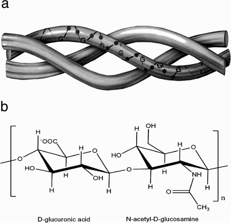
Hyaluronic acid
Hyaluronic acid or hylauronan is a linear polysaccharide that consists of alternating units of repeating disaccharide, β-1,4-
Hyaluronic acid is also an attractive biological molecule for a wide spectrum of biological applications, e.g. tissue engineering, drug delivery, ocular surgery, viscosupplementation for arthritis and wound healing.89,90 However, its uncontrolled degradation rate and poor mechanical strength are the limiting factors against its utility.
91
Chemical modification of HA provides means to control its degradation and mechanical properties. In addition, it also provides a way to tailor the molecule for a specific application.91,92 Interestingly, the solution to this problem lies in its own structure. The HA molecule has potential sites for chemical modification on its backbone (as shown in Fig. 2b).
(i) —COOHgroup: (ii) –OH group: (iii) –NHCOCH3 group:
The –COOH group is usually modified by amidation [carbodiimide, 2-chloro-1-methylpyridinium, 2-chloro-dimethoxy-1,3,5-triazine, carbonyldiimidazole], Ugi condensation, ester formation [using alhyl halides, tosylate activation, diazomethane, epoxides].
95
Other functionalisation routes include modification with dialdehyde
92
and disulphide.
96
Methacrylamide modified carboxylic groups have been crosslinked by photo crosslinking technique.
97
This group can be modified into ether (epoxide,
98
divinylsulphone
99
and ethylene sulphide
100
), hemiacetal,
101
ester (octenyl succinic anhydride, methacrylated anhydride and activated compounds
95
) and cabamate.
102
In addition, the OH group can be oxidised with sodium periodate.
103
An amino group can be yielded by deacetylation of N-acetyl group. This amino group is capable of reacting with acid leading to the similar modifications to amidation.
104
Each of the above set of reactions has a specific mechanism and yields a different HA derivative. Hyaluronic acid molecule has extraordinary potential to be remodelled into versatile biomaterials ranging for a wide spectrum of clinical applications.
Crosslinking routes
Physical crosslinking
Simple thermal treatment, dehydrothermal treatment (DHT) and photo crosslinking methods are widely utilised techniques for the physical crosslinking of proteins. Conventional heat treatment in a simple drying oven at 100°C is capable of introducing crosslinks in collagen structure. This is shown by the increase in Young's modulus of heat treated collagen films. 105 Dehydrothermal treatment involves treating the collagen at a certain temperature over a specific period of time under vacuum. 106 Experimental parameters such as temperature, exposure time and vacuum can be optimised to get the desired properties in collagen–GAG scaffolds. It has been found that an increase in either temperature or exposure time enhances the compressive strength up to twofold and tensile strength up to fourfold. The increase in temperature also improves crosslinking density proportionally. Despite being a promising technique to alter the mechanical properties of collagen, DHT involves the risk of denaturation of collagen structure at high temperatures. 107
Photo crosslinking techniques employ a light source such as gamma or UV light to introduce crosslinks in the irradiated collagen. Light intensity and exposure time are the key parameters to control in determining the degree of crosslinking. The use of gamma rays has been abandoned because the intensity of radiation is high enough to destroy the protein structure. 108 The UV treated collagen films are found to be mechanically robust (improved tensile strength and stiffness) and resistant to enzymatic degradation. The extent of improvement in these properties is highly dependent on the exposure time at a given intensity level. For example, mechanical and biological properties reach maxima at exposure time of ∼4 h at intensity of 1.66 mW cm− 2. However, prolonged exposure times induce deterioration of these characteristics due to the denaturation of collagen structure. 109 In addition to collagen, UV irradiation technique is also well established for the crosslinking of photo crosslinkable glycidalmethacrylated coupled HA. 110 As compared to DHT, photo crosslinking technique has certain advantages in terms of close control of parameters and much shorter exposure times, e.g. 15 min of exposure to UV source can produce the same effect that is achieved with DHT within 3–5 days. 111
The freeze–thawing technique can be categorised as a non-thermal physical crosslinking of collagen. It has been found that traces of secondary polymer such as polyvinyl acetate as well as ice crystals formed during the process of freezing act as small crystallites (physical interaction) in the freeze–thawed collagen marix. 112
Chemical crosslinking
Chemical crosslinking of collagen based materials can be carried out in a number of ways. Glutraldehyde is the most commonly used crosslinking agent for the stabilisation of biological prosthesis for heart valves as well as synthetic collagen scaffolds for tissue engineering applications.
The mechanism of crosslinking collagen with GA switches from intrafibrillar to interfibrillar as the concentration increases up to 1.35 mM gcol− 1. 113 Glutraldehyde introduces Schiff base structures while reacting with the amino acids of proteins, which enhance thermal (shrinkage temperature increases from 56 to 78°C) and mechanical stability (elastic modulus increases from 20 to 60MPa) of the collagen. 114 Furthermore, enhanced physical properties of GA crosslinked HA hydrogels such as dimensional stability in physiological solutions and controlled degradation have been shown. 114 The mechanism of GA crosslinking reaction is fast and complex, but poorly understood. 114 In addition, an undesirable side reaction imparts a pale yellow to brown colour in collagen, and toxic byproducts are difficult to control.115,116 Genipin is a plant derived non-toxic substance recently explored for the crosslinking of protein structures with promising results.117,118 Similar to GA, it imparts an undesirable dark colouration to the crosslinked construct.119,120.
Carboimide chemistry is also a very popular route for crosslinking.93,121 In this family, EDC/NHS has been extensively studied for the crosslinking collagen and HA with different EDC/NHS ratios. In a typical reaction, EDC/NHS attacks the carboxylic group in a molecule in an acidic media. An increase in either the EDC/NHS ratio or incubation time for a given ratio results in an increase in crosslinking density. Various studies have confirmed that the EDC/NHS treated biomaterials are superior in mechanical, thermal and biological properties. These are resistant against enzymatic degradation, and have high cell attachment potential and controlled swelling characteristics with less cytotoxicity as compared to GA.66,122–124
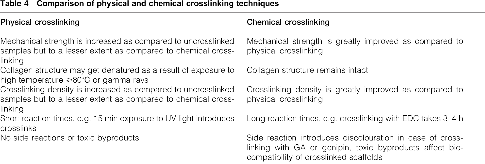
Combinations of physical and chemical crosslinking methods have been attempted to maximise the advantages of both techniques. Typically, DHT treatment of collagen may be followed by chemical crosslinking with either GA 125 or EDC/NHS.126,127 The sequence of application of physical and chemical crosslinking techniques does not seem to affect the results. For example, DHT treatment followed by EDC treatment produces the same crosslinking effect as the EDC treatment followed by DHT treatment. 128 Such a hybrid technique could be potentially very attractive for producing multiple crosslinking densities in hydrogels as drug delivery vehicle for a controlled drug release or in scaffolds for tissue culture. A comparison of physical and chemical crosslinking techniques has been given in Table 4.
Comparison of physical and chemical crosslinking techniques
Enzymatic crosslinking
Enzymes are capable of catalysing the chemical reaction of polymers. 129 For instance, horseradish peroxidase facilitates the crosslinking of phenol groups in the presence of hydrogen peroxide (H2O2) to yield tissue friendly biomaterials. 129 Horseradish peroxidase has been applied to yield an HA–tyramine (HA-Tyr) network by Lee et al. The network was further combined by simultaneous crosslinking with fibrin to yield an IPN with better mechanical properties. 55 Enzymatic crosslinking offers advantage as compared to physical and chemical crosslinking in terms of use of non-toxic substances. However, enzymatic crosslinking yields polymers with inferior mechanical properties and rapid degradation, which limit their application as injectable hydrogels only.130,131 A recent study by Zhang et al. has solved this problem to some extent using a newly devised bienzymatic crosslinking technique for the development of gelatin/chitosan IPN system. 132 The results indicated an improvement in hydrogel stiffness, degradation behaviour and swelling ratios. Biocompatability of gelatin/chitosan IPNs in above study was confirmed by the L929 cell attachment and proliferation. Although no enzymatically crosslinked IPN based on combination of collagen and HA has been reported to date, the above studies suggest that both collagen and HA have potential functional groups for enzymatic crosslinking. Enzymatic crosslinking can be successfully applied to any biopolymer, which will further extend the existing library of biomaterials.
Summary
This review has assessed HA–collagen polymer systems as potential biodegradable scaffold for guided tissue regeneration of diseases heart valves. Biological and mechanical prosthesis with their characteristics and limitations were then reviewed from a clinical prospect. A snapshot of key literature as well as recent research related to potential utilisation of HA–collagen combination in tissue engineering has been presented. The HA–collagen system has found applications in reconstruction of different tissues in the body such as bone, skin and eye. The fundamentals of this approach have been extended to utilise HA–collagen for the development of novel TEHV.
The principles of different approaches towards the design of IPNs were studied in detail. Structure–property relationship of HA–collagen based IPNs made by various routes was explored. Current techniques are largely based on physical crosslinking techniques such as DHT and photo assisted crosslinking. These techniques yield prostheses with low crosslinking densities and require careful handling of the processing parameters to avoid the alteration in protein structures. A few studies have partly used chemical route of crosslinking for making sIPNs with little success. Enzymatic crosslinking is quite a new route for making natural polymer based IPNs, however once well established, has excellent potential.
Animal trials so far with the TEHVs have failed after a few months of implantation due to poor in vitro mechanical stability. Chemical crosslinking of HA–collagen system to hybrid full IPN not only provides a pathway for empowering the weak link of mechanical stability in physiological environment but also synchronises the degradation rate with tissue growth rate. The aim of this review is to understand the basic principles involved and extract the recent scientific developments related to HA–collagen based IPNs for tissue engineering in general and heart tissue engineering in particular. Hence, it is highly desired to formulate a unique experimental route based on chemical crosslinking to fabricate the HA–collagen based full IPN capable of as closely mimicking the healthy heart valve as possible in terms of mechanical and in vitro biological compatibility.
Footnotes
Acknowledgements
I would like to thank my supervisor Prof. Jan T. Czernuszka for his generous guidance and valuable feedback throughout the writing of this review.