
Abstract
AISI type 304 stainless steel (SS) specimens in solution annealed and cleaned and passivated conditions were exposed to laboratory culture of Leptothrix sp. Epifluorescence studies and total viable count analysis of these specimens showed that bacterial density and biofilm coverage were the least on cleaned and passivated SS surfaces. To confirm whether MnS inclusions play a role in this, adhesion studies at applied potentials, surface characterisation studies using SEM and EDAX analysis and pitting studies with ferric chloride were carried out. These studies clearly revealed the removal of MnS inclusions. This removal leads to a significant reduction in the adhesion of Leptothrix sp. The significance of the present study is that pretreatment of SS by cleaning and passivation inhibited the adhesion of Leptothrix sp. This reduction in adhesion is responsible for the increased corrosion resistance of SS under biofilms in aqueous environments.
Introduction
Austenitic stainless steels (SSs) are prone to microbiologically influenced corrosion in natural waters. This happens in cooling water systems where ideal conditions like highly oxygenated water, good exposure to sunlight and air, temperature in the range of 27–60°C and a pH between 6 and 9 favour the growth of microorganisms.1 The microbial colonisation of a surface leads to biofilm formation. This biofilm can increase the corrosion potential of the SS to a higher value, and this process is called ennoblement.2 This ennoblement can easily lead to localised corrosion initiation at anodic sites.3 Material factors play a very important role in the onset of pitting corrosion. In AISI type 304 SS, where there is 1·55%Mn and 0·03%S, the role of manganese is to tie up residual sulphur as manganous sulphide.4 It is well known that MnS inclusions present on the SS surface act as initiation sites of pitting corrosion.5 – 8 This is due to the fact that these inclusions are more anodic and active on the passive SS surface. These inclusions dissolve more rapidly, leading to locally aggressive solution compositions, which cause the breakdown of passive films of SSs.6,9 Dissolution is known to be localised to the inclusion/metal interface because of the low electrical conductivity of the inclusion relative to the metal. This results in a crevice, which in turn leads to pit initiation. Sustained pitting and absence of repassivation happens when inclusions are polarised to potentials where they dissolve, releasing metal ions into the crevice. In biofilm forming environments, such ennoblement and consequent pitting corrosion are reported by many workers.10,11
Microbial colonisation of the metal surface is considered to be a crucial step in initiating microbiologically influenced corrosion. Bacterial adhesion is known to be affected by a number of factors, including the properties of the substratum, the liquid phase12,13 and the biological properties of the bacterial species, e.g. the physiological stage and the cell surface characteristics.14,15 The authors’ earlier studies16 have shown that electrochemically active surfaces favour the adhesion of Pesudomonas sp. The Leptothrix sp. used in this study is a sheath forming bacteria that show high affinity for metal cations. The exopolysaccharide (EPS) produced by the bacterium consists of polysaccharides with abundant carboxyl and hydroxyl groups that provide potential binding sites for metal ions. In addition to EPS, they also secrete a Mn oxidising enzyme and hence called manganese oxidising bacteria (MOB). The biogenic Mn oxide particles formed due to the enzymatic activity have in turn high affinity for trace nutrient elements (such as Zn, Cd and Co).17 Many workers have reported the ennoblement under Leptothrix sp. biofilms on SS and localised corrosion initiations.10,18 However, so far, no work is reported on the influence of MnS inclusions on the adhesion of MOB on the SS surface. Hence, it is important to examine whether the initial attachment of Leptothrix sp. is influenced by the presence of MnS inclusions on the metal substratum. This will help in the selection of appropriate surface modification measures to prevent biofilm formation in cooling water systems. Thus, the main objective of the present study is to know the effect of pretreatment (removing of MnS inclusions) of SS surfaces on the adhesion of manganese oxidising Leptothrix sp. The present study also aims to provide a method for preventing localised corrosion initiation on 304 SS in biofilm forming environments.
Experimental
Specimen preparation
The AISI type 304 SS is employed in this study [Fe–0·07C–0·48Si–18·5Mn–18·5Cr–9·3Ni–0·045P–0·03S (wt-%)].The solution annealed (1383 K for 30 min and water quenched) specimens of dimension 30×15×1·6 mm were ground to diamond finish and then cleaned with detergent solution, degreased with ethanol and air dried. One set of these specimens was cleaned of MnS inclusions and passivated (50%HNO3+2% sodium dichromate) at 50°C for 30 min as per the standard method.19
Isolation and identification studies of Leptothrix sp.
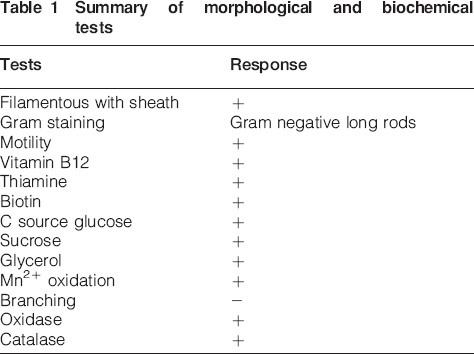
The AISI type 304 SS frames (450×300×7 mm) with tungsten inert gas welded joints exposed in a fresh water reservoir at a depth of 1·5 m for 5 years developed several brown coloured biofilms. The biofilm on the frame was removed using sterile cotton plugs and dispersed in sterile buffer by ultrasonication.16 Mn agar (beef extract, 1 g; yeast extract, 0·075 g; manganous carbonate, 2 g; ferrous ammonium sulphate, 0·15 g; sodium citrate, 0·15 g; and agar 12 g in 1 L distilled water) was used to isolate Leptothrix sp. from the biofilm suspension. Manganous carbonate provided Mn2+ ions, which get oxidised, and is responsible for the black brown colouration of the filamentous colony.20 Genus level identification was performed 21,22 based on morphological and physiological characteristics and by biochemical tests. The ability of the isolate to oxidise manganese was tested by culturing in nutrient broth amended with 50 ppm of MnCl2 for a period of 72 h. The culture was then centrifuged at 15 000 rev min−1 for 40 min, and the remaining Mn in the supernatant was measured using inductively coupled plasma–atomic emission spectroscopy.
Laboratory exposure studies for evaluation of adhesion and corrosion characteristics
For laboratory exposure studies of solution annealed and cleaned and passivated 304 SS specimens, Leptothrix sp. was cultured in a mineral medium [0·07 g Ca(NO3)2, 0·008 g K2HPO4, 0·024 g MgSO4, 0·05 g Na2SiO3, 0·0002 g FeSO4, 0·00001 g MnCl2 and 0·005 g Na2EDTA] modified by adding a minimum amount of carbon source (1 mL glycerol) and growth factors of Leptothrix sp. (0·1 g thiamine HCl, 0·5 μg biotin and 0·5 g L−1 vitamin B12 ). This medium is henceforth referred to as modified mineral medium. Specimens exposed for short term studies were withdrawn after 3 and 6 days for evaluation of adhesion characteristics.
The solution annealed 304 SS specimens were exposed for 1 year in laboratory culture of Leptothrix sp. in modified mineral medium to study the corrosion characteristics. Every week, 25 mL old culture medium is removed using sterile 25 mL glass pipette, and 25 mL of new medium was added to keep it as a semicontinuous culture. The chloride content of the medium was analysed after exposure studies using argentometric method as per APHA (1989).21
Evaluation of presence/absence and distribution of MnS inclusions on different SS surfaces
Using the potential–pH diagram, Sedriks4 showed that, at pH from 6 to 8, as the potential of SS is raised above −200 mV in 0·1M NaCl solutions, MnS will dissolve, and Mn can exist as Mn2+ ions. Hence, the solution annealed SS specimens mounted in araldite with electrical connection was anodically polarised to +200 mV(SCE) in the electrolyte (24 h culture of Leptothrix sp. in modified mineral medium with 0·01M NaCl) for 4 h to study the adhesion rate of this species on the SS surface due to MnS dissolution by applied potential. Scanning electron microscope (SEM, XL30 ESEM, M/s Philips) was used for evaluating the morphology of the surface of the specimens in the different conditions after coating with a film (∼200 Å thick) of gold/palladium (60∶40). The presence or absence of MnS was understood by EDAX analysis. In order to clarify the distribution of MnS on the 304 SS surfaces, pitting corrosion test was carried out in FeCl3 solution as per the procedure described by Tsutsumi et al.23 The solution annealed 304 SS samples were completely immersed in 10%FeCl3 solution for 15 min and then cleaned with distilled water. The pits developed were counted using TCS SP2-RS (M/s Leica, Germany) confocal laser scanning microscope (CLSM) having a scan speed of 7·4 frames/s, equipped with a 488 nm Ar ion laser. The number of pits corresponds to the number of inclusions. The immersion was repeated three times, and the surface was observed every time. The cleaned and passivated specimens were also immersed and observed.
Post-exposure evaluation
The density of MOB on the specimen surface after various durations of exposure was estimated using Mn agar plating. Three specimens of each metallurgical condition (triplicate experiments) were used for total bacterial density estimation. One specimen of each metallurgical condition exposed in medium was used for direct microscopic observation. The methodology for plate counting and slide preparation for microscopy is explained elsewhere.16 The pits developed on SS specimens exposed for 1 year in bacterial culture were also observed using CLSM. All the images are captured in the reflection geometry using a ×100/1·3 numerical aperture objective lens.24
Statistical analysis of the data was carried out using MYSTAT software. Three replicates were analysed for each experimental condition. A Tukey–Kramer multiple comparison test was performed to assess the significance between bacterial counts on different surfaces.
Results
Isolation and identification of Leptothrix sp.
Brown coloured colonies developed on Mn Agar plates from the brown biofilms on SS frames were streaked on glass slides and observed under an optical microscope (Fig. 1). The presence of filaments having a diameter of about 1–2 μm, the occurrence of irregular rusty metal encrustations and the presence of empty sheaths indicated the species to be Leptothrix sp. The manganese oxidising ability of the isolate was analysed using inductively coupled plasma–atomic emission spectroscopy. It was observed that the isolated organism was capable of bringing significant reduction (final concentration of 0·06 ppm) in the Mn2+ content of the culture broth (initial concentration 50 ppm). Biochemical tests confirmed the genus (Table 1).
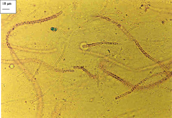
Optical micrograph showing sheathed brown coloured filamentous organism isolated from brown biofilmed area
Summary of morphological and biochemical tests
Laboratory exposure for adhesion and corrosion characteristics
Total viable count analysis showed (Fig. 2) the least adhesion on cleaned and passivated SS specimens. Epifluorescence microscopic observations further confirmed these results (Fig. 3). The Tukey–Kramer multiple comparison tests showed that the probability p value for cleaned and passivated versus solution annealed specimens is equal to 0·01, which was a statistically significant variation.
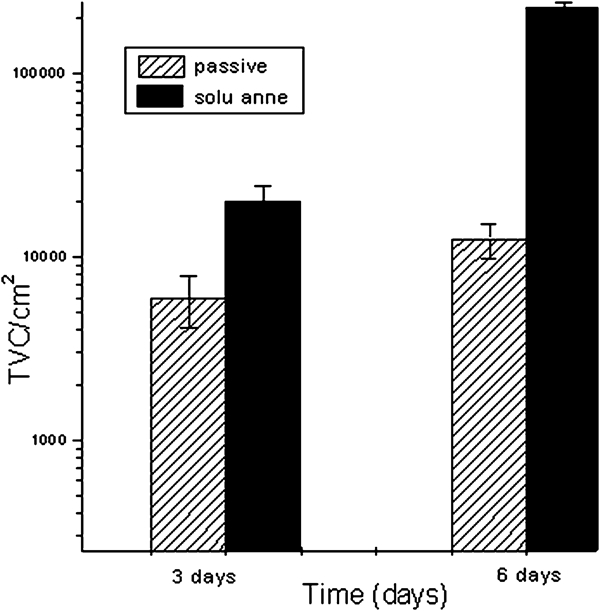
Total viable count of Leptothrix sp. on 304 SS with different microstructures in short term exposure studies (bars = SD of mean)
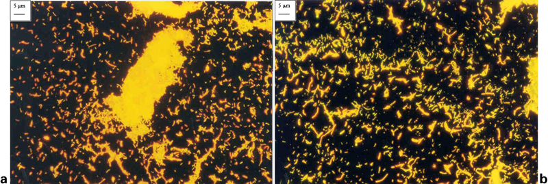
Epifluorescence micrographs showing adhesion of Leptothrix sp. on 304 SS specimens a solution annealed and b cleaned and passivated specimens exposed for 6 days in pure culture
Epifluorescence micrographs of the 304 SS samples withdrawn after 1 year showed a dense biofilm of Leptothrix sp. on SS, and CLSM observations showed pitting corrosion of the specimen. The chloride content of this medium was analysed by titration and was found to be 29·2 ppm. The three-dimensional reconstructed CLSM image (Fig. 4) from 23 optical slices shows the localised corrosion of 304 SS specimen exposed to Leptothrix culture. The volume of the specimen scanned was 62·5×62·5×7·09 μm, where the white regions represent the material surface, and the dark regions represent the corroded areas. Several pits as large as 19·8 μm and micropits as small as 0·9 μm were observed.
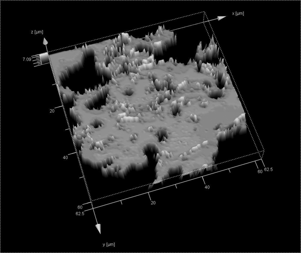
Three-dimensional reconstructed CLSM image of corroded 304 SS specimen exposed to Leptothrix sp. for 1 year
Evaluation of presence and distribution of MnS inclusions on different SS surfaces
Epifluorescence micrographs of 304 SS specimens anodically polarised to +200 mV(SCE) in a 24 h culture of Leptothrix sp. in modified mineral medium for 4 h showed accelerated adhesion of Leptothrix sp. The SEM images and the EDAX analysis have clearly revealed the presence of many MnS inclusions on the surface of the solution annealed 304 SS (Fig. 5). However, on the passivated surfaces, inclusions and cavities from where inclusions were removed were observed. The EDAX analysis showed the presence of S and lesser Mn (Fig. 6).
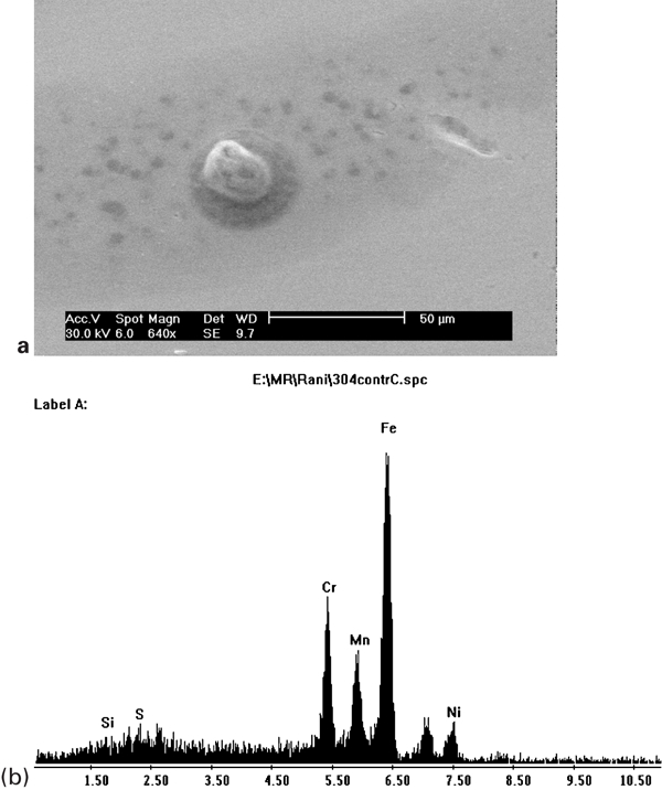
a SEM picture showing presence of inclusions and b EDAX results corresponding to above picture showing presence of manganese and sulphur in these inclusions on solution annealed (control) 304 SS specimen surface
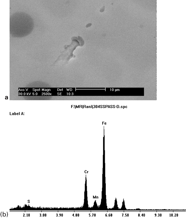
a SEM picture showing areas from where inclusions are removed during passivation treatment and b EDAX analysis of this area confirms significant removal of manganese on passivated 304 SS specimen surface
After pitting corrosion test in FeCl3 of the solution annealed 304 SS, the specimens were observed in CLSM. Each imaged region corresponds to an area of 44×104 μm2. Figure 7 shows the CLSM image of the specimen with numerous pits after exposing it to FeCl3 solution for 15 min. The average pit density for this specimen was found to be 3·6±1·8 μm2 with an average pit diameter of 114 μm. The pit density was found to remain the same for the specimen even after two to three times of exposure to FeCl3 solution. However, a noticeable change in the diameter of the pits has been observed under CLSM (225 and 260 μm for the two and three times of exposures respectively). The cleaned and passivated specimens have not developed pits after exposing to FeCl3 solution.
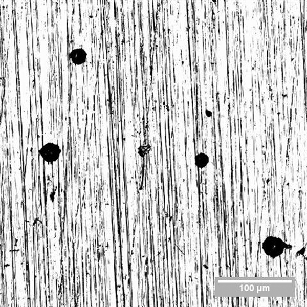
Image (CLSM) of 304 SS specimen showing pits after 15 min of exposure to FeCl3 solution
Discussion
The SS is used for diverse applications in industries owing to their corrosion resistance. Many failures occurring in aggressive environments due to pitting corrosion have been reported.25,26 The presence of MnS inclusions is known to be one of the main reasons leading to pitting corrosion.27,28 The present study is carried out to understand the influence of cleaning and passivation of SS by the way of removing MnS inclusions on the attachment of a MOB Leptothrix sp. This bacterium is known to be the culprit in the localised corrosion of SSs, especially AISI type 304 SS.
First, the study involved the isolation of the manganese oxidising Leptothrix sp. from fresh water biofilms on SS and the identification of the species to generic levels by morphological, physiological and biochemical tests. Then, the manganese oxidising ability of the species was confirmed with inductively coupled plasma–atomic emission spectroscopy studies. Second, exposure studies of specimens in cultures were performed for comparative adhesion studies. The total bacterial density estimated by culturing techniques and studies on the morphology of biofilms with epifluorescence microscopy have revealed reduced adhesion of the manganese oxidising species on the cleaned surfaces.
Adhesion studies at applied potential (electrochemical polarisation) were carried out to prove that the surfaces of 304 SS favoured the adhesion of Leptothrix sp. due to the release of Mn2+ ions. Epifluorescence micrographs of these specimens clearly showed accelerated adhesion of Leptothrix sp. compared to specimens at rest potential. The SEM images and EDAX analysis have clearly revealed the presence of many MnS inclusions on the surface of the solution annealed AISI type 304 SS. However, on the cleaned and passivated surfaces of specimens, inclusions were lesser or nearly absent. Ferric chloride immersion helped in removing the MnS inclusions from the surface of the solution annealed specimens, and subsequent pits helped in the evaluation of the density of MnS on this steel. Repeated immersion only increased the pit depth but not the pit number. Thus, as reported by Tsutsumi et al.,23 the FeCl3 immersion studies clearly helped in evaluating the density of MnS inclusions. The absence of pits on the cleaned and passivated specimen immersed in FeCl3 solution further confirmed these results.
Once they attach on to the surface, the Leptothrix sp. biologically catalyse Mn2+ to Mn4+ under neutral pH in aquatic conditions.29 The deposition of Mn4+ ions under the biofilms of Leptothrix sp. can lead to the ennoblement of SSs, and this can further accelerate the propagation of pitting corrosion.5,6 The evidence of this localised corrosion initiation was found on specimens exposed in the pure culture of Leptothrix sp. in the laboratory for more than a year, where the size of the micropits more or less corresponded with the size of inclusions, as seen in the CLSM images. Thus, this study also confirmed the potential of this species to initiate localised corrosion even in low chloride environments.
The significance of this study is that proper surface preparation of SSs by the way of cleaning and passivation can inhibit the adhesion of Leptothrix sp., and to provide additional benefit for the corrosion resistance properties of SS even under biofilms in aqueous environments.
Conclusion
The significant conclusions from the present study are as follows.
A 1 year old biofilm of Leptothrix sp. (MOB) initiated localised corrosion on 304 SS even in low chloride environments.
Bacterial density and biofilm coverage were least on the cleaned and passivated SS specimens compared to the solution annealed specimens.
The SEM, EDAX and pitting studies in ferric chloride solutions confirmed the removal of MnS inclusions on the cleaned and passivated surface compared to the solution annealed specimens.
Thus, this study confirmed that the pretreatment of cleaning and passivation of AISI type 304 SS by way of removing MnS inclusions inhibited the adhesion of MOB Leptothrix sp.
Footnotes
Acknowledgements
The authors sincerely acknowledge Dr B. Raj, Director, Indira Gandhi Centre for Atomic Research, and Dr T. Jayakumar, Director, MMG, for their keen interest in the study and constant encouragement. The authors also acknowledge the assistance of M. Radhika for SEM and EDAX studies.