
Abstract
The cytocompatibility and haemocompatibility of three zirconium based films, i.e. Zr, ZrC and ZrCN, deposited on NiTi shape memory alloy by magnetron sputtering are investigated and compared to those of electropolished NiTi shape memory alloy. The Zr(C,N) series films have deteriorated wettability but have positive effects on the blood compatibility and cytocompatibility of NiTi. Better haemolysis resistance and thromboresistant properties are observed. There are more living cells on the Zr(C,N) series films, and the cells show a higher relative growth rate value than those on the electropolished NiTi. The Zr(C,N) series films act as barrier layers and promote the proliferation of fibroblasts by blocking the leaching of toxic nickel ions from NiTi.
Introduction
Zirconium based films are important materials found in many industrial applications, such as nuclear fuel particle coatings, field emitting coatings and thermophotovoltaic coatings, because of their outstanding properties, such as high hardness, melting point, corrosion resistance and abrasion resistance.1, 2 In addition, the Zr metal has good biocompatibility.3 Proper incorporation of nitrogen and carbon can enhance the surface chemistry and promote fast osteointegration, and so it has been used in the fabrication of multifunctional biocompatible films on biomedical implants.4, 5 Another potential application of Zr(C,N) films is to improve the biological safety of biomedical NiTi shape memory alloy (SMA).6 Although NiTi SMAs are used in biomedical devices,7 – 9 the high Ni content of ∼50 at-% causes health concern.10, 11 The Zr(C,N) series films can serve as barrier layers to block the out-diffusion of nickel from the materials into body tissues and fluids. However, the cytocompatibility and haemocompatibility of these coatings are crucial to clinical applications, but there are only scattered and incomplete data in the literature concerning the biocompatibility of Zr(C,N) films. The purpose of this study is to evaluate the haemocompatibility and cytocompatibility of three different Zr(C,N) films, namely Zr, ZrC and ZrCN, by scanning electron microscopy (SEM), X-ray photoelectron spectroscopy (XPS), contact angle measurement, haemolysis test, platelet adhesion test and methyl tetrazolium (MTT) test and to compare the results to those obtained from electropolished NiTi SMA.
Experimental
Fabrication of Zr(C,N) films
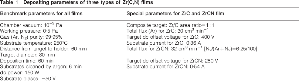
Biomedical NiTi SMAs containing 50··8 at-%Ni samples were electropolished at a constant voltage of 10 V for 6 min at room temperature in an electrolytic cell with an electrolyte consisting of 21 vol.-% perchloric acid (HClO4, 70–72%) and 79 vol.-% acetic acid (CH3COOH, 99··5%). They were then divided into two groups, with the first group being the control (EP-NiTi). The second group was used as substrate for further deposition of Zr, ZrC and ZrCN films using a reactive direct current (dc) magnetron sputtering system (JGP450A2).12 The deposition parameters for the three kinds of Zr(C,N) films are listed in Table 1. The chemical compositions of the Zr(C,N) films were determined by XPS on a VG Scientific ESCALAB-5 spectrometer with monochromatic Al Kα (1486··6 eV) X-ray radiation.
Depositing parameters of three types of Zr(C,N) films
Contact angle, haemolysis and platelet adhesion tests
Surface contact angles were measured using the liquid drop method on a contact angle goniometer (JC2000B, China). Each contact angle was the average of 10 measurements.
In the haemolysis test, 8 mL of fresh blood was collected from a rabbit and diluted with 10 mL of 0··9% saline. Each sample was put into a test tube with 10 mL saline and incubated at 37°C for 30 min. Afterwards, 0··2 mL of diluted blood was added to each test tube and incubated for another 60 min. The suspension was then centrifuged at 2500 rev min−1 for 5 min, and the absorbance of the supernatant fluid was measured by spectrophotometry (UV240, China). The positive control was a blood–deionised water mixture, and the negative control was a blood–saline mixture. The haemolysis ratio (HR) was calculated using the procedures described in our previous paper,13 and each haemolysis data point was the average of three measurements.
In the platelet adhesion test, a 3··8 wt-% citrate acid solution was added to fresh rabbit blood at a blood to citrate acid ratio of 9∶1. The solution was centrifuged to form a platelet rich plasma (PRP) and erythrocyte, and then 0··1 mL of the PRP was added to each well. After incubation at 37°C for 3 h, the PRP was taken out of the wells. A phosphate buffered saline solution was added to the wells and gently rinsed two to three times to get rid of the platelets that adsorbed loosely on the surface. The samples were then soaked in 2··5% glutaraldehyde at room temperature for 12 h to fix the adhered platelets, followed by dehydration in 50, 75, 90 and 100% ethanol for 10 min sequentially. After dehydration, the residual alcohol was removed in 50, 75, 90 and 100% isoamyl acetate aqueous solutions for 10 min. After critical point drying, the samples were coated with gold, and the distribution and morphology of the platelets were examined by SEM.
Methyl tetrazolium test
In the MTT test, fibroblasts L929 were cultured in Dulbecco's minimal essential media supplemented with 15% (v/v) foetal calf serum at 37°C under 5%CO2 and 95% air. The cleaned samples were fixed on the bottom of a 24-well tissue culture plate, and 1 mL of the cell suspension consisting of 105 cells was seeded onto each surface with the wells without cell samples as the control. After culturing at 37°C under 5%CO2 and 95% air for 24 h, the medium was replaced with 100 μL of 0··5%MTT and 400 μl Dulbecco's minimal essential media followed by incubation for four additional hours. Afterwards, the cells were dissolved in dimethylsulphoxide and agitated using a shaker for 10 min. The optical absorbance of the fluid was monitored at a wavelength of 575 nm (620 nm reference). The relative growth rate (RGR) of the fibroblasts on the sample surface was calculated as described in our previous paper.14 The experimental results were expressed as mean values±standard deviation, and Student t test was adopted for statistical analysis. Following culturing, the cells were washed twice with a phosphate buffered saline solution. The morphology of the fibroblasts L929 in the vicinity of the samples was microscopically photographed using an inverted microscope (IX70; Olympus, Japan) equipped with a digital camera (DSC-P72, SONY).
Results
Chemical compositions
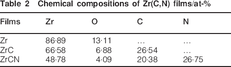
Table 2 lists the chemical compositions of the Zr(C,N) films as determined by XPS. The Zr film has the highest oxygen content of ∼13··11 at-% among the three types, and it can be explained by the strong affinity of oxygen to Zr.15, 16 In comparison, the O contents in the ZrC and ZrCN films are ∼6··88 and 4··09 at-% respectively. The lower O concentrations can be attributed to the characteristics of magnetron sputtering because the high temperature resulting from the glowing discharge can lead to the deoxidisation reactions of zirconium oxide by C.16 The Zr/C atomic ratio in the ZrC film is calculated to be ∼2··5, which is a little different from the projected ratio of the sputtering yield ratio of Zr to C. Under the sputtering power used in the process, the sputtering yield of C should be only ∼1/3 of that of Zr.17 In contrast, the ZrCN film has the expected stoichiometry with (C+N) to Zr of ∼1 to 1. The C/N atom ratio in the ZrCN film is also very close to 1.
Chemical compositions of Zr(C,N) films/at-%
Blood compatibility
Figure 1 shows the HRs of the Zr(C,N) films as well as EP-NiTi SMA. The HRs determined from the four samples are <5%, suggesting that all samples can meet the requirements of biomedical implants. A higher HR (1··4%) means that more haemolysis can occur on the surface of the EP-NiTi SMA, whereas the smaller HR (0··61%) of the pure Zr film shows that it has the best haemolysis resistance among the Zr(C,N) films.
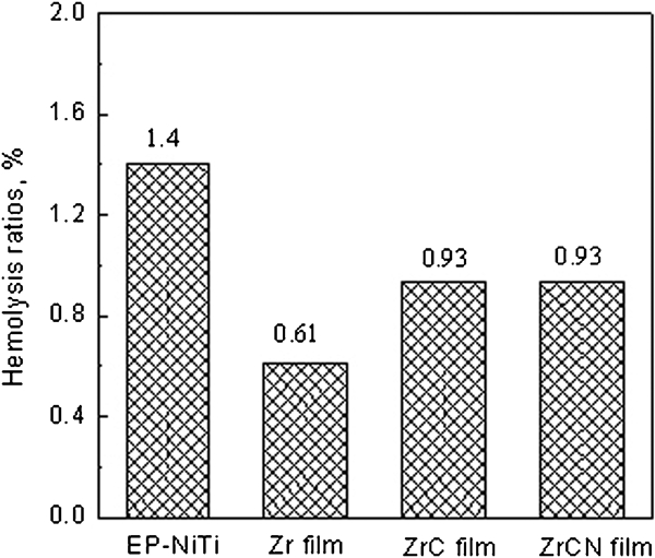
Haemolysis ratios of Zr(C,N) films as well as EP-NiTi SMA
The morphology of the adhered blood platelets on the four samples after 3 h of incubation is depicted in Fig. 2. In general, the lesser the number of adherent platelets and the smaller the deformation of the adherent platelets, the better the blood compatibility. As shown in Fig. 2, the number of adherent platelets on the Zr film is the smallest, and there is no sign of accumulation and only slight pseudopodia. Hence, the Zr film has the best thromboresistance among the samples. There are no big differences in the number of adherent platelets among the other three samples. The platelets on these surfaces form a single layer and are isolated. However, many platelets on the EP-NiTi surface are flat, and the enchylema spread among the pseudopodia. Although the platelets on the ZrC and ZrCN films exhibit more pseudopodia, the enchylema shows no spread among the pseudopodia. It can be inferred that the ZrC and ZrCN films have better blood compatibility than EP-NiTi.
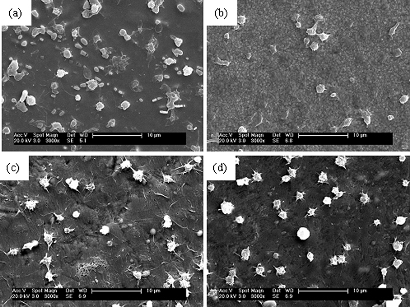
Images (SEM) showing platelet adhesion on surface:
Cytocompatibility
The cell morphology on the EP-NiTi SMA and ZrCN film after culturing with fibroblasts L929 for 7 days is displayed in Fig. 3. The fibroblasts exhibit a normal morphology and are attached to the surface of both samples, suggesting that the samples are well tolerated by fibroblasts. A closer examination reveals that the fibroblasts on the ZrCN film have a higher density and a better pike shape configuration than those on the EP-NiTi SMA.
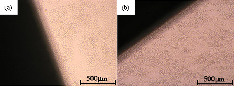
Microscopic views on vertical sites of samples after culturing with fibroblasts L929 for 7 days (dark parts in images are samples):
The MTT reagent is a pale yellow substance that is reduced to a dark blue formazan product when incubated with viable cells, and the production of formazan can be utilised to gauge cell viability. Figure 4 shows the optical density of the cells in the MTT tests from different samples after culturing with fibroblasts L929 for 1, 3 and 7 days. The Zr film shows the highest optical density, whereas the lowest value is observed from the EP-NiTi SMA, indicating that there are more living cells in the cultured fluids on the Zr(C,N) films. Figure 5 shows the RGRs of fibroblasts on the different samples after culturing for 1, 3 and 7 days. The cells on the Zr(C,N) films show higher RGR values than those on EP-NiTi SMA. Among the three coatings, the cells cultured on the Zr film have the highest RGR value. All in all, the ZrCN film can promote the proliferation of fibroblasts and improve the cytocompatibility.
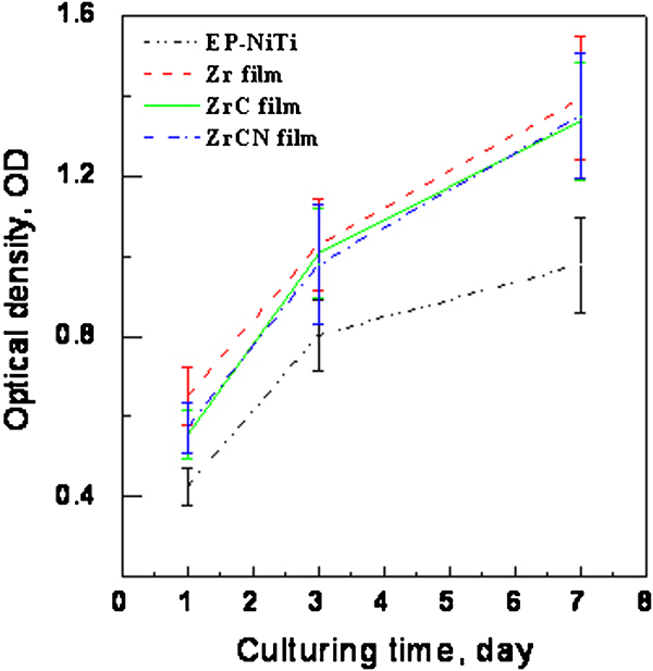
Optical density of cells fluid in MTT tests determined from different samples
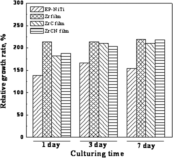
Relative growth rate of fibroblasts on sample surfaces
Discussion
The most apparent feature of NiTi SMA is that its flexibility (also known as superelasticity) is 10 times better than that of stainless steel. It is thus more convenient and safe to use in some artificial stent applications, such as the carotid and femoral arteries, where the vessels may be subjected to outside pressure that may cause conventional stainless steel stents to collapse.18 However, the high Ni content (50 at-%) in NiTi SMA is of great health concern, and the toxicity, carcinogenicity and allergic hazards associated with Ni have been reported.10, 11 In vitro and in vivo biocompatibility studies on NiTi have yielded inconsistent results, 19 19,20 and NiTi alloy has been reported to promote the formation of thrombus.21 In order to gain acceptance in cardiovascular products, we developed Zr(C,N) series films in this work to improve the blood compatibility and cytocompatibility of NiTi SMA.
During the deposition of Zr(C,N) series films in this work, the NiTi substrate was heated to 250°C. It is well known that significant aging occurs above 300°C for NiTi SMA, which may have effects on the shape memory effect of the NiTi substrate.22, 23 Thus, the deposition parameter of Zr(C,N) series films in this work may have little effect on shape memory effect of the NiTi substrate. By the way, the mechanical behaviour of Zr(C,N) series films deposited on NiTi SMA by magnetron sputtering will be studied and reported in other literature.24 The purpose of this study is to evaluate the haemocompatibility and cytocompatibility of Zr(C,N) series films and to compare the results to those obtained from electropolished NiTi SMA.
Obviously, the Zr(C,N) series films, including Zr, ZrC and ZrCN, deposited by magnetron sputtering have better haemocompatibility and cytocompatibility than the electropolished NiTi SMA. They have better haemolysis resistance and thromboresistance. There are more living cells on the Zr(C,N) films, and the RGR values are higher than that on the NiTi SMA. The Zr(C,N) series films act as barrier layers to block the leaching of toxic Ni from the NiTi substrates and consequently promote the proliferation of fibroblasts. In general, the surface chemical composition and wettability play important roles in the cytocompatibility and haemocompatibility of biomedical materials.25
It is well known now that there is a complex dependence between platelet adhesion and wettability. Ikada26 reported that the number of platelets that adhered on various polymers went up with increasing contact angles up to 70° but decreases above 70°. Figure 6 shows the contact angles determined from Zr(C,N) films and EP-NiTi SMA. The EP-NiTi SMA has the best wettability with a small contact angle of ∼45··3°, whereas the contact angle on the ZrCN film is 74··3°. Hence, the Zr(C,N) films deteriorate the wettability, but our other data show enhanced blood and cytocompatibility. Hence, the overall effect is a composite effect encompassing many factors. Among the three types of Zr(C,N) films studied here, the Zr film possesses the highest blood compatibility and cytocompatibility. Zr has a strong affinity to oxygen (chemisorption), and a bioinert ZrO2 phase has indeed been identified in the Zr film. Moreover, the outermost surface of the Zr film can readily be oxidised upon exposure to air, forming a thin ZrO2 film that has good chemical stability, corrosion resistance and compatibility. In conclusion, our data indicate that the Zr(C,N) series films can be safely used as barrier layers on NiTi biomedical implants.
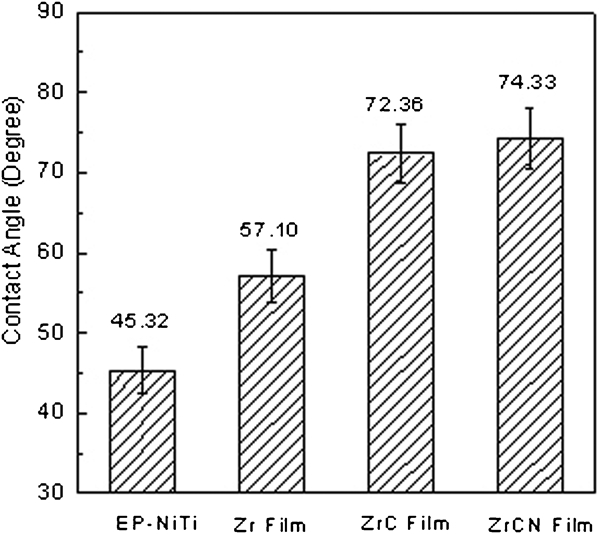
Contact angles determined from Zr(C,N) films and EP-NiTi
Conclusion
The haemocompatibility and cytocompatibility of Zr, ZrC and ZrCN films deposited on NiTi SMA are investigated and compared to those of electropolished NiTi SMA. The Zr(C,N) films deteriorate the wettability but enhance both the blood and the cytocompatibility of NiTi, especially the Zr film. There are more living cells, and the cells show higher RGR values than those on the EP-NiTi. Our data suggest that the Zr(C,N) series films can be safely used as barrier layers on NiTi biomedical implants.
Footnotes
Acknowledgements
The work was jointly supported by the Program for New Century Excellent Talents (NCET-06-0464) in the University of Ministry of Education of China, the National Natural Science Foundation of China (project no. 50501007), the Natural Science Foundation of Jiangsu Province (project no. BK2007515), the National High-Tech Program-863 Projects of China (project no. 2006AA03Z445) and the Hong Kong Research Grants Council General Research Funds (no. CityU 112510).