
Abstract
In this study, silver particles were embedded in Ni–P matrix by electroless deposition on medium carbon steel substrates to produce Ni–P–Ag composite coating. The structure of as plated and heat treated coatings were evaluated by X-ray diffraction analysis. Tribological properties of the coatings were investigated by pin on disc test method using 52100 steel pin as counter body at room and high temperatures. Three-dimensional optical profiler was employed to determine wear rate of the deposits. Surface morphology, cross-section of coatings and wear scars were studied by using scanning electron microscopy equipped with EDS analysis. The results showed that nanocrystalline Ni–P–Ag composite coating had a self-lubricating property at room and high temperatures. It was concluded that diffusion and nucleation of silver on the surface of Ni–P–Ag coating and also formation of a silver thin layer on the sliding surface led to decrease the friction coefficient of Ni–P–Ag coating.
Introduction
In the past decades, several efforts have been performed on mechanical and tribological properties of protective coatings in various conditions. Most of these investigations have been focused on wear resistant and lubricant coatings which have been used in both of low and high temperatures.1 – 5 For instance, Erdemir et al.6 has outlined a historical review on solid lubricant coatings. Dellacorte has described advanced coatings containing solid lubricants for engine and aerospace applications. He mentioned that conventional types of solid lubricants such as graphite and MoS2 are degraded above ∼350°C, whereas advanced solid lubricants, e.g. metal oxide (NiO, MoO3), inorganic fluorides (LiF2, CaF2, BaF2) and soft noble metals (Ag, Au, Pt) can be used at high temperatures due to their high stability and low shear strength.7 Electroless composite coatings have been widely developed in sliding contact conditions in recent years.8 – 11 They have been classified into lubricant and wear resistant coatings with soft and hard particles.12 – 15 Silver as a new and specific lubricant material is a good candidate for lubrication at low and high temperatures. Xiong and co-workers found that friction coefficient decreases with increasing amount of silver in Ni based composite bulk materials produced by powder metallurgy.16 Also, Mulligan et al.17 evaluated the self-lubricating properties of CrN–Ag hybrid coatings produced by magnetron sputtering. Although different investigations on tribological properties of electroless composite coatings at room temperature have been done,8 – 15 but there is no information about high temperature tribological properties of Ni–P coatings containing silver particles. The aim of this work is to investigate the tribological behaviour of nanocrystalline Ni–P–Ag composite coatings at room and high temperatures.
Experimental procedures
Disc shape samples were made from medium carbon steel with 40 mm in diameter and 5 mm in thickness. Before deposition, surface preparation of the substrates was carried out by grinding with SiC paper up to 600 grade, degreasing in alkaline bath and acetone with intermediate deionised water rinses respectively. The commercial nickel phosphorous electroless solution (MacDermid NiKlad ELV 811) including 6 g L−1 nickel sulphate, 30 g L−1 NaH2PO2 and suitable values of additive and stabiliser was used. The agitation of plating bath was done by a magnetic stirrer and a PTFE coated magnet with 80 mm length and 10 mm in diameter (∼350 rev min−1). The electroless plating process was implemented in a 1000 mL double wall beaker connected to the thermo stated circulating water. The plating temperature and pH were adjusted to 90±2°C and 4·8±0·1 respectively. Before entering silver particles (with 2–7 μm in size and concentration of 30 mg L−1) into the plating bath, the particles were well dispersed in some electroless solution containing suitable amounts of cationic surfactant [cetyl trimethyl ammonium bromide (CTAB)] by using an ultrasound for 1 h. Co-deposition of the particles was done for 2 h. Heat treatment of the coatings was performed isothermally at 400°C for 1 h. The hardness of the coatings was measured using a Leitz Wetzlar microhardness tester with a Vickers diamond indenter under a 50 g load. The structure of Ni–P and Ni–P–Ag coatings was evaluated by X-ray diffraction (XRD) analysis using Cu Kα radiation in 40 kV. Pin on disc wear tests were done by using CSM tribometer at room and high temperature (∼500°C) under normal force of 10 N and sliding speed of 0·05 m s−1. The wear tests were accomplished against AISI 52100 steel pins (with 10 mm in diameter) in dry conditions in an air with 40% relative humidity. The surface morphology, cross-section and wear scars of the coatings were studied by using scanning electron microscopy (JEOL JSM-5410 SEM) equipped with EDS analysis. Wear track and wear rate of the coatings were also investigated by using Wyko 1100NT 3D optical profiler.
Results and discussion
Structural characterisation
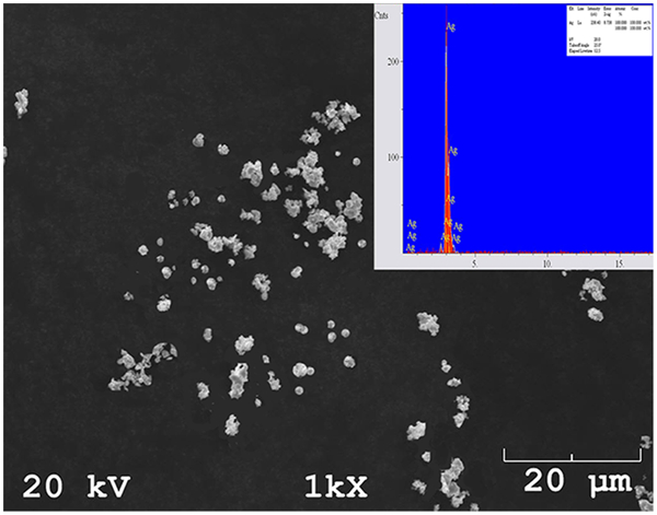
Figure 1 shows the particle size and spherical shape of silver powder. These particles are co-deposited within Ni–P matrix by electroless composite plating to produce Ni–P–Ag composite coating. As shown in Fig. 2, there are a few loose bonding particles on the surface of the composite coating whereas they could not be embedded completely in Ni–P coating. By growth of Ni–P coating, silver particles with poor bonding are fully interlocked in Ni–P matrix. From viewpoint of co-deposition mechanism, silver microsized particles have high surface electric charge in chemical solution and the reduction of nickel ions can be quickly taken place on the surface of silver particles during composite plating. The size, shape and type of particles have the significant influences on the mechanism of entrapment. The co-deposition process in electroless composite coatings has been correlated to Langmuir physical adsorption and Temkin chemical adsorption by Balaraju et al.18 Surfactants can also modify the surface electric charge of the particles and subsequently, the homogeneous dispersion of particles without any agglomeration in the coating.19, 20 CTAB as a cationic surfactant could adjust the surface electric charge and uniform distribution of silver particles in the plating bath. Spot EDS analysis of Ni–P matrix showed high phosphorous content in Ni–P based coating (Fig. 3). As illustrated in Fig. 4, there is a wide and broad peak in XRD patterns of as plated coatings at ∼45° to validate amorphous structure of these coatings. Heat treatment of composite coating at 400°C leads to precipitation hardening in Ni–P matrix with nucleation and growth of nickel phosphide secondary phases and also changing the structure of Ni–P coating from amorphous to nanocrystalline. The formation of nanocrystalline structure and also Ni3P intermetallic compounds were predictable by heat treatment at 400°C as observed in other electroless coatings.21 – 24
Analysis (SEM and EDS) of silver particles
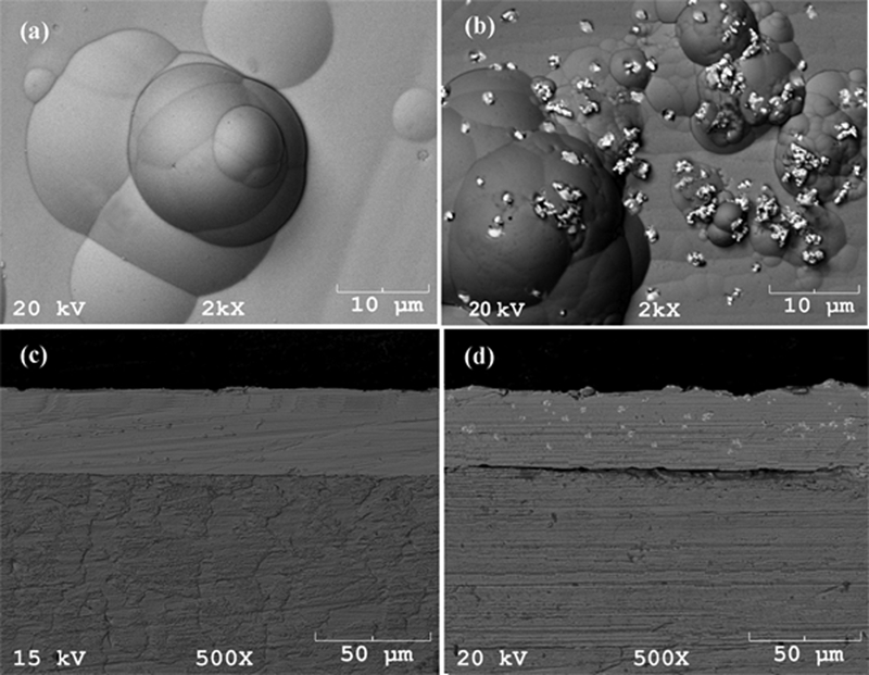
Surface morphology of a Ni–P and b Ni–P–Ag coatings and cross-section micrograph of c Ni–P and d Ni–P–Ag coatings
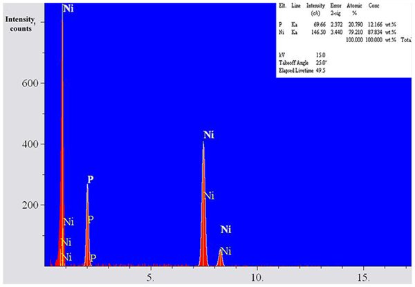
Spot EDS analysis of Ni–P matrix
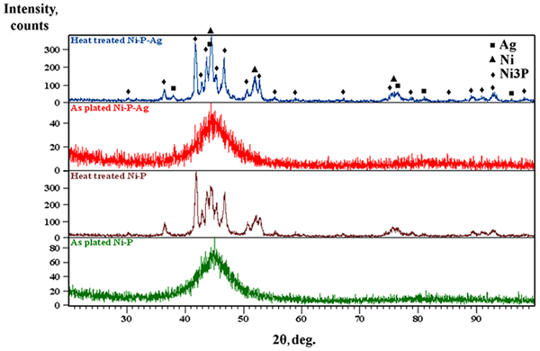
Comparison between XRD patterns of as plated and heat treated Ni–P and Ni–P–Ag coatings
Hardness, lubrication and wear resistance
Microhardness data in Fig. 5 show that Ni–P–Ag coating has lower hardness than Ni–P coating. Low hardness of silver particles (∼251 HV) leads to easier plastic deformation during microhardness test. On the other hand, heat treatment can improve the hardness via precipitation hardening. Therefore, heat treated coatings will have higher hardness than as plated coatings. The similar results have also been obtained in Ni–P–PTFE, Ni–P–MoS2 and Ni–P–hBN lubricant electroless composite coatings.12, 15, 23 Figure 6 shows a comparison between wear rate of Ni–P and Ni–P–Ag composite coatings at room and elevated temperatures. High temperature wear rate of Ni–P coating is ∼10 times more than room temperature wear, but this value reduces to about two times for nanocrystalline Ni–P–Ag composite coating. It seems that the entrapment of silver particles in Ni–P matrix could improve the wear resistance of Ni–P coating at high temperature. On the other hand, the average friction coefficients of Ni–P and Ni–P–Ag coatings are about 0·46 and 0·32 at room temperature respectively, but these quantities have been increased to 0·58 and 0·37 at high temperature conditions as presented in Fig. 7. Hu et al. and co-authors demonstrated that silver could be deformed and/or flattened during sliding due to its low shear strength. They observed that the diffusion of silver from the inside of composite bulk to the surface and subsequently, the formation of a few lubricant islands affects the self-lubricating characteristics of YSZ–Ag–Mo nanocomposite coatings at high temperature sliding.25 Low friction coefficient and formation of silver nodules on the surface of coating (Fig. 8) imply the similar self-lubricating behaviour of silver particles in Ni–P–Ag composite coatings at high temperature wear conditions. It seems that silver particles could diffuse from the inside of the coating to the surface and then, they could deform and flatten in the wear scars during sliding at elevated temperature to form a mixed thin film of Ni–P and silver. This self-lubricating layer could be the main reason for the improvement of lubrication in nanocrystalline Ni–P–Ag composite coatings at room and high temperatures. Furthermore, evaluation of wear tracks by optical profilometry and electron micrographs showed that there was a microploughing shape around wear scars of Ni–P and Ni–P–Ag coatings to indicate abrasive wear mechanism (Figs. 9 and 10). Staia and co-workers have also reported a mixed mechanism of adhesive and fatigue wear in Ni–P–BN(h) electroless composite coating at elevated temperature.23 As shown in Fig. 9, wear depth and wear rate of Ni–P–Ag coating were more than Ni–P coating at room temperature. Similar results were also achieved at elevated temperature. Besides, some materials accumulation had been created after high temperature sliding in Ni–P–Ag composite coating. Electron micrograph in BSE mode demonstrated high quantities of aggregated materials around wear scars as dark regions in Fig. 10b. The EDS analysis demonstrated the presence of iron element as well as Ni, P, Ag and O elements (Fig. 10c). It could be as the result of iron transfer from steel counter body to the interface of sliding. High temperature oxidation of steel pins and adhesion between two mating surfaces can cause the high fluctuations in the curve of friction coefficient and also high quantity of iron oxide around wear scars in Ni–P–Ag composite coating.
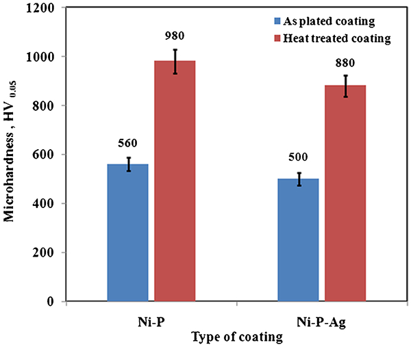
Microhardness data of Ni–P and Ni–P–Ag coatings
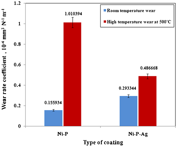
Wear rate of Ni–P and Ni–P–Ag coatings at room and high temperatures
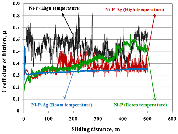
Friction coefficient of Ni–P coatings with or without silver particles at room and high temperatures
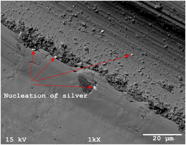
Diffusion and nucleation of silver on wear scars of Ni–P–Ag composite coating during high temperature sliding
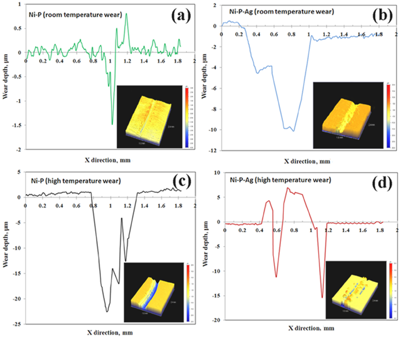
Two-dimensional profiles of worn scars of Ni–P and Ni–P–Ag coatings: a Ni–P coating at room temperature; b Ni–P–Ag coating at room temperature; c Ni–P coating at high temperature; d Ni–P–Ag coating at high temperature
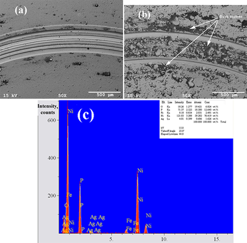
Images (SEM) of high temperature wear tracks of a Ni–P and b Ni–P–Ag coatings and c EDS analysis of dark region around wear scar of Ni–P–Ag coating
Conclusion
Entrapment of silver particles in Ni–P matrix could increase the lubrication and wear resistance of Ni–P coating at room and elevated temperatures.
Silver could diffuse and nucleate on the surface of Ni–P–Ag composite coating during high temperature sliding wear and it could also act as a self-lubricant material in nanocrystalline Ni–P–Ag composite coating.
Abrasive wear was dominant mechanism in degradation of heat treated Ni-P and Ni–P–Ag coatings.
Silver particles could reduce the fluctuations of friction coefficients and also, they decrease the average values of friction coefficients at room and high temperatures. So, Ni–P–Ag composite coating has the suitable capabilities in high temperature sliding applications such as aerospace industries.
Footnotes
Acknowledgements
The authors would like to thank Dr Kursat Kazmanli, Farideh Tabatabaei, Seyhan Atik and Sevgin Türkeli for helpful assistances in this work.