
Abstract
Using a plasma electrolytic oxidation (PEO) process, this study investigated how the pore size of the coating surface influences the growth of osteoblast cells and their morphology in pure titanium. With this aim, PEO coatings were applied using four different electric and electrolyte conditions to vary the mean pore sizes of the coatings from ∼2 to ∼5 μm. In vitro examinations showed that the osteoblast cells on the surface of the coating with ∼2 μm pores had grown healthily with good attachment by multiple pseudopodia connections; cells were highly proliferated, compared to those of other samples. In addition, the coating with ∼2 μm pores showed the highest cell viability among the samples.
Introduction
Pure titanium and its alloys have been widely used for orthopaedic and dental implants due to their high specific strength, excellent corrosion resistance and good biocompatibility in the human body. 1 Nevertheless, a passive layer of titanium in nature is bioinert, which leads to delayed bone/implant interlocking after surgical treatment. 2 Therefore, substantial attention has been paid to study the formation of pores and/or roughness on titanium implants; this structure plays a favourable role in enhancing osseointegration. 3 In this regard, surface modification techniques such as chemical etching, sand blasting, anodising, etc., have been extensively studied. 4 Among them, plasma electrolytic oxidation (PEO) coating has been highlighted as an eco friendly process that, by controlling the alkaline electrolytes, current density, coating time, waveform, etc., allows the formation of rough and porous oxide layers with complex geometry with ease.5,6
There has been much interest in porous biomaterials, which provide anchorage by favouring ingrowth of mineralised tissue and bone cells and also promote the cell orientation along the grooves and ridge. 7 In this context, numerous attempts have been made to demonstrate the effect of pore size on the bone growth and cell proliferation on scaffolds whose pore sizes are normally in the range of hundreds of micrometres. 8 However, little attention has been paid to cell responses to the pores, of which the size is in the range of a few micrometres, although micrometre size pores are very important for the cell extension; the pores, whose size is suitable for the interlocking of pseudopodia and adhesion of cell body, can readily promote cell proliferation and differentiation by its rapid growth. 9 Furthermore, no studies have so far been carried out to consider the pore size effect of the coating on the cell response in titanium via a PEO process. Therefore, in this study, four different porous coatings containing pores with ∼2, ∼3, ∼4 and ∼5 μm mean sizes were fabricated by PEO process; the variables were finely distinguished due to very subtle responses of cells to the microstructure. The present paper is devoted to the study of determining cellular performances of the micrometre pores by the in vitro cell examinations using MC3T3-E1 osteoblast bone cells.
Experimental
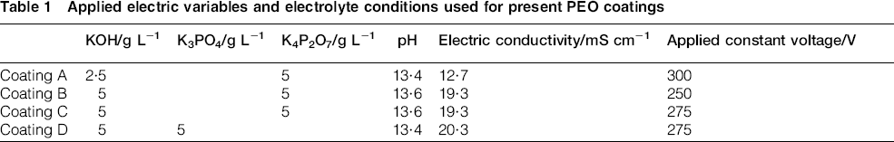
Commercially pure titanium (grade II) plates with size of 20 × 30 × 2 mm3 were used as substrate. Before PEO coating, titanium plates were ground with #200–1000 abrasive papers and cleaned in acetone. In order to consider solely the pore size effect avoiding the influences of coating thickness, roughness and porosity, we carefully selected PEO conditions among our experimental database, and the applied electric variables and electrolyte conditions are tabulated in Table 1. The AC power supply was connected to an electrolyte bath consisting of an acrylic vessel container with a sample holder and a cathode composed of stainless steel. To stabilise the electrochemical conditions during PEO coatings, the electrolyte temperature was maintained at 20–30°C.
Applied electric variables and electrolyte conditions used for present PEO coatings
The surface morphologies of each sample were observed by utilising a field emission scanning electron microscope (HITACHI S-4800). The phase components of the samples were measured by X-ray diffraction (XRD, RIGAKU D/MAX 2500) with a step size of 0.05° and a scan range of 20 to 60°. In vitro cell tests using osteoblast precursor cells (MC3T3-E1, ATCC, USA) were performed to examine the cell viability and proliferation. The precultured cells were seeded on samples with a cell density of 2 × 10⁴ mL− 1 and incubated in a humidified incubator in an atmosphere containing 5% CO2 at 37.5°C for up to 12, 24 and 48 h. Minimum essential medium (Gibco, USA) with 10% fetal bovine serum (Gibco, USA) was used as the culturing medium. To observe the cells attached to the samples, the cultured samples were placed in Dulbecco's phosphate buffered saline (Gibco, USA) and rinsed quickly. Then, samples were fixed in 4% glutaraldehyde at 4°C for 4 h. After cleaning three times in Dulbecco's phosphate buffered saline, the samples were dehydrated in a graded series of ethanol. The samples were dried in air and coated with gold to observe the SEM images.
Results and discussion
Figure 1 presents SEM images revealing the surface morphologies of coatings A, B, C and D. The mean pore sizes of each coating were measured to be about ∼2, ∼3, ∼4 and ∼5 μm respectively, while no appreciable distinction in the surface roughness and porosity was observed (Table 2). It is of interest to note that the circular pores and their distribution became irregular with increasing the pore size. This might be due to the fact that the microarcs tend to locally converged to reduce the electrical stress when the size of microdischarge channels, which trace to the pores in the coating, increases. 10 As listed in Table 1, the electric conductivity of the electrolyte producing coating D was nearly double of that producing coating A, so that, in the case of coating D, the electron avalanche at the interface between the electrolyte and coating was sustained for a relatively long time where microarcs once occurred. Moreover, highly intense microdischarges irregularly generated at local weak points of the coating to find the way through. As a result, the pores with irregular shape and size were distributed on the surface of the coating D, while the circular pores formed uniformly thorough out the surface of the coating A.
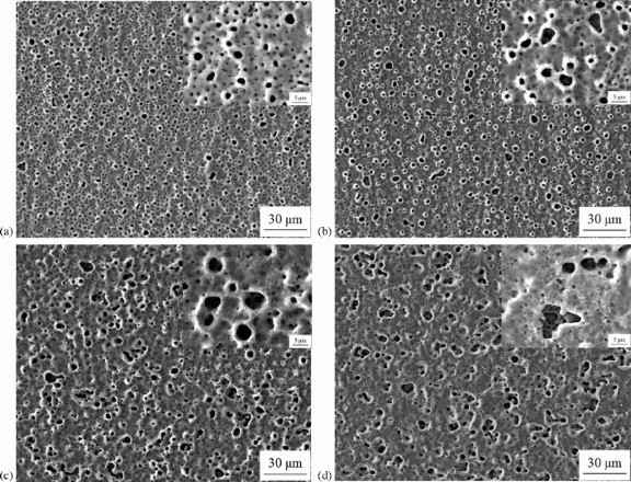
Images (SEM) showing surface morphologies of a coating A, b coating B, c coating C and d coating D; insets indicate high magnification images
Coating thickness, surface roughness and porosity of coatings A, B, C and D
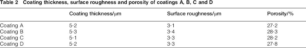
Figure 2 displays the XRD patterns of the coatings A, B, C and D. All coatings consisted of mainly titanium dioxide (anatase and rutile) and titanium, which were generally formed when the PEO coating is carried out in the phosphate electrolyte. 11 Here, peaks of titanium originated from titanium substrate due to relative thin coatings, which X-ray could penetrate with ease. If there are no specific metallic additives in the electrolyte except for the complexing agent such as potassium phosphate, titanium dioxide is formed in the coating by electrochemical reactions because the component of the coating solely depends on the electrolyte, regardless of applied voltage, current density, pH and coating time. Therefore, no remarkable difference was observed in patterns.
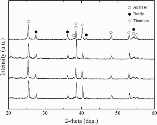
Patterns (XRD) of coatings A, B, C and D
Figure 3 shows the results of in vitro examinations with MC3T3-E1 osteoblast cells cultured for 12 h displaying cell morphology and distribution on the coating surfaces. According to the dark field images, the matured osteocyte cells seemed to adhere more firmly and conform to a larger extent on the surfaces of coatings A and B (Fig. 3a and b) revealing ∼2 and ∼3 m mean pore sizes respectively, whereas the elongated cells together with rounded cells were found on surfaces of coatings C and D (Fig. 3c and d). A close look at insets showing the bright field SEM images with high magnification reveals that the polygonally flattened cells with the firm attachment by multiple pseudopodia connections were extended over the surface of the coating A (Fig. 3a). Compared to the other samples, the cells cultured on the surface of the coating A exhibited the more spread out morphology, highly connected with the surface, and the pseudopodia were much more observable and longer. In the meantime, viewed in the light of the cell growth and spreading, though a part of cells cultured on the coating B can be seemingly regarded as having similar condition to the proliferated osteocyte cells on the coating A, their less pseudopodia connections and spreading morphology were observed in the high magnification images (Fig. 3b). On the other hand, the elongated thin cells with the immature growth spread over the surfaces of coatings C and D (Fig. 3c and d). It is mainly attributed to the fact that multiple pores with size of ∼2 μm are favourable for the growth, attachment and spreading of cells on the surface.
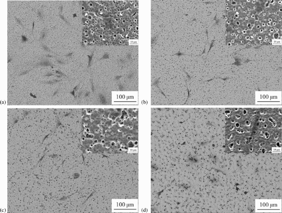
Dark field SEM images showing cell distributions of a coating A, b coating B, c coating C and d coating D; insets and arrows indicated bright field images with high magnification and pseudopodia respectively
Figure 4 exhibits the morphology of the cell cultured on the surface of the coating B where various size pores were coexisted. It is here important to note that the extension of pseudopodia was facilitated mainly along the ∼2 μm pores (Fig. 4a). It may be for this reason that the diameter of the end of pseudopodium was ∼2 μm so that the pseudopodium could readily stick into the pores to grow and spread of the cell body, as can be seen in Fig. 4b.
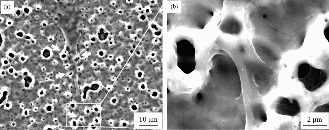
a images (SEM) showing cell morphology on coating B (arrows indicate pseudopodium), and b high magnification image of pseudopodium
Figure 5 shows the viability of cells cultured on the four different coatings. The cell viability of the coating A was the highest (P < 0.01) among coatings, and it decreased steadily with increasing the mean pore size. This strongly suggests that the fabrication of the coating containing ∼2 μm pores with uniform distribution on the surface plays a critical role in enhance the bioactivity of titanium coating via PEO process due to the suitable pore size for the attachment of pseudopodia, as aforementioned. Over all, what is important particularly from the in vitro point of view would be the coating manipulation of the mean pore size to be ∼2 μm with the uniform distribution for enhancing the osseointegration properties of the titanium implant.
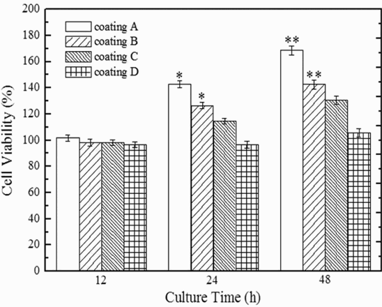
Viability of cells cultured on four different coatings for 12, 24 and 48 h; error bars mean standard deviation for n = 5, *P < 0.05, **P < 0.01
Conclusion
The influence of the pore size of the coating surface on osteoblast cell growth and morphology via PEO process was studied. The in vitro examinations resulted in the fact that, compared to the other samples, the osteoblast cells were well proliferated with multiple pseudopodia connections on the coating surface containing ∼2 μm pores distributed uniformly. These pores caused a significant increase in cell viability because they provided suitable site for the pseudopodia attachment to grow and proliferate of cells with ease. Thus, it is reasonable to conclude that the manipulation of the coating to form ∼2 μm pores on the surface uniformly is of particular significance to enhance bioactive properties. Furthermore, this result will be exploited to provide influential information in the fabrication of porous biomaterials using surface treatments forming micro pores as well as PEO technique.
Footnotes
Acknowledgements
This work was supported by the BK21 Plus project of the National Research Foundation of Korea Grant.